
Structure and Function
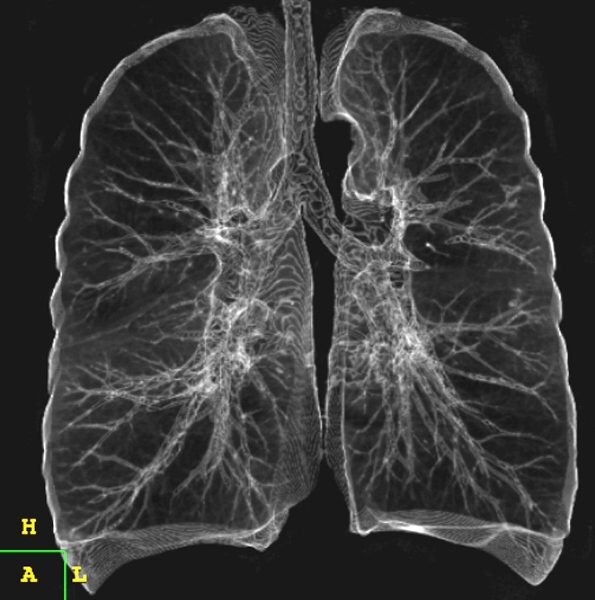
Pulmonary system provides the most intimate interface with the external environment . epresents the largest body surface area exposed to the environment
TheCommonVein.net 32679
Parts and Bonds
Ashley Davidoff MD
Size
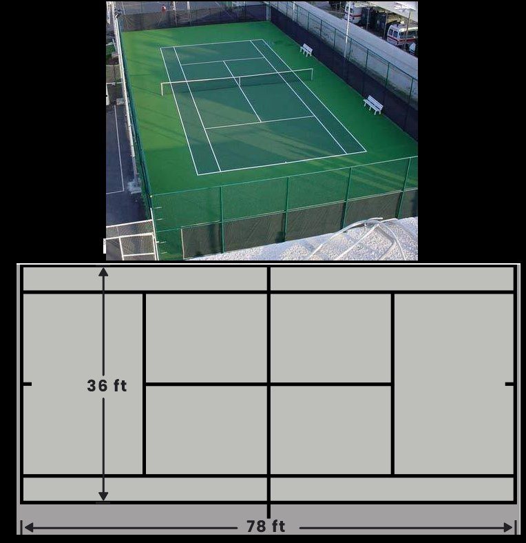
The surface area of the alveoli is equal to about 1/2 a tennis court. A tennis court is 78 by 36 feet equal to an area of 2,808 sq feet. Estimates for the surface area for the alveoli are between 800-1100 sq feet.
The skin in comparison of an adult is approximately 22 square feet
Each day, the lungs are exposed to 7,000 L of air and all it contains.
- At the level of the alveoli where gas exchange occurs, the biological barrier presents as an extremely attenuated interface composed of the
- surfactant
- 1 cell layer thick membranes
- alveolar lining and its lamina base
- endothelial lining and its fused basal lamina.
-

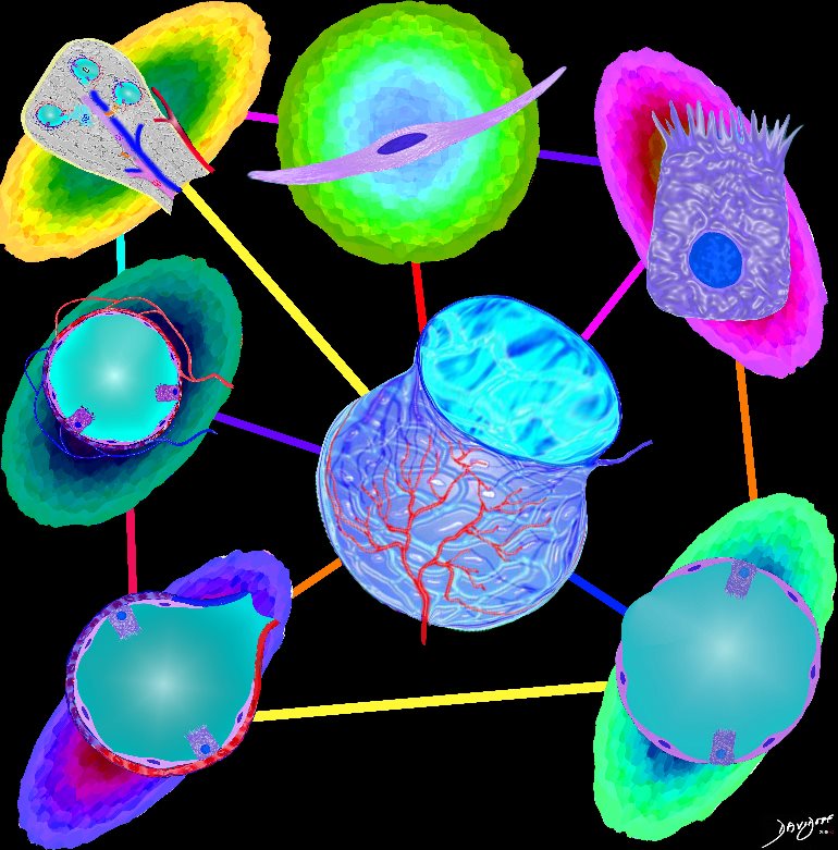
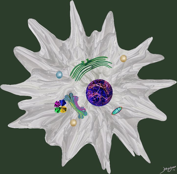
Cellular Makeup of the Normal Alveolus
The diagram shows the lining of the normal alveolus composed of type 1 pneumocyte squamous in nature and the cuboidal cell (type pneumocyte) which rest on a lamina propria, and basement membrane (not shown) shared with the inner endothelial layer of the capillary. Intra-alveolar macrophage lies within the alveolar lumen
Ashley Davidoff
TheCommonVein.net
- Surfactant
- first line of defense against immunological, biological and non-biological threats
- thickness
- less than 0.1 ?m
- major components are
- phospholipids – 90%
- proteins
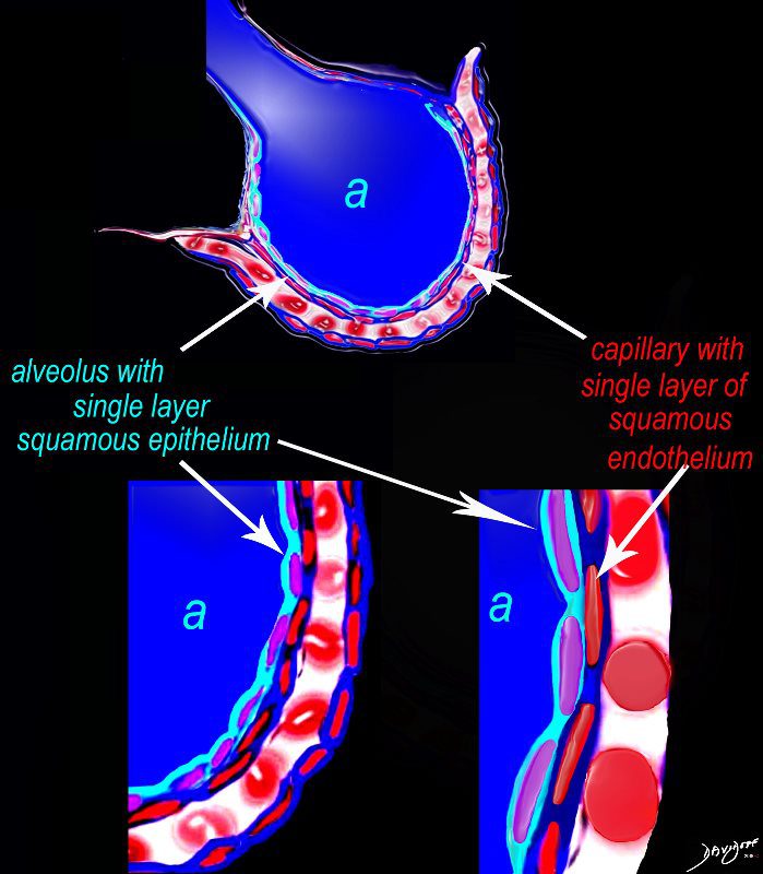
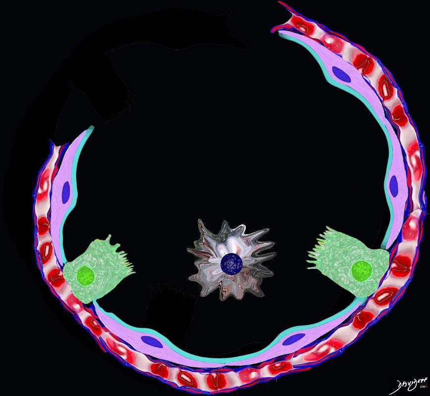
The diagram shows an alveolus (a) above, lined by a single layer of squamous cells, surrounded by a capillary with red cells which is also lined by a single layer of squamous endothelial cells . The images below show progressive magnification of the alveolar wall demonstrating the two thin layer of the alveolar membrane .
Courtesy Ashley Davidoff 2019
lungs-0028-low res
Cellular Environment
In order to protect this thin surface from the chemical and biological environment, the immune mechanisms at the interface have to be quite sophisticated There are about 40 different cell types have been described in the lining tissues, bronchial tree and respiratory tissues
Ashley Davidoff MD
The Buck Ends Here
The alveolus is lined by a simple epithelium ? one cell layer thick. There are two types of lining cells; Type 1 pneumocytes are squamous cells that cover 90% of the surface of the inner lining of the lung , and type II cuboidal pneumocytes that are in fact much more numerous than Type I. They are involved in the production of surfactant . In the lumen there are resident macrophages which play a crucial role in the immune system. The mucosa is grounded by a basement membrane and a lamina propria, and connected to the lamina propria and basement membrane of the surrounding capillary. The alveolus is lined by a thin layer of surfactant. (teal blue)
Ashley Davidoff
TheCommonVein.net