The Inverted Chest ? A Wineglass Ashley Davidoff TheCommonVein.net . 22071b01.800
The apex, also called the cupola is dome shaped, fitting snuggly into the space created by the soft tissue confluence of the mediastinal parietal pleura with the costal parietal pleura and the bony frame formed by the clavicle and first rib. It is positioned 2-3cm superior to the medial third of the clavicle where it projects through the superior thoracic inlet.
Cupola – shape of the apex This cupola or dome was photographed in the church of the Villa Melzi gardens in Bellagio, Italy. If you imagine yourself in the chest cavity and you look up towards the neck, this is what you will see ? the dome shaped structure of the apex of the lung and pleura. Ashley Davidoff TheCommonVein.net 78115pb01
Parts – Ribs and Spine
Kyphosis
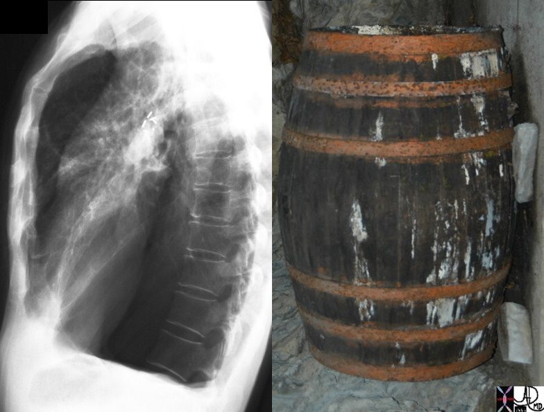
We are Going and You are Coming
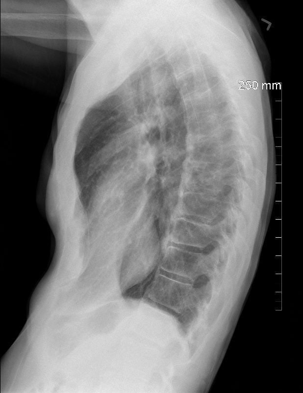
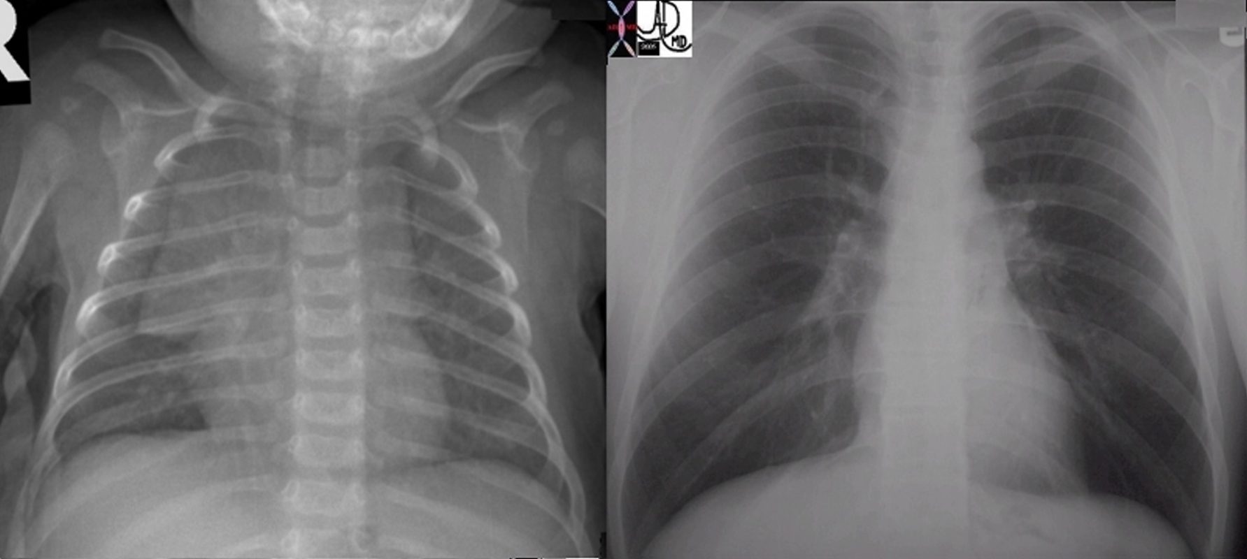
The top row of images from left to right reflect, A-P examination of the immature lumbar spine of very young patient, juxtaposed with the lateral examination of a normal thoracic of a normal young adult and lastly the lateral examination of a severely kyphotic elderly patient. The photograph was taken in Italy showing ages ranging from the youngest child in a stroller perhaps 2 years in age, her brother of about 5 or 6, their mother in her late twenties or early thirties and an elderly couple both suffering from the wraths of aging bones ? osteoporosis and severe kyphosis. (the kyphosis couple)
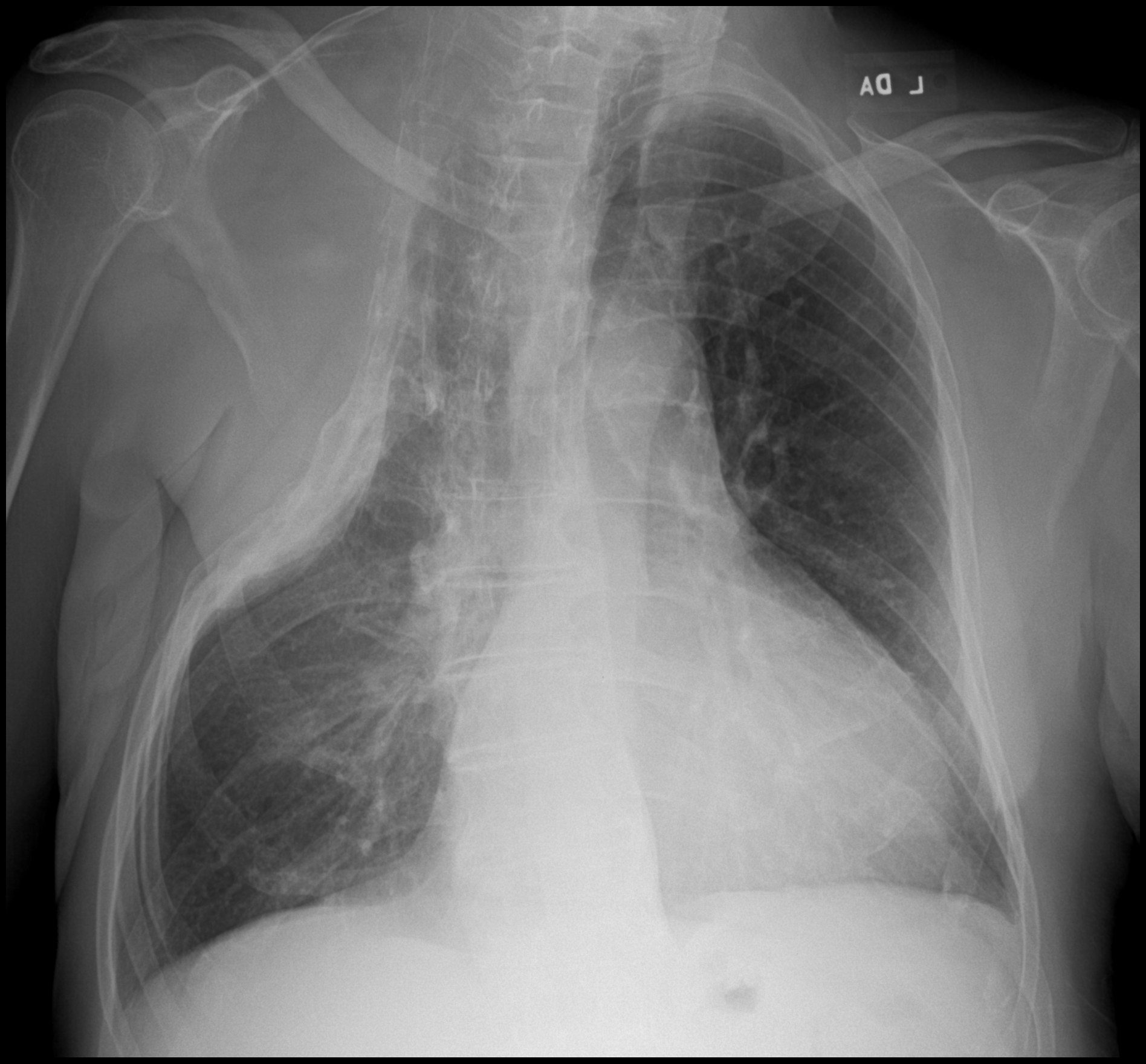
Frontal CXR – Miliary TB 60-year-old immunocompromise female presents with a cough and weight loss CXR shows a diffuse miliary pattern. Final diagnosis was mycobacterium tuberculosis. Associated findings include healed right sided rib fractures and surgical clips in the left axilla Ashley Davidoff MD TheCommonVein.net 265Lu 136197
Parts Lungs
Size Narrow -AP
Normal Thoracic Spine
73290c01 bone spine thoracic spine spinous processes normal interspinous ligament spine vertebral bodies CTscan Courtesy Ashley Davidoff MD
Narrow A-P and Spontaneous Pneumothorax
CXR Spontaneous Pneumothorax 20-year-old female presents with acute left sided chest pain. She has a narrow A-P diameter exemplified in the lateral projection (below) and the asthenic build raises the suspicion for spontaneous pneumothorax. Frontal CXR shows a small subtle pneumothorax characterised by a thin pleural line and relative lucency of the left apex compared to the right Ashley Davidoff MD TheCommonVein.net 117246c CXR Spontaneous Pneumothorax 20-year-old female presents with acute left sided chest pain. She has asthenic build which raises the suspicion for a spontaneous pneumothorax. Frontal CXR shows a small subtle pneumothorax characterised by a thin pleural line (b, white arrowhead) and relative lucency of the left apex Ashley Davidoff MD TheCommonVein.net 117246c01
Shape
Severe Pectus Excavatum
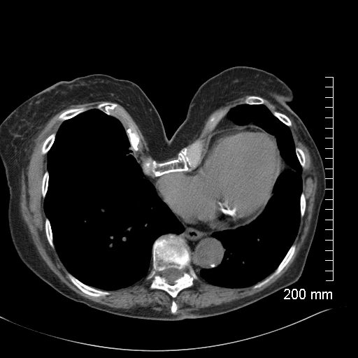
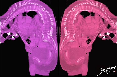
CXR Severe Pectus Excavatum and Dextrocardia 68-year-old female presents with a severe pectus excavatum. CXR in the frontal view shows horizontal orientation of the ribs and distortion of the right heart border Ashley Davidoff MD TheCommonVein.net 270Lu 121391 CXR Severe Pectus Excavatum 68-year-old female presents with a severe pectus excavatum. CXR in the lateral view shows significant depression of the sternum Ashley Davidoff MD TheCommonVein.net 270Lu 121392 CT – Severe Pectus Excavatum and Dextrocardia 68-year-old female presents with severe pectus excavatum. CT in the axial plain shows significant depression of the sternum, and dextrocardia Ashley Davidoff MD TheCommonVein.net 270Lu 121393 CT – Severe Pectus Excavatum 68-year-old female presents with severe pectus excavatum. CT in the sagittal plain shows significant depression of the lower sternum, compression fracture of a midthoracic vertebra and surgical hardware in a proximal vertebra Ashley Davidoff MD TheCommonVein.net 270Lu 121396 CT – Severe Pectus Excavatum 68-year-old female presents with severe pectus excavatum. 3D CT in the RAO projection shows significant depression of the sternum Ashley Davidoff MD TheCommonVein.net 270Lu 121397
Pectus Excavatum and Pectus Carinatum
Lateral CXR Shows Pectus Excavatum and Pectus Carinatum 66 year malnourished immunodeficient male with right upper lobe aspergilloma in the lung Lateral CXR shows pectus excavatum and pectus carinatum with flattened hemidiaphragms Ashley Davidoff TheCommonVein.net
Position Character
Time
Shape Changes with Time keywords lung chest thymus baby adult mediastinum normal anatomy applied biology CXR chest X-ray plain film time Ashley Davidoff MD TheCommonVein.net 6663c01
Associated Findings
Infection
DEFORMITY OF THE CHEST WALL FOLLOWING THERAPY FOR RUL TB 82 year old man s/p thoracoplasty for treatment of right upper lobe TB. Associated finding is left ventricular enlargement Ashley Davidoff MD TheCommonVein.net 42020
Pectus Carinatum Hyperinflation from Cystic Fibrosis –
19 year old female
Cystic Fibrosis and Bronchiectasis 19 year old female with cystic fibrosis and bronchiectasis CT scan through the upper lung fields shows multiple thickened and mucin filled subsegmental bronchi of the posterior segment of the right upper lobe Courtesy Priscilla Slanetz MD MPH TheCommonVein.net
Pectus Excavatum and Carinatum
Lateral CXR Shows Pectus Excavatum and Pectus Carinatum 66 year malnourished immunodeficient male with right upper lobe aspergilloma in the lung Lateral CXR shows pectus excavatum and pectus carinatum with flattened hemidiaphragms Ashley Davidoff TheCommonVein.net Frontal CXR with Right Upper Lobe Nodule 66 year malnourished immunodeficient male with right upper lobe aspergilloma in the lung Frontal CXR with Right Upper Lobe Nodule Ashley Davidoff TheCommonVein.net
Barrel Shaped Chest Pectus Carinatum
The lateral examination of the chest shows the classical barrel shape to the chest with an increase in the retrosternal air space and flattening of the #signs in medicine Ashley Davidoff MD TheCommonVein.net
DOMElement Object
(
    [schemaTypeInfo] =>
    [tagName] => table
    [firstElementChild] => (object value omitted)
    [lastElementChild] => (object value omitted)
    [childElementCount] => 1
    [previousElementSibling] => (object value omitted)
    [nextElementSibling] => (object value omitted)
    [nodeName] => table
    [nodeValue] =>


Normal Thoracic Spine


73290c01 bone spine thoracic spine spinous processes normal interspinous ligament spine vertebral bodies CTscan Courtesy Ashley Davidoff MD


    [nodeType] => 1
    [parentNode] => (object value omitted)
    [childNodes] => (object value omitted)
    [firstChild] => (object value omitted)
    [lastChild] => (object value omitted)
    [previousSibling] => (object value omitted)
    [nextSibling] => (object value omitted)
    [attributes] => (object value omitted)
    [ownerDocument] => (object value omitted)
    [namespaceURI] =>
    [prefix] =>
    [localName] => table
    [baseURI] =>
    [textContent] =>


Normal Thoracic Spine


73290c01 bone spine thoracic spine spinous processes normal interspinous ligament spine vertebral bodies CTscan Courtesy Ashley Davidoff MD


)
DOMElement Object
(
    [schemaTypeInfo] =>
    [tagName] => td
    [firstElementChild] =>
    [lastElementChild] =>
    [childElementCount] => 0
    [previousElementSibling] =>
    [nextElementSibling] =>
    [nodeName] => td
    [nodeValue] => 73290c01 bone spine thoracic spine spinous processes normal interspinous ligament spine vertebral bodies CTscan Courtesy Ashley Davidoff MD
    [nodeType] => 1
    [parentNode] => (object value omitted)
    [childNodes] => (object value omitted)
    [firstChild] => (object value omitted)
    [lastChild] => (object value omitted)
    [previousSibling] => (object value omitted)
    [nextSibling] => (object value omitted)
    [attributes] => (object value omitted)
    [ownerDocument] => (object value omitted)
    [namespaceURI] =>
    [prefix] =>
    [localName] => td
    [baseURI] =>
    [textContent] => 73290c01 bone spine thoracic spine spinous processes normal interspinous ligament spine vertebral bodies CTscan Courtesy Ashley Davidoff MD
)
DOMElement Object
(
    [schemaTypeInfo] =>
    [tagName] => td
    [firstElementChild] => (object value omitted)
    [lastElementChild] => (object value omitted)
    [childElementCount] => 1
    [previousElementSibling] =>
    [nextElementSibling] =>
    [nodeName] => td
    [nodeValue] => Normal Thoracic Spine
    [nodeType] => 1
    [parentNode] => (object value omitted)
    [childNodes] => (object value omitted)
    [firstChild] => (object value omitted)
    [lastChild] => (object value omitted)
    [previousSibling] => (object value omitted)
    [nextSibling] => (object value omitted)
    [attributes] => (object value omitted)
    [ownerDocument] => (object value omitted)
    [namespaceURI] =>
    [prefix] =>
    [localName] => td
    [baseURI] =>
    [textContent] => Normal Thoracic Spine
)
DOMElement Object
(
    [schemaTypeInfo] =>
    [tagName] => td
    [firstElementChild] => (object value omitted)
    [lastElementChild] => (object value omitted)
    [childElementCount] => 1
    [previousElementSibling] =>
    [nextElementSibling] =>
    [nodeName] => td
    [nodeValue] => 75675c04
    [nodeType] => 1
    [parentNode] => (object value omitted)
    [childNodes] => (object value omitted)
    [firstChild] => (object value omitted)
    [lastChild] => (object value omitted)
    [previousSibling] => (object value omitted)
    [nextSibling] => (object value omitted)
    [attributes] => (object value omitted)
    [ownerDocument] => (object value omitted)
    [namespaceURI] =>
    [prefix] =>
    [localName] => td
    [baseURI] =>
    [textContent] => 75675c04
)
DOMElement Object
(
    [schemaTypeInfo] =>
    [tagName] => table
    [firstElementChild] => (object value omitted)
    [lastElementChild] => (object value omitted)
    [childElementCount] => 1
    [previousElementSibling] => (object value omitted)
    [nextElementSibling] => (object value omitted)
    [nodeName] => table
    [nodeValue] =>


We are Going and You are Coming


The top row of images from left to right reflect, A-P examination of the immature lumbar spine of very young patient, juxtaposed with the lateral examination of a normal thoracic of a normal young adult and lastly the lateral examination of a severely kyphotic elderly patient. The photograph was taken in Italy showing ages ranging from the youngest child in a stroller perhaps 2 years in age, her brother of about 5 or 6, their mother in her late twenties or early thirties and an elderly couple both suffering from the wraths of aging bones ? osteoporosis and severe kyphosis. (the kyphosis couple)
Ashley Davidoff Copyright 2011 75578c01.8s


    [nodeType] => 1
    [parentNode] => (object value omitted)
    [childNodes] => (object value omitted)
    [firstChild] => (object value omitted)
    [lastChild] => (object value omitted)
    [previousSibling] => (object value omitted)
    [nextSibling] => (object value omitted)
    [attributes] => (object value omitted)
    [ownerDocument] => (object value omitted)
    [namespaceURI] =>
    [prefix] =>
    [localName] => table
    [baseURI] =>
    [textContent] =>


We are Going and You are Coming


The top row of images from left to right reflect, A-P examination of the immature lumbar spine of very young patient, juxtaposed with the lateral examination of a normal thoracic of a normal young adult and lastly the lateral examination of a severely kyphotic elderly patient. The photograph was taken in Italy showing ages ranging from the youngest child in a stroller perhaps 2 years in age, her brother of about 5 or 6, their mother in her late twenties or early thirties and an elderly couple both suffering from the wraths of aging bones ? osteoporosis and severe kyphosis. (the kyphosis couple)
Ashley Davidoff Copyright 2011 75578c01.8s


)
DOMElement Object
(
    [schemaTypeInfo] =>
    [tagName] => td
    [firstElementChild] => (object value omitted)
    [lastElementChild] => (object value omitted)
    [childElementCount] => 2
    [previousElementSibling] =>
    [nextElementSibling] =>
    [nodeName] => td
    [nodeValue] =>
The top row of images from left to right reflect, A-P examination of the immature lumbar spine of very young patient, juxtaposed with the lateral examination of a normal thoracic of a normal young adult and lastly the lateral examination of a severely kyphotic elderly patient. The photograph was taken in Italy showing ages ranging from the youngest child in a stroller perhaps 2 years in age, her brother of about 5 or 6, their mother in her late twenties or early thirties and an elderly couple both suffering from the wraths of aging bones ? osteoporosis and severe kyphosis. (the kyphosis couple)
Ashley Davidoff Copyright 2011 75578c01.8s

    [nodeType] => 1
    [parentNode] => (object value omitted)
    [childNodes] => (object value omitted)
    [firstChild] => (object value omitted)
    [lastChild] => (object value omitted)
    [previousSibling] => (object value omitted)
    [nextSibling] => (object value omitted)
    [attributes] => (object value omitted)
    [ownerDocument] => (object value omitted)
    [namespaceURI] =>
    [prefix] =>
    [localName] => td
    [baseURI] =>
    [textContent] =>
The top row of images from left to right reflect, A-P examination of the immature lumbar spine of very young patient, juxtaposed with the lateral examination of a normal thoracic of a normal young adult and lastly the lateral examination of a severely kyphotic elderly patient. The photograph was taken in Italy showing ages ranging from the youngest child in a stroller perhaps 2 years in age, her brother of about 5 or 6, their mother in her late twenties or early thirties and an elderly couple both suffering from the wraths of aging bones ? osteoporosis and severe kyphosis. (the kyphosis couple)
Ashley Davidoff Copyright 2011 75578c01.8s

)
DOMElement Object
(
    [schemaTypeInfo] =>
    [tagName] => td
    [firstElementChild] => (object value omitted)
    [lastElementChild] => (object value omitted)
    [childElementCount] => 2
    [previousElementSibling] =>
    [nextElementSibling] =>
    [nodeName] => td
    [nodeValue] =>
We are Going and You are Coming

    [nodeType] => 1
    [parentNode] => (object value omitted)
    [childNodes] => (object value omitted)
    [firstChild] => (object value omitted)
    [lastChild] => (object value omitted)
    [previousSibling] => (object value omitted)
    [nextSibling] => (object value omitted)
    [attributes] => (object value omitted)
    [ownerDocument] => (object value omitted)
    [namespaceURI] =>
    [prefix] =>
    [localName] => td
    [baseURI] =>
    [textContent] =>
We are Going and You are Coming

)


 75675c04
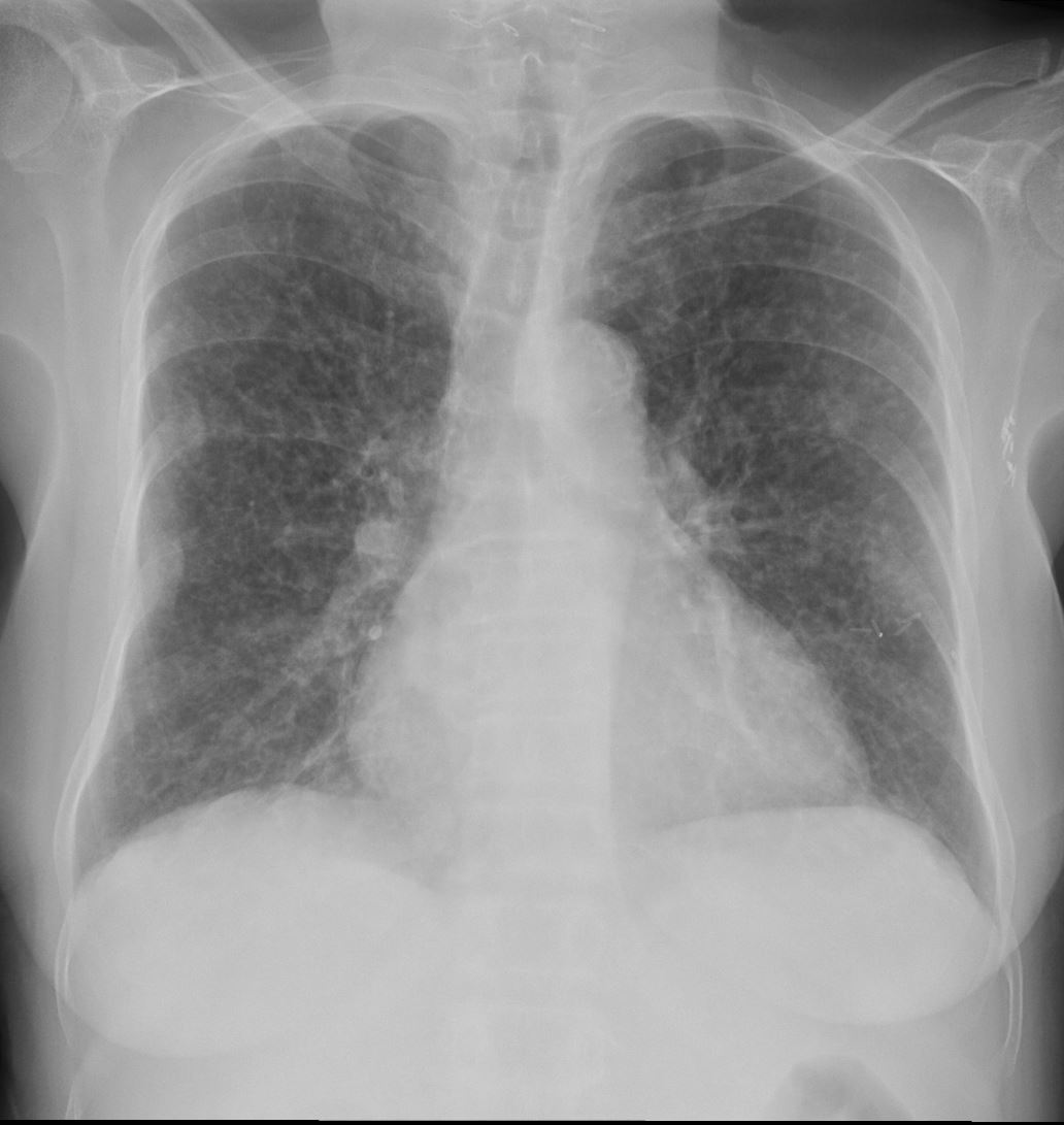
 Normal Thoracic Spine