
- 65-year old male with smoking history presents with
- chest/back pain, myalgias, night sweats
- History
- prior HCV/HBV, COPD, heavy EtOH use, anxiety, depression
CXR Mediastinal Widening and Hilum Overlay Sign
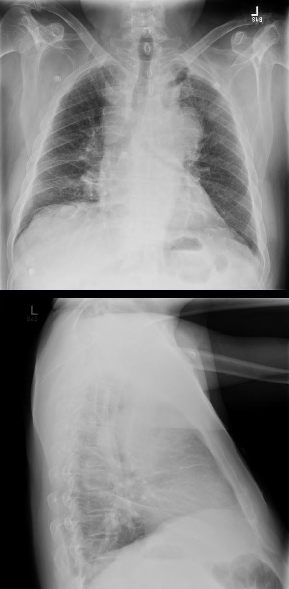
65 year old male presents with a history of chest pain, back pain, myalgias, and night sweats
Frontal CXR shows a left sided mass that silhouettes the aortic knob and the main pulmonary artery indicating that the mass arises from middle mediastinal tumors, hilar adenopathy, or a large pericardial effusion.
The lateral exam suggests an anterior mediastinal soft tissue fullness.
A diagnosis of a large mediastinal small cell carcinoma of the lung was diagnosed
Ashley Davidoff MD TheCommonVein.net 297Lu 136689

65 year old male presents with a history of chest pain, back pain, myalgias, and night sweats
Frontal CXR shows a left sided mass that silhouettes the aortic knob and the main pulmonary artery indicating that the mass arises from middle mediastinal tumors, hilar adenopathy, or a large pericardial effusion – hilum overlay sign.
A diagnosis of a large mediastinal small cell carcinoma of the lung was diagnosed
Ashley Davidoff MD TheCommonVein.net 297Lu 136690c

65 year old male presents with a history of chest pain, back pain, myalgias, and night sweats
Frontal CXR shows a left sided mass that silhouettes the aortic knob and the main pulmonary artery (white arrowhead, b) indicating that the mass arises from middle mediastinal tumors, hilar adenopathy, or a large pericardial effusion. The mass also compresses and displaces the left mainstem (teal arrowhead b) and confirmed in d.
The coronal CT scan (d) confirms the presence of a large mediastinal mass that extends beyond the aortic knob (white arrowhead)
A diagnosis of a large mediastinal small cell carcinoma of the lung was diagnosed
Ashley Davidoff MD TheCommonVein.net 297Lu 136690cL

65 year old male presents with a history of chest pain, back pain, myalgias, and night sweats
Frontal CXR shows a left sided mass that silhouettes the aortic knob and the main pulmonary artery (white arrowhead, b) indicating that the mass arises from middle mediastinal tumors, or hilar adenopathy. The mass also compresses and displaces the left mainstem bronchus (teal arrowhead b).
The axial CT (c) shows th extent of the mass and the coronal CT scan (d) confirms the presence of a large mediastinal mass that extends beyond the aortic knob (white arrowhead)
A diagnosis of a large mediastinal small cell carcinoma of the lung was diagnosed
Ashley Davidoff MD TheCommonVein.net 297Lu 136691cL
-
- CT
- Large heterogeneously enhancing mass centered on the left
hilum measuring approximately 8.1 x 11.6 x 6.4 cm - extends into the pre-vascular, AP window, pre-tracheal, subcarinal, and right hilar spaces.
- encases the left main pulmonary artery with
narrowing of the left upper and lower lobar pulmonary arteries. - area of narrowing in the right apical/anterior segmental pulmonary arteries.
- Adenopathy:
- Supraclavicular lymphadenopathy bilaterally,
- Left axillary lymphadenopathy measuring up to 1.7 cm.
- Large heterogeneously enhancing mass centered on the left
- CT

65 year old male presents with a history of chest pain, back pain, myalgias, and night sweats
CT scan in multiple planes shows a left sided mass that extends from the left hilum to the mediastinum and to theright hilum. The mass compressesthe SVC, (blue arrowhead a), narrows rthe left mainstem bronchus (teal arrowhead, a) and encases the left pulmonary artery.the left mainstem bronchus (teal arrowhead b).
A diagnosis of a large mediastinal small cell carcinoma of the lung was diagnosed
Ashley Davidoff MD TheCommonVein.net 297Lu 136692cL

65 year old male presents with a history of chest pain, back pain, myalgias, and night sweats
CT scan in multiple planes shows a left sided nodule (green arrowhead b, c, and d) with associated ground glass changes, that was thought to represent the primary tumor.
However the PET scan shows no activity in the nodule (green arrowhead f).
A diagnosis of a large mediastinal small cell carcinoma of the lung was diagnosed
Ashley Davidoff MD TheCommonVein.net 297Lu 136693cL
-
- Stage IV SCLC
- Mets to many LN, bone marrow, pleura, and >25 brain lesions

65 year old male presents with a history of chest pain, back pain, myalgias, and night sweats
PET CT scan in multiple planes shows extensive hypermetabolic activity throughout the mediastinum and extending into the right cervical chain, subpectoral region and axilla on the left.
A diagnosis of a large mediastinal small cell carcinoma of the lung with extensive regional metastases was diagnosed
Ashley Davidoff MD TheCommonVein.net 297Lu 136696

65 year old male presents with a history of chest pain, back pain, myalgias, and night sweats
MRI of the brain in the axial plane shows smal multifocal enhancing lesions throughout the brain on the T1 FLAIR post gadolinium images (upper panels) and on the T2 Flair sequence (lower image)
A diagnosis of a large mediastinal small cell carcinoma of the lung metastatic to the brain was diagnosed
Ashley Davidoff MD TheCommonVein.net 297Lu 136697

65 year old male presents with a history of chest pain, back pain, myalgias, and night sweats
Sagittal CT of the spine shows smal multifocal blastic metastases
A diagnosis of a large mediastinal small cell carcinoma of the lung metastatic to bone was diagnosed
Ashley Davidoff MD TheCommonVein.net 297Lu 136697b
Chemotherapy

Coronal CT of the chest through the mediastinum at the level of the carina before (a) and 7months following chemotherapy, shows significant reduction of the extensive mediastinal and hilar tumor
Ashley Davidoff MD TheCommonVein.net 297Lu 136698
911 Call Found down in the field.

CT of the brain in the axial projection shows a large intracerebral hemorrhagein the right parietal lobewhich was a terminal event for this 65 year old male with metastatic stage IV small cell carcinoma of the lung