
- 46 year healthy F, never smoker
- Clinical
- with cough, dyspnea
- without hypoxemia
- afebrile
- in the setting of
- URI symptoms 2-3 weeks prior
- (COVID Ag neg x3 one home testing).
- viral prodrome prior
- subjective fevers, cough, myalgias)
- improving after which she developed DOE and chest pressure 2-3 days prior to presentation.
- Labs prior
- Prior
- peripheral eosinophilia (> 1k),
- negative infectious work up (PCRs for Flu, COVID and RSV, negative PCT,
- without leukocytosis.
- Prior
- BAL studies:
– cell count 576, 2000 RBC, 37% lymph, 32% eos
– cytology no tumor cells, abundant macrophages and eosinophils
– bacterial, AFB, fungal cx neg
– CD4/CD8 ratio 2.22. -
- Imaging
- CXR and CT chest imaging with diffuse consolidative opacities
- peripheral,
- UL predominant and
- septal thickening
- CXR and CT chest imaging with diffuse consolidative opacities
- Imaging
- URI symptoms 2-3 weeks prior
- Clinical
CXR from 6-months-ago at presentation showing -upper-lobe-peripheral-infiltrates- left more involved than the right

Subsequent diagnosis by BAL of chronic eosinophilic pneumonia (CEP)
Ashley Davidoff TheCommonVein.net
CT from 6-months-ago at presentation showing -upper-lobe-peripheral-infiltrates- left more involved than the right

CT scan in the coronal performed 6 months ago at the time of clinical presentation shows upper lobe predominant peripheral infiltrates with small left lower lobe peripheral infiltrate Subsequent diagnosis by BAL of chronic eosinophilic pneumonia (CEP)
Ashley Davidoff TheCommonVein.net
Interlobular Septal Thickening

CT scan in the axial plane performed 6 months ago at the time of clinical presentation shows apical interlobular septal thickening, a finding characteristic of chronic eosinophilic pneumonia (CEP) in the appropriate clinical setting. Subsequent diagnosis by BAL of chronic eosinophilic pneumonia (CEP)
Ashley Davidoff TheCommonVein.net
Interlobular Septal Thickening Some Centrilobular Nodules and Peripheral Upper Lobe Consolidation

CT scan in the axial plane performed 6 months ago at the time of clinical presentation, shows upper lobe interlobular septal thickening, and peripheral consolidations which are findings characteristic of chronic eosinophilic pneumonia (CEP) in the appropriate clinical setting. Subsequent diagnosis by BAL of chronic eosinophilic pneumonia (CEP) was made
Ashley Davidoff TheCommonVein.net

CT scan in the axial plane performed 6 months ago at the time of clinical presentation, shows upper lobe interlobular septal thickening, and peripheral consolidations which are findings characteristic of chronic eosinophilic pneumonia (CEP) in the appropriate clinical setting. Note of prominent centrilobular nodule likely reflects small airway involvement Subsequent diagnosis by BAL of chronic eosinophilic pneumonia (CEP) was made
Ashley Davidoff TheCommonVein.net

CT scan in the coronal performed 6 months ago at the time of clinical presentation shows upper lobe predominant peripheral infiltrates. Subsequent diagnosis by BAL of chronic eosinophilic pneumonia (CEP)
Ashley Davidoff TheCommonVein.net
Interlobular Septal Thickening Some Centrilobular Nodules Peripheral Upper Lobe Consolidation and Air Bronchograms

CT scan in the coronal performed 6 months ago at the time of clinical presentation shows upper lobe predominant peripheral infiltrates more prominent in the left upper lobe. Subsequent diagnosis by BAL of chronic eosinophilic pneumonia (CEP) was made
Ashley Davidoff TheCommonVein.net

CT scan in the coronal performed 6 months ago at the time of clinical presentation shows upper lobe predominant peripheral infiltrates more prominent in the left upper lobe. Subsequent diagnosis by BAL of chronic eosinophilic pneumonia (CEP) was made
Ashley Davidoff TheCommonVein.net

CT scan in the coronal performed 6 months ago at the time of clinical presentation shows upper lobe predominant peripheral infiltrates more prominent in the left upper lobe. Subsequent diagnosis by BAL of chronic eosinophilic pneumonia (CEP) was made
Ashley Davidoff TheCommonVein.net
Interlobular Septal Thickening Some Centrilobular Nodules Peripheral Upper Lobe Consolidation and Bronchial Wall Thickening


CT scan in the coronal performed 6 months ago at the time of clinical presentation shows upper lobe predominant peripheral infiltrates more prominent in the left upper lobe and bronchial wall thickening best visualized in the right upper lobe. Subsequent diagnosis by BAL of chronic eosinophilic pneumonia (CEP) was made
Ashley Davidoff TheCommonVein.net
Upper Lobe Peripheral Infiltrates Left Greater than Right

CT scan in the coronal performed 6 months ago at the time of clinical presentation shows upper lobe predominant peripheral infiltrates more prominent in the left upper. Subsequent diagnosis by BAL of chronic eosinophilic pneumonia (CEP) was made
Ashley Davidoff TheCommonVein.net

CT scan in the coronal performed 6 months ago at the time of clinical presentation shows upper lobe predominant peripheral infiltrates more prominent in the left upper. Subsequent diagnosis by BAL of chronic eosinophilic pneumonia (CEP) was made
Ashley Davidoff TheCommonVein.net
Smaller Right Lobe Subsegmental Infiltrate and No Infiltrates in the Left Lower Lobe

CT scan in the coronal performed 6 months ago at the time of clinical presentation shows a subsegmental left lower lobe peripheral infiltrate. Subsequent diagnosis by BAL of chronic eosinophilic pneumonia (CEP) was made
Ashley Davidoff TheCommonVein.net
Mediastinal Adenopathy

CT scan in the axial plane performed 6 months ago at the time of clinical presentation shows an enlarged A-P window lymph node likely reactive. Subsequent diagnosis by BAL of chronic eosinophilic pneumonia (CEP) was made
Ashley Davidoff TheCommonVein.net
1 Month Later Following Medical Therapy

CXR- in the frontal projection shows improved but persistent upper-lobe-peripheral infiltrates, left more involved than the right following 1 month of therapy
Ashley Davidoff TheCommonVein.net
6 Months After Initial Presentation Following Medical Therapy

CT scout in the AP projection shows resolution of infiltrates 6 months after medical therapy
Diagnosis: Treated chronic eosinophilic pneumonia (CEP)
Ashley Davidoff TheCommonVein.net
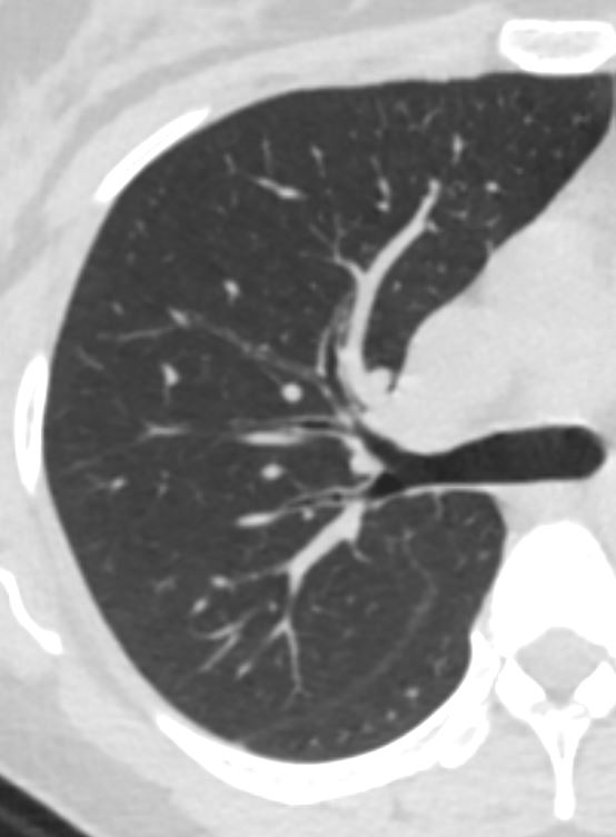
Normal CT of the Upper Lobes 6 Months Later Following Medical Therapy

CT scan in the axial projection at the level of the upper lobes shows resolution of infiltrates 6 months after medical therapy
Diagnosis: Treated chronic eosinophilic pneumonia (CEP)
Ashley Davidoff TheCommonVein.net

CT scan in the coronal projection showing upper middle and lower lobes revealing resolution of infiltrates 6 months after medical therapy
Diagnosis: Treated chronic eosinophilic pneumonia (CEP)
Ashley Davidoff TheCommonVein.net
Residual Bronchial Wall Thickening
CT scan in the axial projection at the level of the upper lobes shows residual mild bronchial w all thickening 6 months after medical therapy
Diagnosis: Treated chronic eosinophilic pneumonia (CEP)
Ashley Davidoff TheCommonVein.net