
- 30 y/o F with
- PMhx of opiate use disorder
- asthma, hep C (treated) who
- presents with s
- chronic productive cough,
- weight loss
- high volume hemoptysis.
- CXR and CT
- #LLL pneumonia with superimposed pulmonary abscess
#Hemoptysis LLL pneumonia with superimposed 2 cm cavitation. - mycotic pseudoaneurysm noted
- likely septic emboli in the context of her history of IVDU.
- #LLL pneumonia with superimposed pulmonary abscess
Cavitating Left Lower Lobe Pneumonia

30-year-old female with a history of IVDU presents with a a fever and hemoptysis.
Chest X-ray in the frontal view (right upper image and magnified in the right lower image) shows left lower lobe pneumonia with a 21 mm cavity in the anterior segment of the right lower lobe (green overlay). The lateral examination (upper left and magnified in lower left image) confirms the presence of a cavitating pneumonia in the anterior segment of the left lower lobe (cavity, green overlay lower image)
Ashley Davidoff MD TheCommonVein.net 281Lu 136523a01
Cavitating Left Lower Lobe Pneumonia
Surrounding Hemorrhage or Edema

30-year-old female with a history of IVDU presents with a fever and hemoptysis.
CT in the axial plane shows a cavitating pneumonia in the anterior segment of the left lower lobe. There are groundglass changes surrounding the consolidation representing either edema or hemorrhage
Ashley Davidoff MD TheCommonVein.net 281Lu 136524
Cavitating Left Lower Lobe Pneumonia with
Mycotic Pseudoaneurysm (PSA)

30-year-old female with a history of IVDU presents with a fever and hemoptysis.
CT in the axial plane shows a cavitating pneumonia in the anterior segment of the left lower lobe (a,b white arrowheads). There are groundglass changes surrounding the consolidation representing either edema or hemorrhage (b pink arrowhead). A focally dilated artery in the consolidation (red arrowheads (c, and d) represents a mycotic aneurysm
Ashley Davidoff MD TheCommonVein.net 281Lu 136526cL

30-year-old female with a history of IVDU presents with a fever and hemoptysis.
CT in the sagittal plane shows a cavitating pneumonia in the anterior segment of the left lower lobe. A focally dilated artery in the consolidation (red arrowheads a, and b) represents a mycotic aneurysm. There are groundglass changes surrounding the consolidation representing either edema or hemorrhage
Ashley Davidoff MD TheCommonVein.net 281Lu 136530cL
Cavitating Left Lower Lobe Pneumonia with Pseudoaneurysm (PSA) Pre and Post Embolization
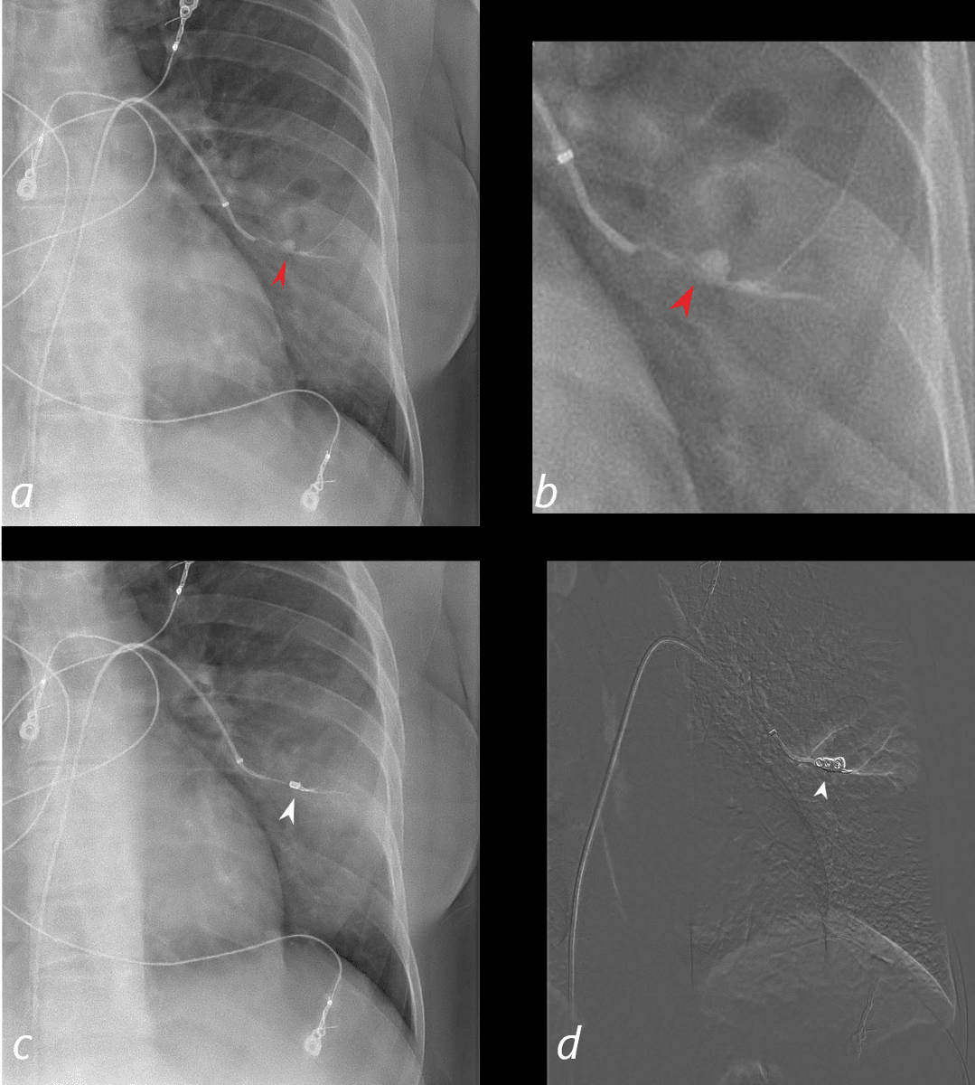
30-year-old female with a history of IVDU presents with a fever and hemoptysis.
Selective angiography of the anterior subsegmental artery of the left lower lobe shows a mycotic pseudoaneurysm (a,b red arrowhead) in the region of a cavitating pneumonia with noted air filled cavity (yellow arrowheads b) reflecting cavitating pneumonia. Images c and d show the embolization coil ((white arrowhead in c and d) with obliteration of the PSA.
Ashley Davidoff MD TheCommonVein.net 281Lu 136532cL