
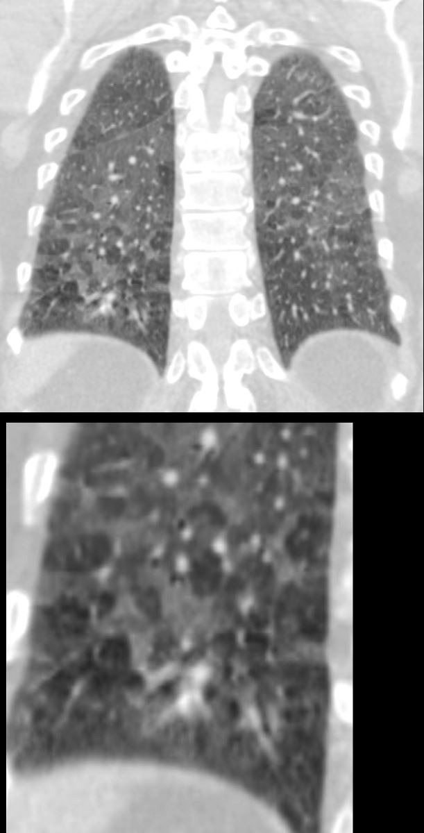
60-year-old male smoker with a history of progressive dyspnea. Coronal CT through the posterior lung fields at the level of the vertebral column shows extensive patchy ground glass changes and mosaic attenuation. A few of the secondary lobules show prominent centrilobular nodules reflecting a small airways component but the predominant pattern is an alveolar pattern
Pathology confirmed a diagnosis of DIP
Ashley Davidoff MD TheCommonVein.net 253Lu 136014c
Emphysema with Basilar Ground Glass Changes

51-year-old female smoker with a history of COPD asthma and pulmonary hypertension presents with progressive dyspnea. Frontal chest Xray shows diffuse bilateral interstitial disease characterised by coarsening of the lung markings and an enlarged main pulmonary artery (MPA). Path confirmed diagnosis of DIP.
Ashley Davidoff MD TheCommonVein.net 252Lu 135957
Mild Patchy Ground Glass Changes
Lower Lobe Predominant

51-year-old female smoker with a history of COPD asthma and pulmonary hypertension presents with progressive dyspnea. Axial CT through the lower lung fields shows patchy ground glass changes in the middle lobe inferior ligula and lower lobes and some regions of mosaicism. Focal regions of interlobular septal thickening are noted left lower lobe (lower panel). Pathology confirmed a diagnosis of DIP
Ashley Davidoff MD TheCommonVein.net 252Lu 135969c

51-year-old female smoker with a history of COPD asthma and pulmonary hypertension presents with progressive dyspnea. Coronal CT through the mid lung shows upper lobe centrilobular emphysematous disease and patchy ground glass changes in the lower lobes
Pathology confirmed a diagnosis of DIP
Ashley Davidoff MD TheCommonVein.net 252Lu 135990

51-year-old female smoker with a history of COPD asthma and pulmonary hypertension presents with progressive dyspnea. Sagittal CT through the lateral right mid lung shows upper lobe posterior and superior segmental centrilobular emphysematous disease and ground glass changes in the anterior segment of the upper lobe and right lower lobe
Pathology confirmed a diagnosis of DIP
Ashley Davidoff MD TheCommonVein.net 252Lu 135998
Crazy Paving

51-year-old female smoker with a history of COPD asthma and pulmonary hypertension presents with progressive dyspnea. Coronal CT through the posterior lungs shows diffuse ground glass changes in the lower lobes with interlobular septal thickening
Pathology confirmed a diagnosis of DIP
Ashley Davidoff MD TheCommonVein.net 252Lu 135997

51-year-old female smoker with a history of COPD asthma and pulmonary hypertension presents with progressive dyspnea. Axial CT through the right posterior recesses at end inspiration (upper panel) and end expiration (lower panel) confirms the presence of air trapping indicating the presence of mild small airway disease
Pathology confirmed a diagnosis of DIP
Ashley Davidoff MD TheCommonVein.net 252Lu 135987