
58-year-old female presents with a cough
Frontal CXR –
Silhouetting Left Heart Border – Lingula Atelectasis

58-year-old female presents with a cough Frontal CXR shows silhouetting of the left heart border with hazy or veiling opacity extending out from the left hilum and fading out inferiorly . The left hilum is pulled superiorly, resulting in an almost horizontal course of the left main bronchus and vertical orientation of the left lower lobe bronchus
Ashley Davidoff MD TheCommonVein.net 257Lu 136109
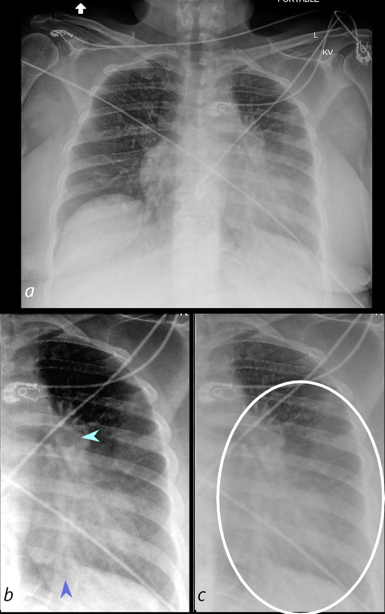
58-year-old female presents with a cough Frontal CXR shows silhouetting of the left heart border with hazy or veiling opacity extending out from the left hilum and fading out inferiorly (white circle c). The left hilum is pulled superiorly (teal arrowhead b) , resulting in an almost horizontal course of the left main bronchus and vertical orientation of the left lower lobe bronchovascular bundle (dark blue arrowhead b)
Ashley Davidoff MD TheCommonVein.net 257Lu 136109cL01
CT Lingula Atelectasis
Silhouetting of the Left Heart Border
Aerated
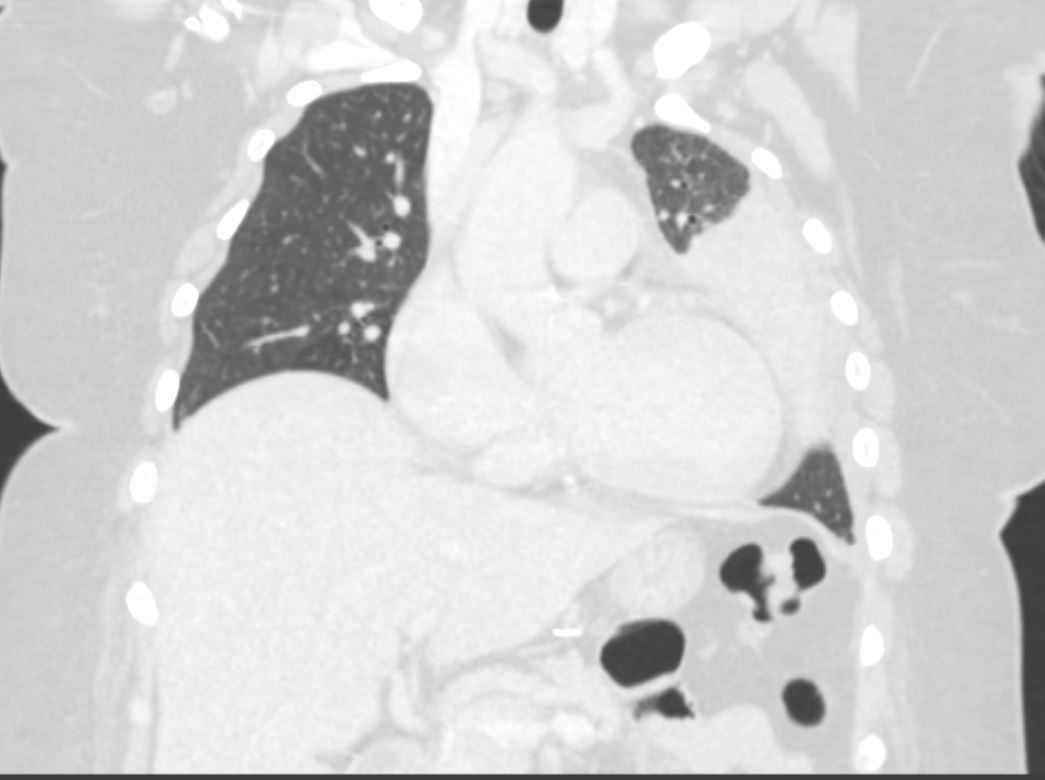
58-year-old female presents with a cough. CT in the coronal plane shows post obstructive atelectasis of the lingula which silhouettes the left heart border. A small portion of aerated left upper lobe is noted in the left apex.
Pathology revealed findings consistent with a carcinoid tumor of the left bronchus.
Ashley Davidoff MD TheCommonVein.net 257Lu 136115
CT Obstructing Nodule in the
More Horizontally Oriented Left Main Stem Bronchus
58-year-old female presents with a cough Coronal CT shows a nodule at the branching of the more horizontally oriented left mainstem bronchus with post obstructive atelectasis of the lingula and mild hyperinflation of the upper lobe segments.
Pathology revealed findings consistent with a carcinoid tumor of the left bronchus.
Ashley Davidoff MD TheCommonVein.net 257Lu 136118
Obstructing Nodule in the Left Main Stem Bronchus with Horizontal Displacement of Left Main Stem

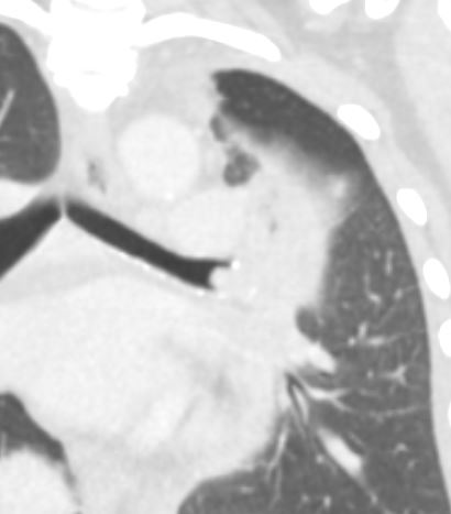
58-year-old female presents with a cough. Coronal CT shows a nodule (green arrowhead, b) at the branching of the more horizontally oriented left mainstem bronchus (teal arrow, a) with post obstructive atelectasis of the lingula (black arrowhead, b) and hyperinflation of the superior aspect of the lower lobe (white arrowhead) which occupies portion of the left apex. (Luftsichel sign)
Pathology revealed findings consistent with a carcinoid tumor
Ashley Davidoff MD TheCommonVein.net 257Lu 136118cL
CT Obstructing Nodule in the Left Main Stem Bronchus,
Lingula Atelectasis
Hyperinflation of the Apical Segment of the Left Lower Lobe, Fissural Displacement,
Aeration of the Upper Segments of the Left Upper Lobe

58-year-old female presents with a cough. CT in the sagittal plane shows a nodule in the left mainstem bronchus of the lung with post obstructive atelectasis of the lingula, a hyperinflated portion of the apical segment of the left lower lobe, superior and anterior migration of the left major fissure and a small portion of aerated left upper lobe anteriorly that appears congested . In the left lower lobe, there is a loculated effusion with compressive atelectasis.
Pathology revealed findings consistent with a carcinoid tumor in the left mainstem bronchus
Ashley Davidoff MD TheCommonVein.net 257Lu 136119
Frontal CXR with Correlative Sagittal CT to
Explain the Findings in the Left Apex
(“Partial Luftsichel Sign”)

58-year-old female presents with a cough Frontal CXR shows silhouetting of the left heart border with hazy or veiling opacity extending out from the left hilum and fading out inferiorly. The left apex (ringed in white, a, and magnified in c) is lucent and reflects a combination of the normal aerated left upper lobe segments and the hyperinflated left lower lobe as explained in the sagittal correlates (b and d) The aerated left upper lobe segments are anterior (purple arrowhead d) and are separated by the superiorly and anteriorly displaced major fissure (yellow arrowhead). The left lower lobe is superiorly displaced and consists of a focally hyperinflated apical subsegment (white arrowhead), and other apical and lower lobe segments (light blue arrowhead). The central portion of the atelectatic lingula is noted (black arrowhead) and just a subtle hint of the tumor in the left mainstem bronchus is appreciated (green arrowhead)
Ashley Davidoff MD TheCommonVein.net 257Lu 136109c02L
CT Lingula Atelectasis, Hyperinflation of the Left Lower Lobe, Fissural Displacement, Subsegmental Atelectasis of the Posterior Subsegment of the Left Upper Lobe Segment and Aeration of the Remaining Upper Segments of the LUL

58-year-old female presents with a cough. CT in the sagittal plane shows post obstructive atelectasis of the lingula (black arrowhead), a hyperinflated portion of the apical segment of the left lower lobe, (white arrowhead), superior and anterior migration of the left major fissure, (yellow arrowhead), subsegmental atelectasis of the posterior segment of the left upper lobe, (teal arrowhead), and a small portion of aerated left upper lobe anteriorly that appears congested, (purple arrowhead). There is a loculated effusion with subsegmental compressive atelectasis of the left lower lobe, (pink arrowhead).
Pathology revealed findings consistent with a carcinoid tumor in the left mainstem bronchus
Ashley Davidoff MD TheCommonVein.net 257Lu 136120cL
CT Lingula Atelectasis, Hyperinflation of the Left Lower Lobe, Fissural Displacement, Anterior and Superior Migration
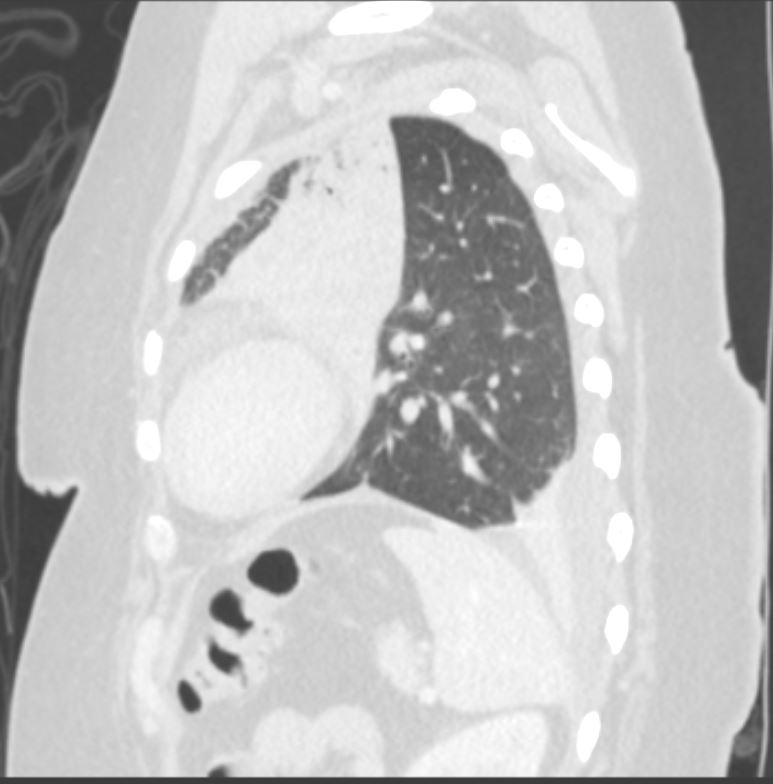
58-year-old female presents with a cough. CT in the sagittal plane shows post obstructive atelectasis of the lingula, hyperinflation of the left lower lobe, superior and anterior migration of the left major fissure, and a small portion of aerated left upper lobe anteriorly. There is a loculated effusion with subsegmental compressive atelectasis of the left lower lobe.
Pathology revealed findings consistent with a carcinoid tumor in the left mainstem bronchus
Ashley Davidoff MD TheCommonVein.net 257Lu 136121
CT Endobronchial Lesion in the Left Main Stem Bronchus and Lingula Atelectasis

58-year-old female presents with a cough. CT in the axial plane shows an obstructing lesion in the left mainstem bronchus of the lung with post obstructive atelectasis of the lingula and a small portion of aerated left upper lobe anteriorly. T he major fissure is displaced anteriorly.
Pathology revealed findings consistent with a carcinoid tumor of the left bronchus.
Ashley Davidoff MD TheCommonVein.net 257Lu 136110

58-year-old female presents with a cough. CT in the axial plane shows an obstructing lesion in the left mainstem bronchus of the lung (green arrowhead) with post obstructive atelectasis of the lingula (black arrowhead) and a small portion of aerated left upper lobe anteriorly (white arrowhead). The major fissure is displaced anteriorly.
Pathology revealed findings consistent with a carcinoid tumor of the left bronchus.
Ashley Davidoff MD TheCommonVein.net 257Lu 136110cL

58-year-old female presents with a cough. CT in the axial plane on soft tissue windows shows an obstructing lesion in the left mainstem bronchus of the lung with post obstructive atelectasis of the lingula (black arrowhead) and a small portion of aerated left upper lobe anteriorly (white arrowhead).
Pathology revealed findings consistent with a carcinoid tumor of the left bronchus.
Ashley Davidoff MD TheCommonVein.net 257Lu 136114

58-year-old female presents with a cough. CT in the axial plane post obstructive atelectasis of the lingula which silhouettes the left heart border.
Pathology revealed findings consistent with a carcinoid tumor of the left bronchus.
Ashley Davidoff MD TheCommonVein.net 257Lu 136113