
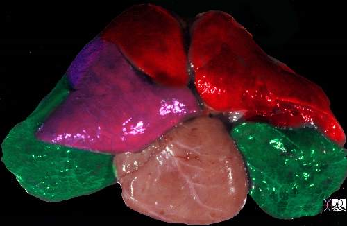
The post mortem specimen is viewed from the anterior aspect showing the upper lobes in red, the right middle lobe in pink and the lower lobes in green.
Ashley Davidoff MD TheCommonVein.net 32558b02

This diagram shows the basic division of the tracheobronchial tree into lobes. The right lung is divided into right upper (RUL) (teal) right middle, (RML pink) and right lower lobe (RLL green). The left lung is divided into left upper (LUL teal), which includes the lingula(dark blue), and left lower lobe (LLL= green). Note that the two mainstem bronchi are of unequal length and size. The right mainstem is short and fat while the left is long and thin. This irregular dichotomous branching pattern is characteristic of the branching pattern of all the conducting systems within the lungs.
Ashley Davidoff
TheCommonVein.net 32686b05
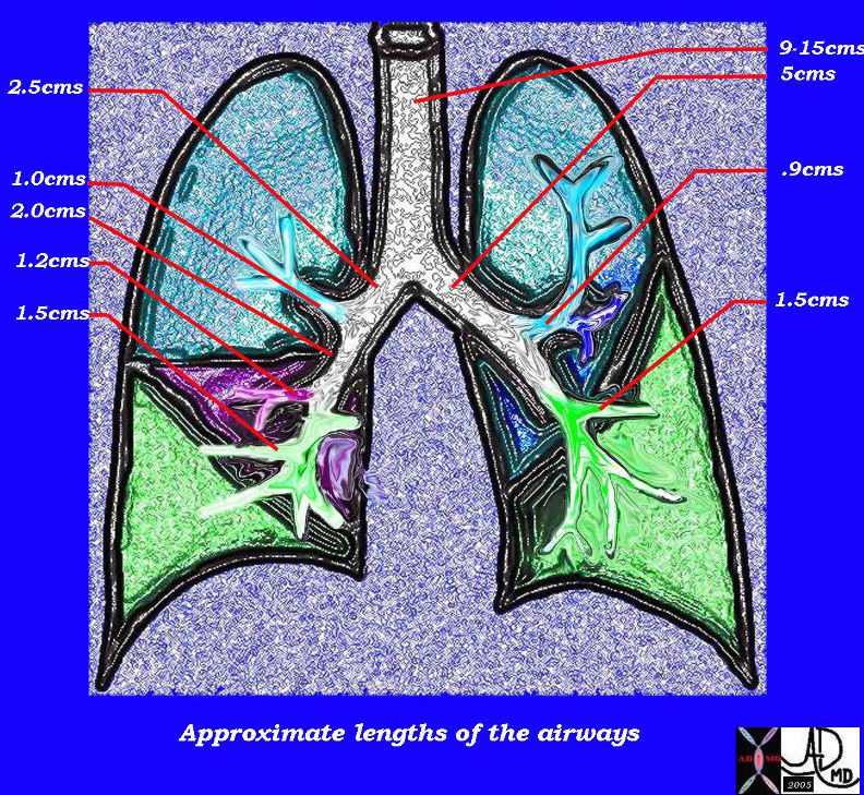
This diagram of the airways reveals the approximate lengths of the airways. Note that the left main stem bronchus is about twice the length of the right whose length is truncated by the take off of the right upper lobe bronchus.
Courtesy Ashley Davidoff MD
TheCommonVein.net
32686b05L04b

Ashley Davidoff TheCommonvein.net 32686b05L segmental bronchi.8
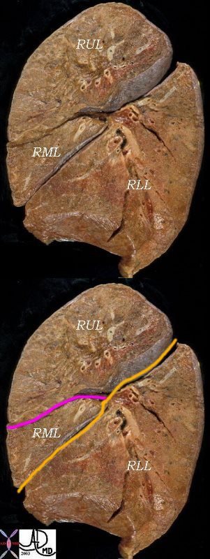
The right lung has a small right upper lobe (RUL) separated from the middle lobe (RML) by the minor fissure (pink,lower image) . Both the RUL and RML are anterior and are separated from the lower lober by the major fissure (orange line)
Ashley Davidoff MD

Ashley Davidoff M.D.
TheCommonVein.net 32682
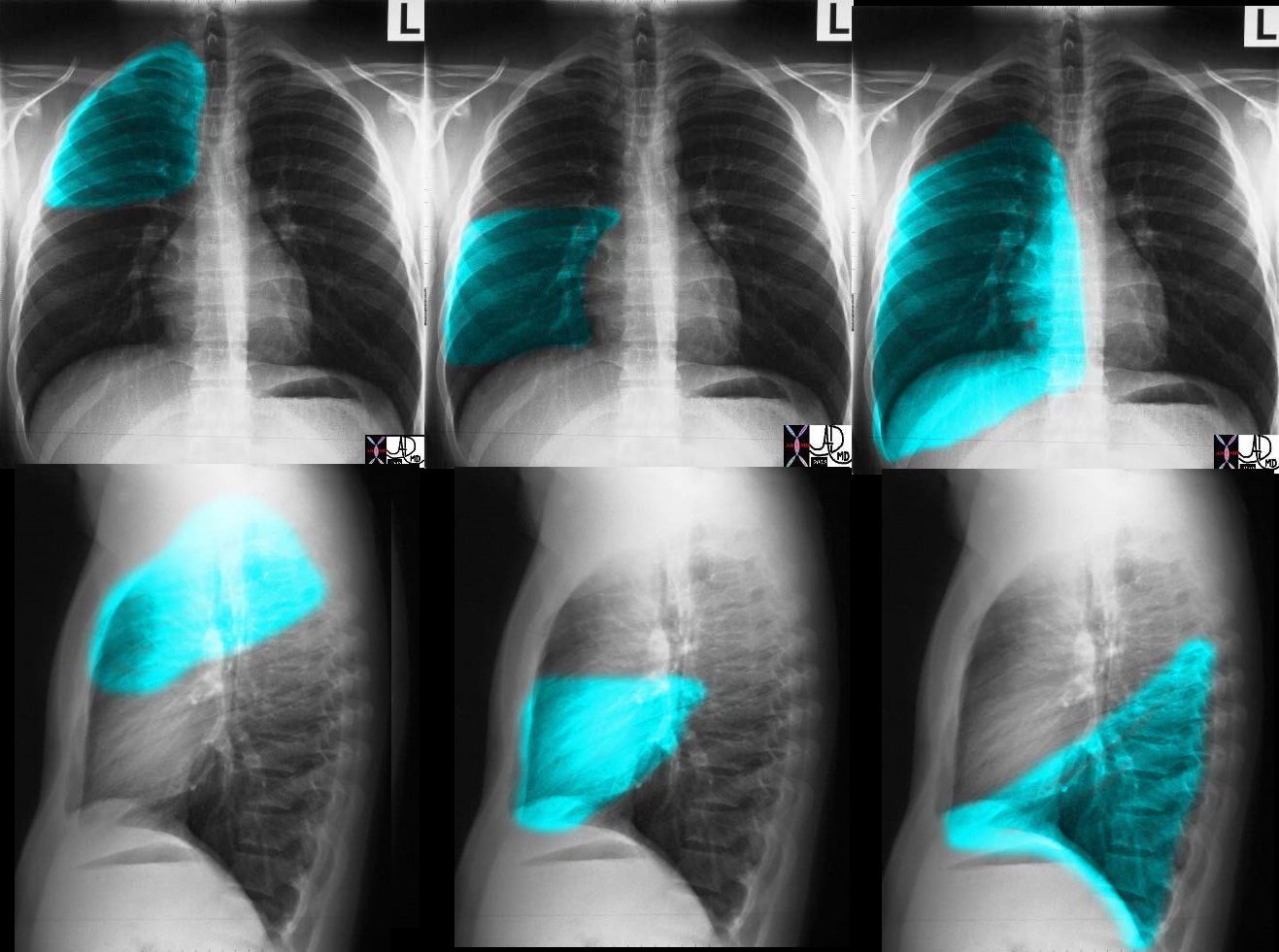
These images (left to right) show the shapes of the RUL, RML and RLL. In Fig 1a and 1b the overlay represents the upper lobe. Image 2a and 2b represent the middle lobe while in 3a and 3b the lower lobe is represented. Note how the middle lobe hugs the right heart border and how much larger the RLL is compared to the RML and the RUL.
Ashley Davidoff TheCommonVein.net 30398c01
Ashley Davidoff MD

The right lung has a relatively small right upper lobe (RUL) separated from the middle lobe (RML) by the minor fissure (pink,lower image). Both the RUL and RML are anterior and are separated from the lower lobe by the major fissure (orange line)
Ashley Davidoff MD. TheCommonVein.net 30398b06b
The secondary lobules, connect, and unite, linked through the airways, blood vessels, lymphatics and nerves, to form segments in the lungs
There are ten bronchopulmonary segments in the right lung: three in the upper lobe, two in the middle lobe, and five in the lower lobe. Some of the segments may fuse in the left lung to form usually eight to nine segments (four to five in the upper lobe and four to five in the lower lobe.
Ashley Davidoff MD
TheCommonVein.net lungs-0015