
CT Pulmonary Embolus Pulmonary Infarction

Patient presented with dyspnea and chest pain. CTPA shows large pulmonary embolus subtending a region of right lower lobe infarction.
Ashley Davidoff MD TheCommonVein.net 19443L
CT Tricuspid Regurgitation Secondary to a
Large Pulmonary Embolus
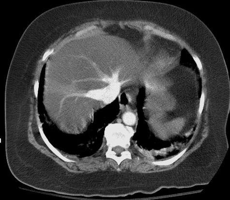
Patient presented with dyspnea and chest pain. CTPA showed a large pulmonary embolus. This image shows filling of hepatic veins prior to the portal venous phase consistent with tricuspid regurgitation secondary to the elevated pulmonary pressures. In addition, there is evidence of severe fatty change (steatosis) of the liver.
Ashley Davidoff MD TheCommonVein.net 19449
Patient had a PFO and as a result of the High Right Atrial Pressure Paradoxical Embolus Resulted
CT Paradoxical Embolus involving the
Aorta and Left Kidney with a
Left Renal Segmental Ischemic Defect
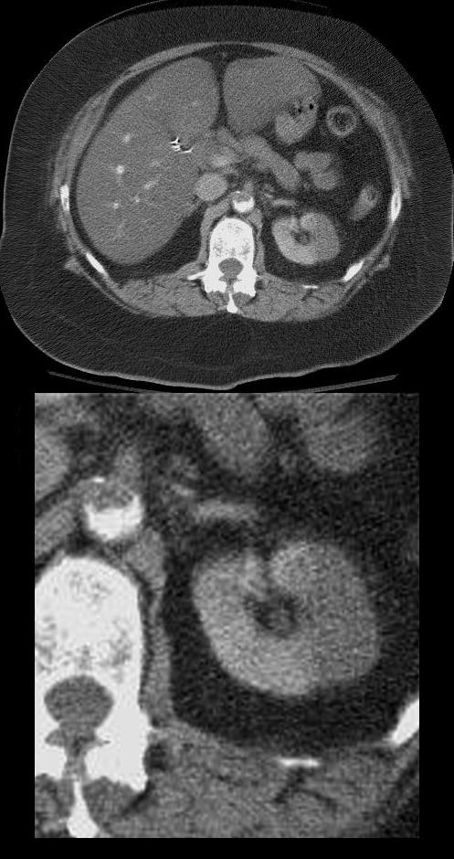
Patient presented with dyspnea and chest pain. A diagnosis of large pulmonary embolus with pulmonary infarction was made on CTPA. This image shows an embolus in the aorta and segmental non-perfusion of the left kidney as a result paradoxical embolic occlusion
Ashley Davidoff MD TheCommonVein.net 19453
CT Paradoxical Embolus Aorta and Right Renal Segmental Ischemia
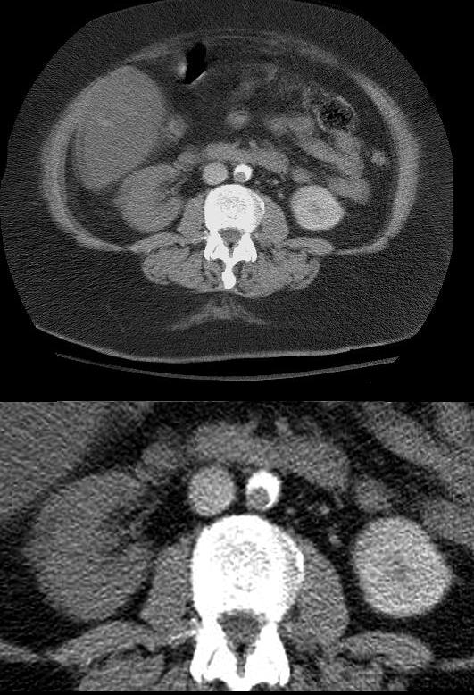
Patient presented with dyspnea and chest pain. A diagnosis of large pulmonary embolus with pulmonary infarction was made on CTPA. In addition, paradoxical emboli were identified in the aorta, with non-perfusion of the right kidney and occlusion of the right iliac artery. This image shows an embolus in the aorta and non-perfusion of the right kidney as a result embolic occlusion
Ashley Davidoff MD TheCommonVein.net 19463
Large Embolus in the Aorta Distally

Patient presented with dyspnea and chest pain. A diagnosis of large pulmonary embolus with pulmonary infarction was made on CTPA. In addition, paradoxical emboli were identified in the aorta, with non-perfusion of the right kidney and occlusion of the right iliac artery. This image shows an embolus in the aorta and non-perfusion of the right kidney as a result embolic occlusion
Ashley Davidoff MD TheCommonVein.net 19464
CT Paradoxical Embolus Right External Iliac Artery

Patient presented with dyspnea and chest pain. A diagnosis of a large pulmonary embolus with pulmonary infarction was made on CTPA. This image shows occlusion of the right external iliac artery (red arrowhead) as a result of the paradoxical embolus. The left external iliac artery (white arrowhead) shows normal contrast opacification.
Ashley Davidoff MD TheCommonVein.net 19453
Reconstitution of Right Femoral Artery

Patient presented with dyspnea and chest pain. A diagnosis of a large pulmonary embolus with pulmonary infarction was made on CTPA with occlusion of the right external iliac artery. This image shows reconstitution of the right femoral artery showing equal opacification of the right and left femoral arteries.
Ashley Davidoff MD TheCommonVein.net 19461