
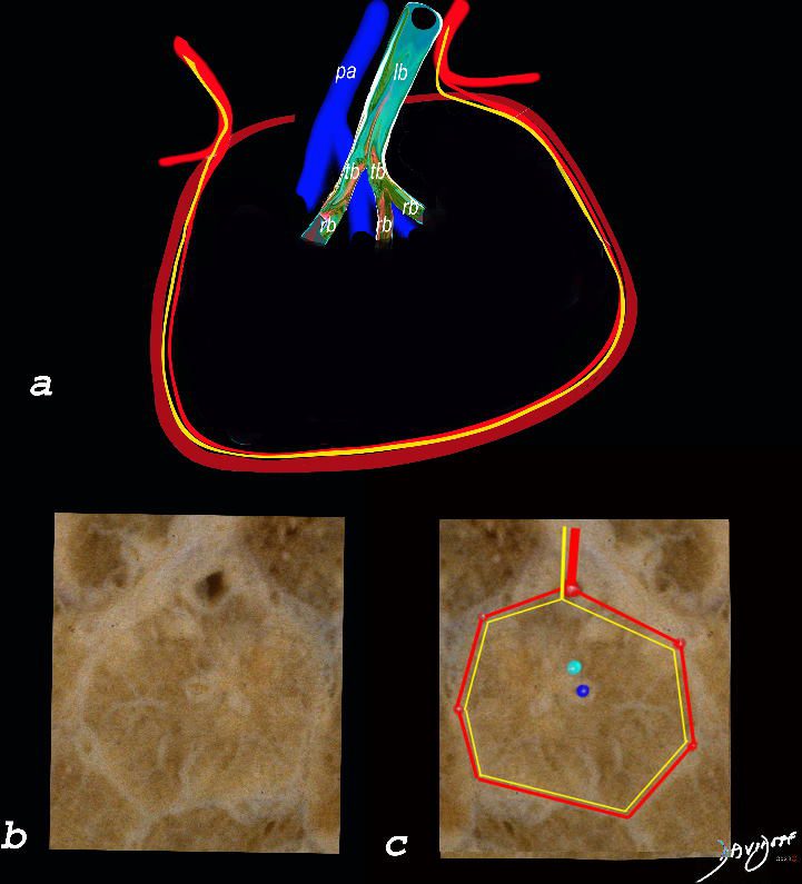
The top image (a) shows an anatomic drawing of a secondary lobule of the lung subtended by a lobular bronchiole (lb) and arteriole (pa). The interlobular septum contains the venule (red) lymphatic (yellow) and septum (maroon)
The anatomical specimen of the lung (b) shows normal intralobular parenchyma while image c shows the centrilobular arteriole (navy blue) and centrilobular bronchiole (teal) and interlobular venule (red) and lymphatics (yellow) The interlobular septum is slightly thickened
Ashley Davidoff TheCommonVein.net
Infection Inflammation
Disease Infection Inflammation
Allergic Bronchopulmonary Aspergillosis (ABPA) Bronchitis and Bronchiectasis

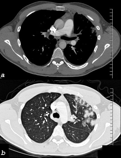
54 year old female with history of asthma, bronchitis, bronchiectasis, ABPA
Current CT scan shows extensive small airway disease in the right lower lobe, magnified in lower image with centrilobular nodules and thickened interlobular septa characterized by ground glass micronodules.
Ashley Davidoff TheCommonVein.net
Allergic Bronchopulmonary Aspergillosis (ABPA) Bronchitis and Bronchiectasis

60 year old male with history of asthma, allergic bronchopulmonary aspergillosis (ABPA)
CT scan shows left upper lobe bronchiectasis and soft tissue/fluid impaction in the anterior segmental and subsegmental airways associated with tree in bud appearance (yellow arrowhead) and centrilobular nodules (red arrowhead) reminiscent of associated small airway disease
Ashley Davidoff TheCommonVein.net
TB

68 year old female presented with malaise night sweats weight loss QuantiFeron gold positive, with a past history of treated TB in her native country as a child. Axial CT images through the upper lobe shows a miliary pattern of disease affecting interlobular septa, centrilobular and tree in bud nodular patterns. Bronchoscopy isolated Mycobacterium complex. She was treated with good result
Ashley Davidoff MD TheCommonVein.net mycobacterium-complex-TB-68-008
Chronic Eosinophilic Pneumonia (CEP)

Alveolar and Interalveolar Interstitial Infiltration with Eosinophils and Inflammatory Exudate – Ground Glass Changes
The ground glass changes are a combination of the cellular and exudative inflammatory response in the small airways, alveoli, interalveolar septa and interstitium, and thickened alveolar septum
The diagram shows the abnormal secondary lobule (a) The involved components include the small airways(b) alveoli and interalveolar interstitium (c) and the thickened interlobular septum (d) surrounding the secondary lobule due to an inflammatory process, cellular infiltrate and congestion of the venules and lymphatics in the septum. An anatomic specimen of a secondary lobule from a patient with thickened interlobular septa and interstitial thickening is shown in image e, and is overlaid in red (f) . A magnified view of an axial CT of the lungs in a patient with acute eosinophillic pneumonia shows thickened interlobular septa and centrilobular nodules (g) The inflammatory changes in the aforementioned structures result in an overall increase in density of the lung manifesting as ground glass changes (g) and overlaid in red (h)
Ashley Davidoff MD The CommonVein.net lungs-0762

CT scan in the coronal performed 6 months ago at the time of clinical presentation shows upper lobe predominant peripheral infiltrates more prominent in the left upper lobe. Subsequent diagnosis by BAL of chronic eosinophilic pneumonia (CEP) was made
Ashley Davidoff TheCommonVein.net

CT scan in the axial plane performed 6 months ago at the time of clinical presentation, shows upper lobe interlobular septal thickening, and peripheral consolidations which are findings characteristic of chronic eosinophilic pneumonia (CEP) in the appropriate clinical setting. Note of prominent centrilobular nodule likely reflects small airway involvement Subsequent diagnosis by BAL of chronic eosinophilic pneumonia (CEP) was made
Ashley Davidoff TheCommonVein.net

CT scan in the coronal performed 6 months ago at the time of clinical presentation shows upper lobe predominant peripheral infiltrates more prominent in the left upper lobe. Subsequent diagnosis by BAL of chronic eosinophilic pneumonia (CEP) was made
Ashley Davidoff TheCommonVein.net
CHF

CT scan at the lung bases shows Kerley B Lines bilateral small pleural effusions, right greater than left. Diagnosis Moderate CHF
Ashley Davidoff TheCommonVein.net

Ashley Davidoff MD TheCommonvein.net 50-007-CT
Pulmonary Venous Congestion and Thrombosis
26 year old male who presents with shortness of breath a few months following RFA ablation for atrial fibrillation. Image a shows the left upper lobe thrombosed and the left lower vein contrast filled
Image b (the lung windows) shows thickening of the interlobular septa and secondary lobules distended with material, porobably representing blood.
following male SOB s/p RFA ablation for atrial fibrillation lung pulmonary let upper lobe pulmonary vein thrombosed secondary lobule interlobular septa are thickened secondary lobule destroyed dx pulmonary vein thrombosis secondary to radiofrequency ablation therapy iatrogenic CTscan Courtesy Ashley Davidoff MD Scott Tsai MD key words
ground glass changes pulmonary infarction venous infarction
TheCommonVein.net 75413c02