
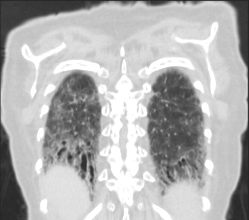
Fibrotic NSIP
The Bronchovascular Disribution in the Lower Lobes
Ashley Davidoff MD
Subpleural Sparing

Ashley Davidoff MD TheCommonVein.net scleroderma-019
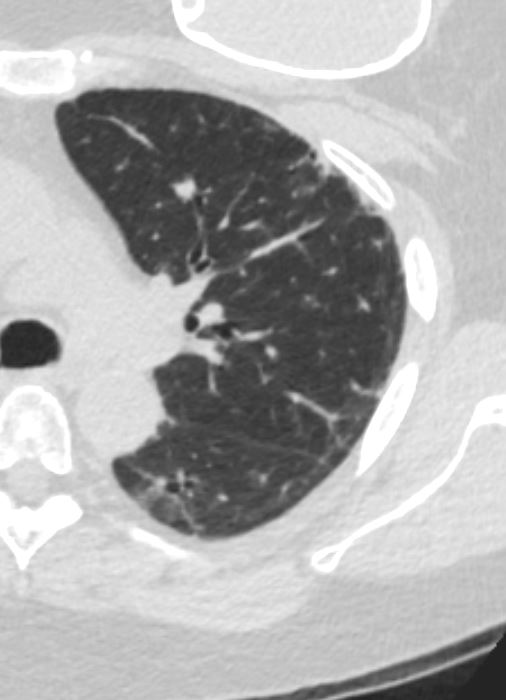
Cellular NSIP
The absence of significant fibrotic component and lack of volume loss in the lower lobes makes cellular NSIP more likely
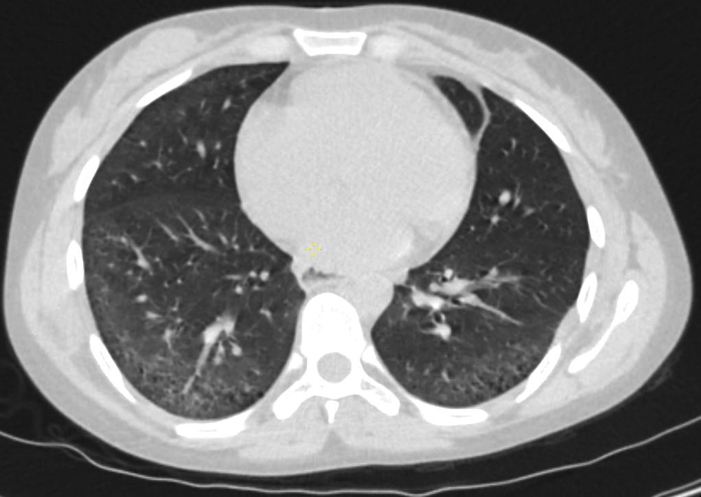
26-year-old female with scleroderma with dyspnea presents for evaluation. Axial CT through the lungs through the lower lungs shows peripherally located, ground glass changes, mild reticulation bronchiolectasis and subpleural sparing. The fissures are normally placed with no obvious loss of volume of the lower lobes. There is no honeycombing
Ashley Davidoff MD TheCommonVein.net 272Lu 136247
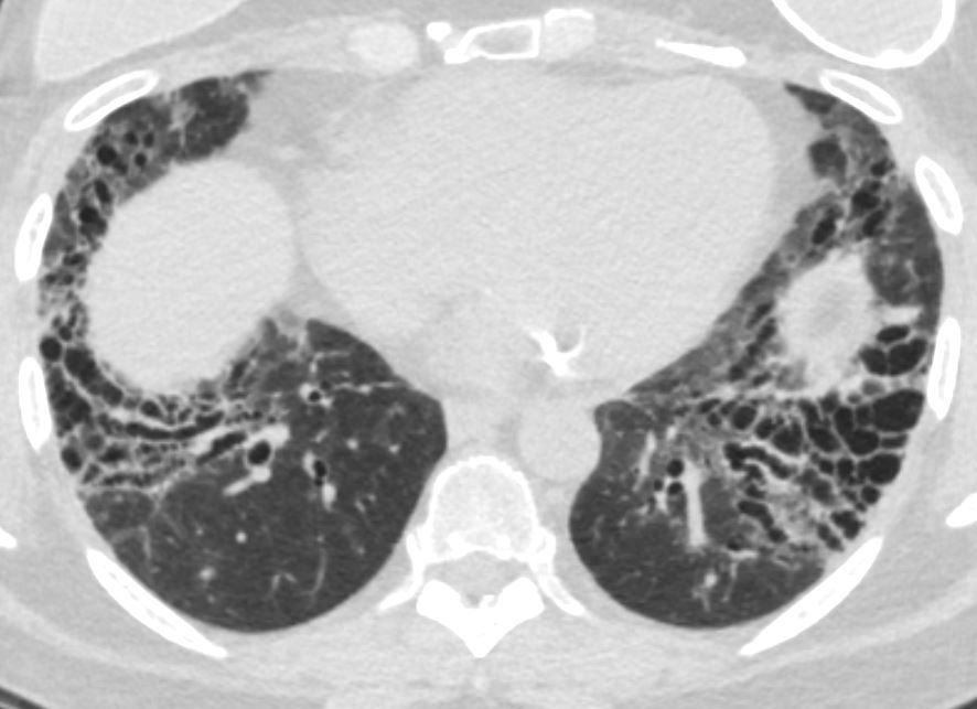
Fibrotic NSIP
Significant Volume Loss
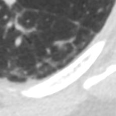
Expansion of the Secondary Lobules in
Regions of Subpleural Sparing
Fibrotic Bands

59-year-old male presents with history of scleroderma, , Raynaud’s disease, and ILD
CXR shows basilar reticular changes and low lung volumes. The heart is enlarged. There is an air bronchogram in the left lower lobe as a result of traction bronchiectasis and fibrotic change surrounding the bronchovascular bundle
Ashley Davidoff MD TheCommonVein.net 110Lu 136589

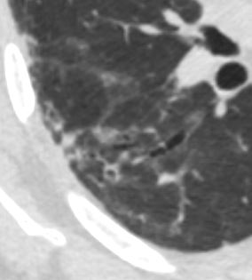
59-year-old male presents with history of scleroderma, Raynaud’s disease, and ILD
Axial CT shows extensive fibrotic change in the apical segments of the lower lobes and the anterior segments of the upper lobe.
There is volume loss architectural distortion, bronchiolectasis and subpleural sparing exemplified in the right lower lobe. peripheral reticular changes, ground glass, bronchiolectasis volume loss and subpleural sparing .
There are bands of fibrosis in the periphery of the right upper lobe with suggestion of intralobular fibrosis.
The spared secondary lobules in the right upper and right lower lobes have also undergone enlargement secondary to the fibrotic process
Ashley Davidoff MD TheCommonVein.net 110Lu 136596
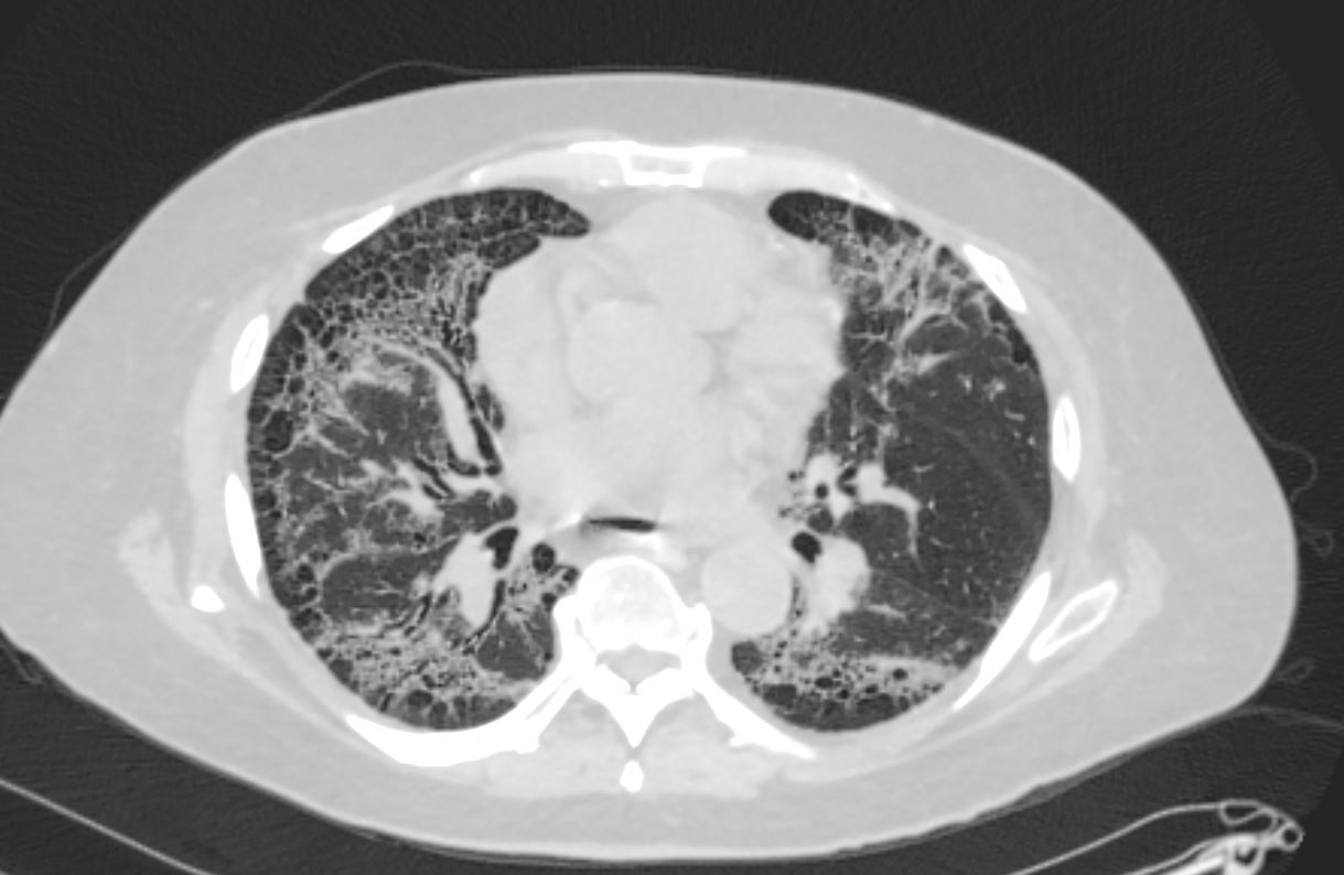
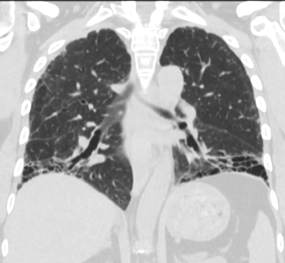
59-year-old male presents with history of scleroderma, Raynaud’s disease, and ILD
Axial CT showsextensive fibrotic change in the apical segments of the lower lobes and the anterior segments of the upper lobes.
There is volume loss architectural distortion, bronchiolectasis and subpleural sparing exemplified in the right lung. In addition there are ground glass changes, and volume loss.
The spared secondary lobules in the right lower lobes have also undergone enlargement secondary to the fibrotic process
Ashley Davidoff MD TheCommonVein.net 110Lu 136597
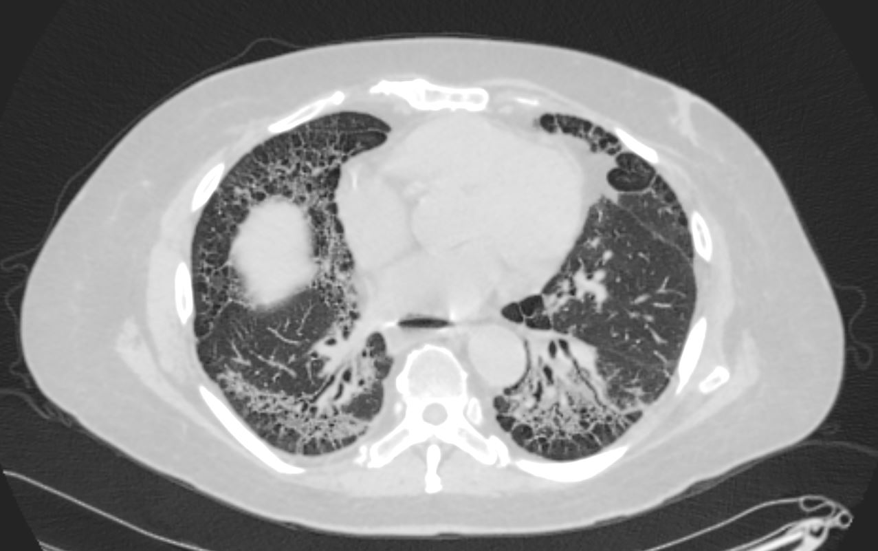
Axial CT shows peripheral reticular changes, ground glass, bronchiolectasis at both lung bases, volume loss with crowding of the bronchovascular bundles posteriorly and subpleural sparing posteriorly. Note air-fluid level in the distended esophagus.
Ashley Davidoff MD TheCommonVein.net 110Lu 136598