
Infection Inflammation Malignancy Mechanical/Atelectasis Trauma Metabolic Circulatory- Hemorrhage Immune Infiltrative Idiopathic Iatrogenic Idiopathic
Infection
Abscess

72-year-old female presents with cough, fever and leukocytosis
The CT confirms a peripheral subsegmental consolidation in the posterior segment of the LUL with cavitation (b and d white arrowheads. There is surrounding ground glass change reflecting surrounding edema (d, red arrowheads). Cultures confirmed bacterial abscess
Ashley Davidoff MD TheCommonVein.net 261Lu 118357cL

72-year-old female presents with cough, fever and leukocytosis
The CT reveals a peripheral subsegmental consolidation in the posterior segment of the LUL with cavitation Biopsy and cultures confirmed bacterial abscess
Ashley Davidoff MD TheCommonVein.net 261Lu 118359
Aspergilloma
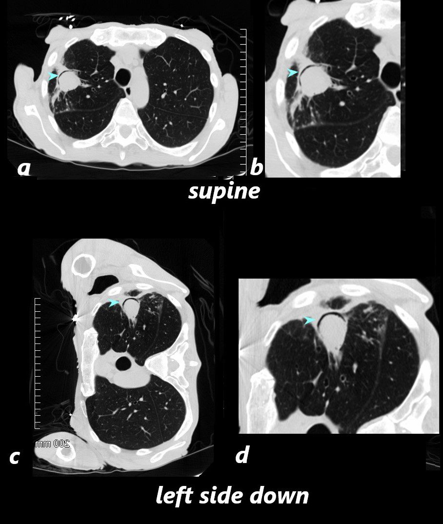
CT scan in the axial projection with the patient in the supine position shows a soft tissue mass in the right upper lung field with an anterior crescent shaped rim of air (blue arrowheads) anterior to the aspergillus fungus ball (a, magnified in b)
Since the fungus ball is movable, when the patient is placed in the the left decubitus position c and d), the fungus ball “sinks” to the dependent position and the air moves to the most superior position (blue arrowheads)
Ashley Davidoff TheCommonVein.net 78612a L
Inflammation

54 year old female presented with painless persistent dry cough, loss of appetite, weight loss, and worsening renal function. Urinary sediment showed white cells suggestive of glomerulonephritis. ANCA test and ANA were negative, ANA negative. Vasculitis was suspected and she was started on solumedrol and cyclophosphamide which improved her symptoms.
She had an uneventful renal biopsy.
The scout film (a) shows two mass like lesions in the left mid and upper lung zones,(white arrowheads) with coronal (b) imaging showing a nodule (black arrowhead) and a mass (red arrowhead) also in the mid and upper left lung. Image c in sagittal projection shows a large mass in the superior segment of the LUL and a second in the anterior segment of the LUL both both with air bronchograms(teal arrowheads). For the large mass like lesions, subacute hemorrhage is a radiological consideration and for the smaller nodules granulomatous nodules of Wgeners seems to be more likely.
Ashley Davidoff MD
Benign Hamartoma Non Calcified

Ashley Davidoff MD TheCommonVein.net benign hamartoma 004 33m
Speckled Calcification

Ashley Davidoff MD TheCommonVein.net hamartoma 003c
Popcorn Calcifications

Ashley Davidoff TheCommonvein.net
hamartoma 0001c01 86f
Heavy Homogeneous Calcification – Hamartoma

and amyloidoma
Ashley Davidoff TheCommonvein.net
hamartoma calcifications 004c stable
Metastases

CT scan through right lower lobe of the lung shows a calcified mass representing a metastasis from a primary uterine leiomyosarcoma.
Ashley Davidoff MD TheCommonVein.net 135680
Castleman’s Disease

The non-contrast CT of the right lower lobe lung mass shows homogeneous soft tissue density . Pathology showed Castleman’s disease
Courtesy Priscilla Slanetz MD MPH
TheCommonVein.net

The contrast enhanced CT of the right lower lobe lung mass in the right lower lobe . I mage a, magnified in b shows mostly homogeneous enhancement with a suggestion of nodular morphology . Image c shows a region anteriorly with slightly greater enhancement , and magnified in d.
Pathology showed Castleman’s disease
Courtesy Priscilla Slanetz MD MPH
TheCommonVein.net
Cancer
Adenocarcinoma

Diagnosis – adenocarcinoma of the lung with extensive necrosis of the tumor
Ashley Davidoff MD The CommonVein.net

Diagnosis – adenocarcinoma of the lung with extensive necrosis of the tumor
Ashley Davidoff MD The CommonVein.net
Congenital Growth Abnormalities

Ashley Davidoff MD TheCommonVein.net intralobar sequestration 008

Ashley Davidoff MD TheCommonVein.net intralobar sequestration 007

Ashley Davidoff MD
TheCommonVein.net intralobar sequestration 001
Pulmonary Hemorrhage Hematoma Fissural Displacement Ground Glass Changes

75-year-old man on blood thinners s/p aortic valve replacement, s/p trauma, presents with hemoptysis. He was afebrile and without an elevated white count
Coronal CT of the posterior lung fields shows inferior displacement of the major fissure by a dense right upper lobe consolidation. The mass effect on the major fissure likely results from a hematoma. Lateral to the consolidation there is a combination of ground glass opacity. There is elevation of the right hemidiaphragm. Left sided pleural effusion is present
Ashley Davidoff MD TheCommonVein.net 165Lu 135860