
Upper Lobes
Perilymphatic
Peripleural Fissural
Peribronchial
Nodules
Upper Lobes

The CXR shows an interstitial process involving the upper lobecharacterised by a reticular pattern
Ashley Davidoff MD TheCommonvein.net lungs sarcoid 001

The coronal CTscan shows an interstitial with thickening and displacement of the fissure and innumerable micronodules some centrilobular and some related to the pleura
Ashley Davidoff MD TheCommonvein.net lungs sarcoid 004
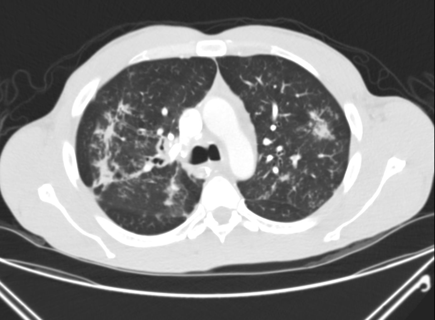
The axial CTscan shows thickening and irregularity of the major fissure, a band of fibrosis in the right upper lobe, thickening of a segmental bronchus in the right upper lobeand bronchocentric fibrosis in the left upper lobeand multiple micronodules
Ashley Davidoff MD TheCommonvein.net lungs sarcoid 002

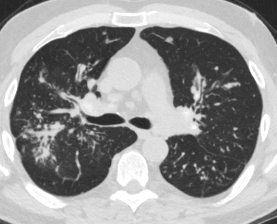
The axial CTscan shows an interstitial innumerable micronodules some centrilobular and some related to the pleura and others probably in the interlobular septa (posterior right upper lobe)
Ashley Davidoff MD TheCommonvein.net lungs sarcoid 003
Peribronchial Tree in Bud Centrilobular and Interlobular
Ashley Davidoff MD The CommonVein.net
sarcoidosis 001 60m
Centrilobular
Alveolar
Hilar and Mediastibal Lymph Nodes