
Frontal CXR of a 98-year-old woman showing a left sided white out secondary to a pneumonectomy. The soft tissue structures of the mediastinum have all shifted into the left hemithorax accounting for the white out. The calcified mitral annulus, calcified coronary arteries and right ventricle (RV pacemaker lead) confirm the diagnosis of acquired dextrocardia. There is hyperinflation of the right lung which crosses the midline associated with a small effusion . A significant dextro-thoracic scoliosis with a compensatory levoscoliosis of the lumbar spine is present
Ashley Davidoff MD TheCommonVein.net 269Lu 136234
Frontal CXR of a 98-year-old woman showing a left sided white out secondary to a pneumonectomy. The soft tissue structures of the mediastinum have all shifted into the left hemithorax accounting for the white out. The calcified mitral annulus (b, white arrowhead), calcified right coronary artery – RCA (b, maroon arrowhead) and left anterior descending (LAD) ? (b, bright red arrowhead) and right ventricle (RV pacemaker lead) confirm the diagnosis of acquired dextrocardia. There is hyperinflation of the right lung which crosses the midline associated with a small right effusion. A significant dextro-thoracic scoliosis with a compensatory levoscoliosis of the lumbar spine is present
Ashley Davidoff MD TheCommonVein.net 269Lu 136234cL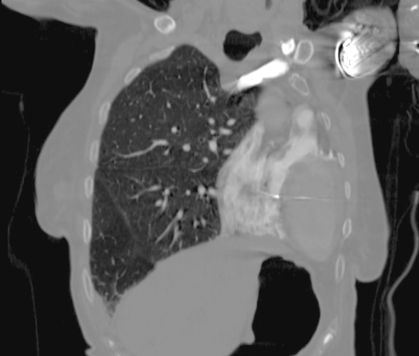
Coronal CT of a 98-year-old woman showing a left sided white out on CXR secondary to a pneumonectomy shows a leftward shift of cardiac structures including the contrast filled right ventricle (RV) and the oval shaped left ventricle (LV) which occupy the left hemithorax. The tip of the pacing lead is noted in the RV septum. There is hyperinflation of the right lung which crosses the midline associated with a small right effusion.
Ashley Davidoff MD TheCommonVein.net 269Lu 136235b
