
- Chronic Eosinophilic Pneumonia (CEP) is an
- inflammatory lung disease
Chronic eosinophilia is characterised by alveolar filling with eosinophils and inflammatory exudates(a) and interalveolar interstitial thickening, (overlaid in red in b). The infiltrates are classically peripherally positioned, usually upper lobes, more commonly bilateral but can be unilateral, and manifest as consolidation and or ground glass opacities. The CT shows bilateral peripheral consolidations in the upper lobes
Ashley Davidoff MD The CommonVein.net lungs-0775e
Chronic Eosinophilic Pneumonia Affects the Alveoli and Alveolar Septal Interstitium
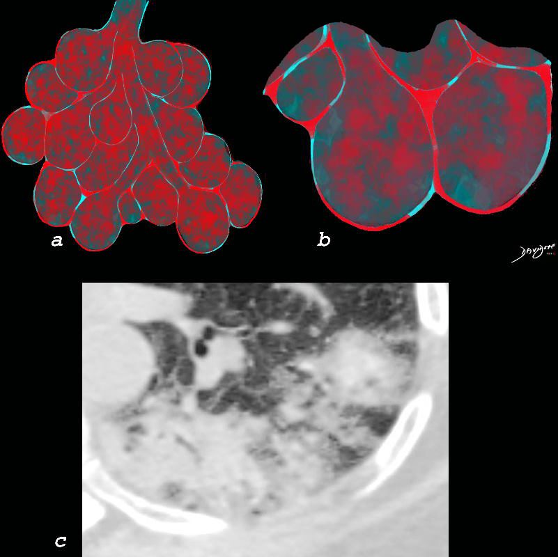
Chronic eosinophilia is characterised by alveolar filling with eosinophils and inflammatory exudates(a) and interalveolar interstitial thickening, (overlaid in red in b). The infiltrates are classically peripherally positioned, usually upper lobes, more commonly bilateral but can be unilateral, and manifest as consolidation and or ground glass opacities. The CT shows a peripheral consolidation in the left upper lobe
Ashley Davidoff MD The CommonVein.net lungs-0764

By Blausen Medical – BruceBlaus
-

Histopathology features of chronic eosinophilic pneumonia. Notes: Alveolar spaces are filled with fibrinous exudate and numerous eosinophils (arrow). Eosinophils and macrophages infiltrate the interstitium (arrow head). H&E stain, ×200.
Crowe M et al Therapeutics and Clinical Risk Management Volume 15:397-403 March 2019 Source Research Gate - characterized by the accumulation of eosinophils in
- alveoli and
- interstitium
- bronchiole
- resulting in damage to lung tissue.
- cause of CEP is
- not fully understood
- Female predominance
- hx of asthma 66%
- hx atopy 50%
- most non smokers (compared to AEP smoking more common)
- may be related to an
- abnormal immune response to a
- unknown trigger or
- certain medications.
- prior radiation for breast cancer
- Symptoms
- Subacute or progressive
- weeks to months
- fatigue
- malaise
- fever
- night sweats
- weight loss
- dyspnea
- cough
- fever
- asthma
- Signs
- wheezes or crackles
- Diagnosis
- Labs
- eosinophilia (20-30% of wcc)
- ESR and C reactive protein
- BAL 12-95% eosinophils
- PFT
- may be restrictive or
- obstructive
- imaging tests
- CXR
- peripheral infiltrates
- upper lobe predominant
- effusions rare
- photographic negative pulmonary edema
- implying that rather that the wings of acute alveolar edema are located peripherally rather than centrally
- CT
- Peripheral opacities almost all cases
- Migratory in 25%
- subpleural regions
- bilateral pulmonary infiltrates
- but can be unilateral
- nonsegmental airspace consolidations
in several segments of the lung (greater than two)) - or GGO
- bronchial wall thickening
- bronchial obstruction, such as mucus plugging,
- bronchiectasis, or
- pleural effusions.
- CXR
- blood tests to measure
- eosinophil levels, and
- may also require a lung biopsy
- Labs
- Treatment usually involves corticosteroid medications
- Prognosis
- is generally good
Peripheral Infiltrates

Crowe M et al Therapeutics and Clinical Risk Management Volume 15:397-403 March 2019 Source Research Gate

Crowe M et al Therapeutics and Clinical Risk Management Volume 15:397-403 March 2019 Source Research Gate

Crowe M et al Therapeutics and Clinical Risk Management Volume 15:397-403 March 2019 Source Research Gate
Links and References
-
- TCV
-
- Eosinophilic lung disease
-
- Systemic
- Eosinophilic granulomatosis with polyangiitis (EGPA) aka Churg Strauss Disease
- Hyper-eosinophilic syndrome
- Lung limited diseases
- Systemic
-
- Eosinophilic lung disease
-
- TCV