
28F-scleroderma-004-hands-2011.jpg


NSIP Probable Cellular Form

CXR shows mild basilar ground glass changes Ashley Davidoff MD TheCommonVein.net scleroderma-007

CXR shows mild basilar ground glass changes Ashley Davidoff MD TheCommonVein.net scleroderma-008
28F-scleroderma-009-2014.jpg
Scleroderma and NSIP (Probable Cellular Form)Barium Sallow shows a wide open GE junction
Ashley Davidoff MD TheCommonVein.net scleroderma-009

Barium Sallow shows a gastroesophageal reflux.
Ashley Davidoff MD TheCommonVein.net scleroderma-009c

Barium Sallow shows a a small hiatus hernia.
Ashley Davidoff MD TheCommonVein.net scleroderma-009b
28F-scleroderma-010-2020.jpg
Scleroderma and NSIP (Probable Cellular Form)CT shows an air fluid level in the esophagus reflecting gastroesophageal reflux.
Ashley Davidoff MD TheCommonVein.net scleroderma-010
28F-scleroderma-010-2020.jpg
Scleroderma and NSIP (Probable Cellular Form)CT shows an air fluid level in the esophagus reflecting gastroesophageal reflux.
Ashley Davidoff MD TheCommonVein.net scleroderma-010
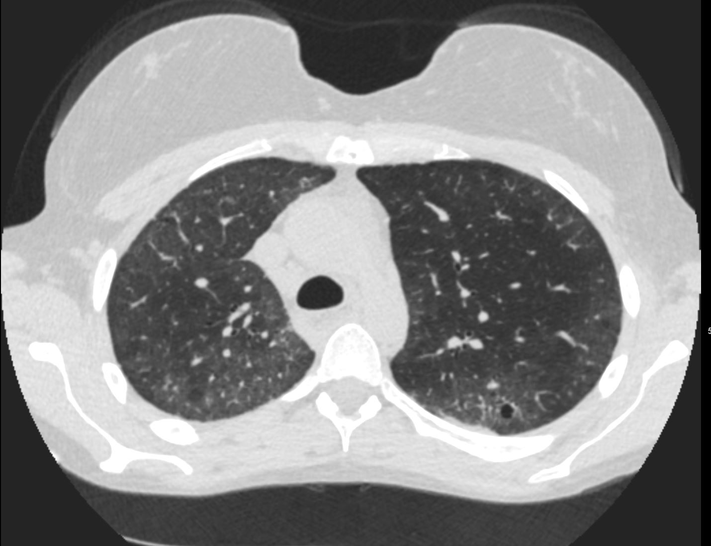
CT shows mild peripheral ground glass changes in the upper lobes with peripheral sparing and minimal reticular change
Ashley Davidoff MD TheCommonVein.net scleroderma-012

CT shows mild peripheral ground glass changes in the upper lobes with peripheral sparing and minimal reticular change
Ashley Davidoff MD TheCommonVein.net scleroderma-013
28F-scleroderma-014-2020.jpg
Scleroderma and NSIP (Probable Cellular Form)CT shows mild peripheral ground glass changes in the upper lobes with peripheral sparing and minimal reticular change
Ashley Davidoff MD TheCommonVein.net scleroderma-014

CT shows diffuse ground glass changes lower lobes and to lesser degree in the lingula and middle lobe with peripheral sparing, minimal reticular change and mild bronchiectasis
Ashley Davidoff MD TheCommonVein.net scleroderma-015

CT shows diffuse ground glass changes lower lobes and to lesser degree in the lingula and middle lobe with peripheral sparing, minimal reticular change and mild bronchiectasis
Ashley Davidoff MD TheCommonVein.net scleroderma-016
28F-scleroderma-017-2020.jpg
Scleroderma and NSIP (Probable Cellular Form)CT shows diffuse ground glass changes lower lobes with peripheral sparing, minimal reticular change and mild bronchiectasis
Ashley Davidoff MD TheCommonVein.net scleroderma-017

CT shows diffuse ground glass changes lower lobes with mild volume loss and mild ground glass change in the upper lobes. There are minimal reticular changes and mild bronchiectasis
Ashley Davidoff MD TheCommonVein.net scleroderma-018

CT shows diffuse ground glass changes lower lobes and to lesser degree in the upper lobes, with minimal reticular change and mild bronchiectasis
Ashley Davidoff MD TheCommonVein.net scleroderma-019
28F-scleroderma-020-2020.jpg
Scleroderma and NSIP (Probable Cellular Form)CT with a posterior coronal projection, shows diffuse ground glass changes in the lower lobes and to lesser degree in the upper lobes, minimal reticular change and mild bronchiectasis
Ashley Davidoff MD TheCommonVein.net scleroderma-020

Axial CT shows no evidence of pulmonary hypertension Pulmonary artery measures 2.9cms
Ashley Davidoff MD TheCommonVein.net scleroderma-021
TCV
-
- Scleroderma Introduction
- NSIP Introduction
- NSIP Cases