Subpleural Sparing

idiopathic, inflammatory, infectious, inhalational, cardiac, traumatic, and bleeding disorders. Specific disorders that can cause subpleural sparing patterns include nonspecific interstitial pneumonia (NSIP), organizing pneumonia (OP), pulmonary alveolar proteinosis (PAP), diffuse alveolar hemorrhage (DAH), vaping-associated lung injury (VALI), cracked lung, pulmonary edema, pneumocystis jirovecii pneumonia (PJP), pulmonary contusion, and more recently, Coronavirus disease 2019 (COVID-19) p


the density and distribution of lymphatics could explain preferential clearing of the subpleural regions during acute injury. T

Infection

PCP PJP COVID

Inflammation 

Inhalational Crack Lung Vaping

Scleroderma NSIP and OP

59-year-old male presents with history of scleroderma, , Raynaud’s disease, and ILD
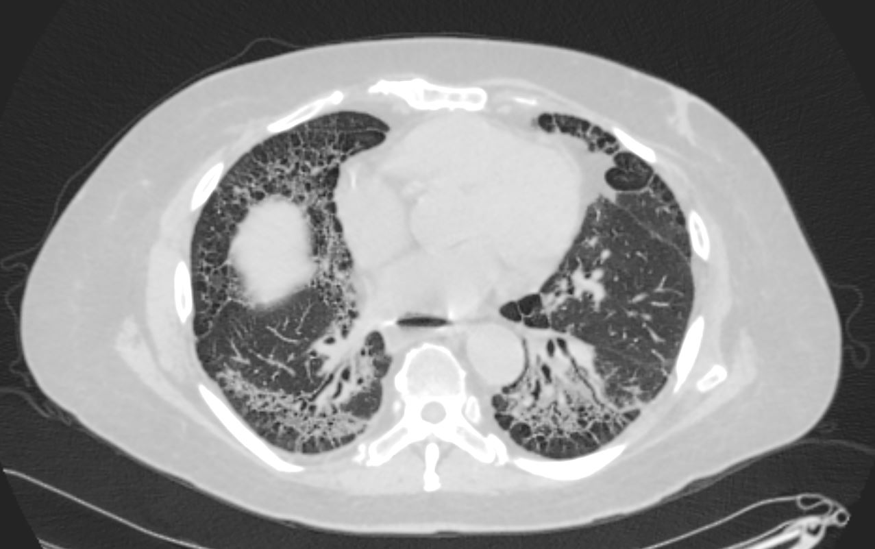
Axial CT shows peripheral reticular changes, ground glass, bronchiolectasis at both lung bases, volume loss with crowding of the bronchovascular bundles posteriorly and subpleural sparing posteriorly. Note air-fluid level in the distended esophagus.
Ashley Davidoff MD TheCommonVein.net 110Lu 136598

Fibrotic NSIP
59-year-old male presents with history of scleroderma, Raynaud’s disease, and ILD
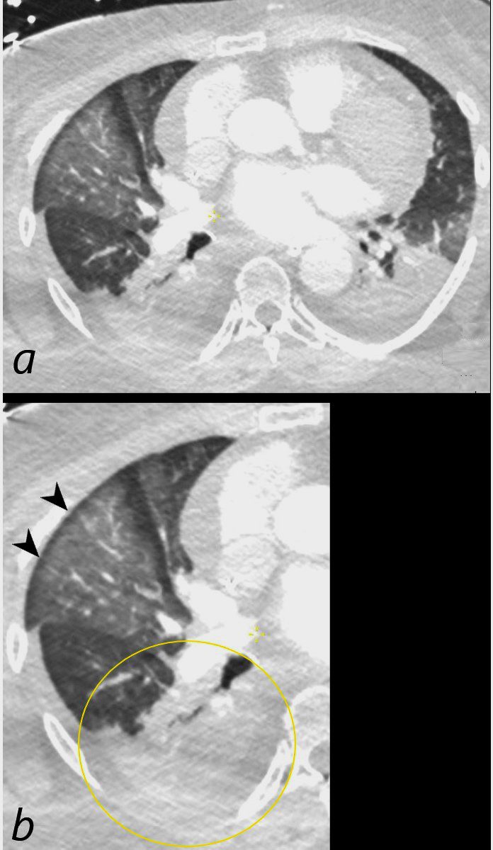
Coronal CT shows bibasilar volume loss, reticular change, ground glass changes, bronchovascular thickening , bronchiectasis, and subpleural sparing, all features characteristic of NSIP
.
The lower image highlights the bronchovascular thickening volume loss bronchiectasis and subpleural sparing. The fibrotic process has resulted in traction of the secondary lobules in the region of subpleural sparing
Ashley Davidoff MD TheCommonVein.net 110Lu 136592c01

   Crack Lung
CT 1month Later Prominent Upper Lobe Ground Glass Parenchymal Changes Crazy Paving  and Subpleural Sparing
55 year old male with substance use disorder presents with progressive and now more severe dyspnea. CT through the upper lung fields shows extensive upper lobe ground glass changes with thickening of the interlobular septa giving the appearance of ?crazy paving?. Thickening and irregularity of the left major fissure is also noted There is bilateral peripheral sparing.
Progressive inhalational pneumonitis from smoking or cocaine inhalation was suspected. DIP and hypersensitivity pneumonitis remained in the differential diagnosis
Ashley Davidoff MD TheCommonVein.net 251Lu 135924
Prominent Upper Lobe and Superior Aspects of the Lower Lobe Ground Glass Parenchymal Changes
55-year-old male with substance use disorder presents with progressive and now more severe dyspnea. Coronal CT through the mid lung fields shows upper lobe predominant ground glass changes with thickening of the interlobular septa and a ?crazy paving? appearance is suggested. The superior segments of the lower lobes are also involved. Thickening and irregularity of the right and left major fissures and the transverse fissure are noted. LVH is suggested.
Progressive inhalational pneumonitis from smoking or cocaine inhalation was suspected. DIP and hypersensitivity pneumonitis remained in the differential diagnosis
Ashley Davidoff MD TheCommonVein.net 251Lu 135934
CT 1month Later Prominent Left Upper Lobe and Superior Segment of the Lower Lobe Ground Glass Parenchymal Changes
55-year-old male with substance use disorder presents with progressive and now more severe dyspnea. Sagittal CT through the left lung field shows ground glass changes in the upper mid and superior segment of the lower lobe. The fissures of the areas of involved lung are focally thickened. There is subpleural sparing.
Progressive inhalational pneumonitis from smoking or cocaine inhalation was suspected. DIP and hypersensitivity pneumonitis remained in the differential diagnosis
Ashley Davidoff MD TheCommonVein.net 251Lu 135941

Circulatory

Aspiration Post Arrest and Resuscitation, Middle Lobe Ground Glass Opacification Subpleural Sparing

CT ? Aspiration Post Arrest and Resuscitation, Middle Lobe Ground Glass Opacification Subpleural Sparing
Axial CT scan at the level of the middle lobe of a 64-year-old female who arrested and required resuscitation is shown. Image a shows diffuse ground glass opacification in the lateral segment of the middle lobe with subpleural sparing (b, black arrowheads) most likely representing edema or hemorrhage. Bibasilar pneumonic infiltrates with bilateral effusions are present, exemplified and magnified in the right lower lobe (b, ringed in yellow)
Ashley Davidoff MD TheCommonVein.net 136192cL

Pulmonary Edema

Hemorrhage Contusion