
Parts
Size Small

CXR Spontaneous Pneumothorax
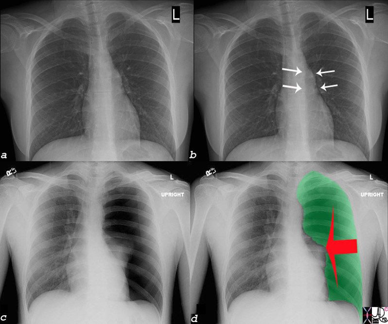
20-year-old female presents with acute left sided chest pain. She has a narrow A-P diameter exemplified in the lateral projection (below) and the asthenic build raises the suspicion for spontaneous pneumothorax. Frontal CXR shows a small subtle pneumothorax characterised by a thin pleural line and relative lucency of the left apex compared to the right
Ashley Davidoff MD TheCommonVein.net 117246c

20-year-old female presents with acute left sided chest pain. She has asthenic build which raises the suspicion for a spontaneous pneumothorax. Frontal CXR shows a small subtle pneumothorax characterised by a thin pleural line (b, white arrowhead) and relative lucency of the left apex
Ashley Davidoff MD TheCommonVein.net 117246c01
Deep Sulcus Sign – Semi-Erect

51 year old male with recent trauma with multiple rib fractures on the left resulting in flail chest. Rib fixation hardware is present on the left. Fractures are also presnt on the right and there are bilaterl chest tubes.
A subpulmonic pneumothoraxis present
Ashley Davidoff TheCommonVein.net 136507

51 year old male with recent trauma with multiple rib fractures on the left resulting in flail chest. Rib fixation hardware is present on the left. Fractures are also present on the right and there are bilateral chest tubes.
A subpulmonic pneumothorax is present with the displaced pleura noted (b, white arrowheads) and the basal pneumothorax (b blue arrowhead). Note the patient is in the semi-upright position and likely more supine than upright based on the position of the pneumothorax
Ashley Davidoff TheCommonVein.net 136507cL
Tension Pneumothorax with Shift of the Heart to the Left

? Tension Pneumothorax
Ashley Davidoff MD TheCommonVein.net
? Tension Pneumothorax
Ashley Davidoff MD TheCommonVein.net
Tension Pneumothorax and the Mediastinum
Shape
SLE PE Bronchopleural Fistula and Loculated Pneumothorax
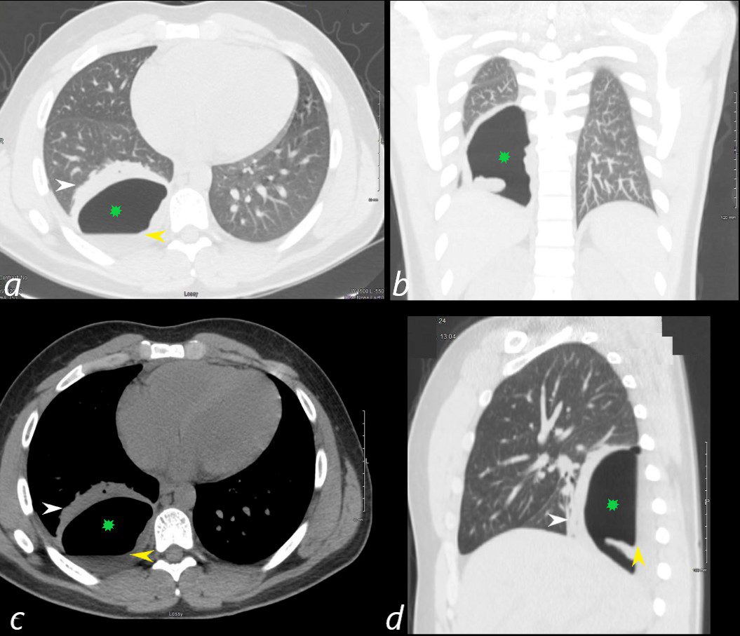
24 year old male with SLE presented with chest pain and dyspnea and initial CT showed occlusive pulmonary emboli to the right lower lobe initially associated with a wedge shaped ground glass region. 2 weeks later this evolved into a bronchopleural fistula, with a loculated pneumothorax in the right lower lobe (green star in a,b,c,d).with an air fluid level (yellow arrowhead in a,c,d) and a region of compressive atelectasis (white arrowhead a,c,d).
Ashley Davidoff MD TheCommonVein.net 130726
Position
Character
Time
Associated Findings
Tension Pneumothorax
Venous Reflux and
Benign Pneumatosis Coli

65 year old male s/p MVA presents in shock. Scout film (top left) shows left sided tension pneumothorax with rightward mediastinal shift. Axial CT through the liver (top right) shows expanded pneumothorax at the left lung base with reflux of contrast into the IVC.. Contrast also refluxes into the right renal vein (bottom left) and into the internal iliac veins (bottom right) Associated pneumatosis intestinalis in the sigmoid colon is present as well and likely secondary to the tension pneumothorax
Ashley Davidoff MD TheCommonVein.net 24153c
Post Cryoablation with Contusion and Pneumothorax

Following biopsy and cryoablation a contusion is noted in the lung associated with a small pneumothorax
Biopsy of the lung confirmed the presence of a squamous cell carcinoma.
Ashley Davidoff MD TheCommonVein.net
Infection
Pneumocystis Carinii Pneumonia and Pneumothorax

Parekh, M et al Review of the Chest CT Differential Diagnosis of Ground-Glass Opacities in the COVID Era Radiology Vol. 297, No. 3 July 2020
Inflammation
Malignancy
Mechanical
Atelectasis
Trauma
Metabolic
Circulatory- Hemorrhage
Catamenial Pneumothorax and Recurrent Hemothorax

30 year old female presented in 2013 with acute pneumothorax with tamponade requiring placement of a chest tube
Ashley Davidoff TheCommonVein.net 130915.8
SLE PE Bronchopleural Fistula and Loculated Pneumothorax
24 year old male with SLE presented with chest pain and dyspnea and initial CT showed occlusive pulmonary emboli to the right lower lobe initially associated with a wedge shaped ground glass region. 2 weeks later this evolved into a bronchopleural fistula, with a loculated pneumothorax in the right lower lobe (green star in a,b,c,d).with an air fluid level (yellow arrowhead in a,c,d) and a region of compressive atelectasis (white arrowhead a,c,d).
Ashley Davidoff MD TheCommonVein.net 130726