

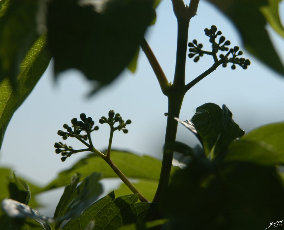
This is a photo taken near Lake Michigan at the end of the Fall in Chicago showing the illuminated base of the naked trees. The dichotomous branching is reminiscent of the tracheal branching into the two mainstem bronchi. Turn them upside down and the similarity is remarkable.
Ashley Davidoff TheCommonVein.net22153e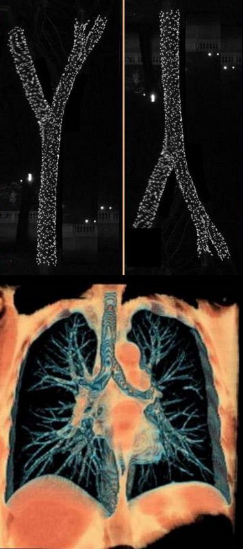
The classical branching pattern of many trees
Ashley Davidoff MD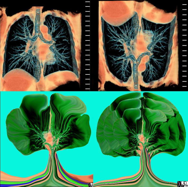
The tracheobronchial tree turned upside down shows it?s similarity to the branching pattern of a tree.
keywords lung bronchus tracheobronchial tree airway tree the common vein applied biology
Ashley Davidoff MD TheCommonVein.net
32620c02.800
Tracheobronchial Tree
Tree, flower, tracheobronchial tree, trachea bronchi lung
Ashley Davidoff TheCommonVein.net 32620b14.800b02p
This diagram shows the basic division of the tracheobronchial tree into lobes. The right lung is divided into right upper (RUL) (teal) right middle, (RML pink) and right lower lobe (RLL green). The left lung is divided into left upper (LUL teal), which includes the lingula(dark blue), and left lower lobe (LLL= green). Note that the two mainstem bronchi are of unequal length and size. The right mainstem is short and fat while the left is long and thin. This irregular dichotomous branching pattern is characteristic of the branching pattern of all the conducting systems within the lungs.
Ashley Davidoff
TheCommonVein.net
32686b05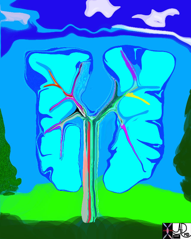
42474b18.800 lung trachea bronchi tracheobronchial tree
Ashley Davidoff MD
TheCommonVein.net 42474b18.800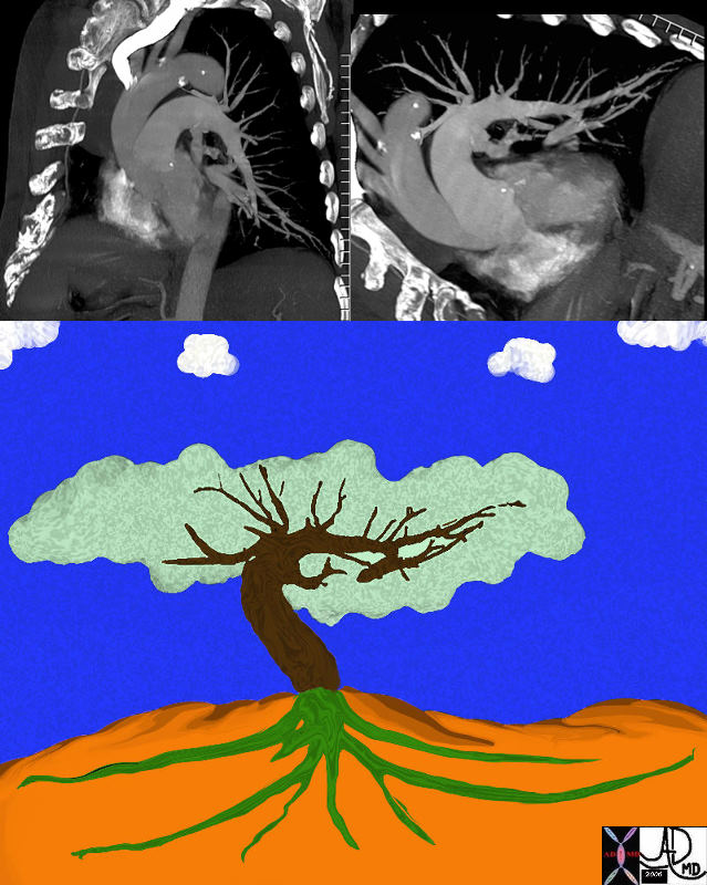
The left pulmonary artery seen in the left upper image is turned 90 degrees anticlockwise to reflect it upright shape of a tree (top left)) then flipped 90degrees and colored over to reveal its tree like morphology. keywords lung pulmonary artery pulmonary trunk
Ashley Davidoff MD TheCommonVein.net 46649c01.800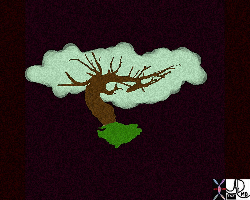
Ashley Davidoff MD
TheCommonVein.net 46649b04b.800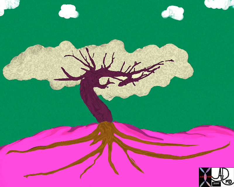

Top left image is sagittal oblique CT scan of the chest and shows a prominent sternum and heavily calcified costochondral junctions. When turned upside down (middle top , a cedar tree is created, then multiplied (middle image) and placed along a river in a mountain under a beautiful summer sun.
Ashley Davidoff MD TheCommonVein.net lungs-0708
Ashley Davidoff MD TheCommonVein.net lungs-0698
Ashley Davidoff MD TheCommonVein.net
lungs-0699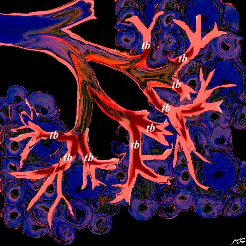
This artistic rendition of the small parts of the lung shows the beginning of the peripheral system just before it enters the acinus. This duct is called the terminal duct and it is the last part of the ductal system that has no ability for gas exchange. After its first division, the bronchioles become the respiratory bronchioles, and they are the first in the system to have an ability to both transport the gases as well as enable gas exchange.
Ashley Davidoff TheCommonVein.net 32645b04b05.8s

This artistic rendition of the small parts of the lung shows the beginning of the peripheral system just before it enters the acinus. This duct is called the terminal duct and it is the last part of the ductal system that has no ability for gas exchange. After its first division, the bronchioles become the respiratory bronchioles, and they are the first in the system to have an ability to both transport the gases as well as enable gas exchange.
Ashley Davidoff TheCommonVein.net
lungs- lo res 0002
Branches and Leaves
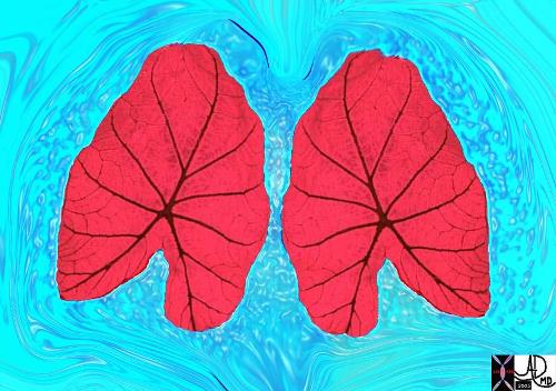
Two leaves of the coleus plant, with a pyramidal or conical shape that reminded the photographer of a set of lungs. The branching system originates from the hilum of the leaf almost at its center, but unlike the tracheobronchial tree it is not irregularly dichotomous.
Ashley Davidoff TheCommonVein.net . 42643
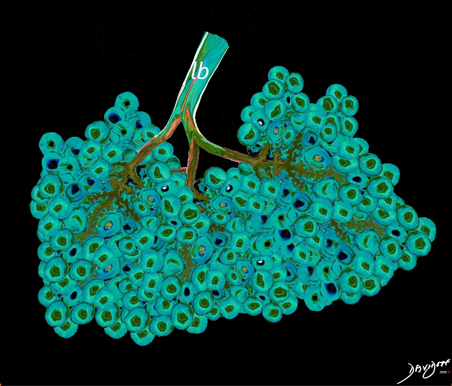
Between 10 and 30 acini combine to form a secondary lobule which is between .5- 2 cms in diameter. It is subtended by a single lobar bronchiole (lb), and is accompanied by arterioles, venules, lymphatics and connective tissue. It is important in clinical radiology since many of the structures can be identified in health, and more particularly in disease, enabling the identification and characterization of many disease processes.
Courtesy Ashley Davidoff MD
lungs-0035-low res

The subsegmental medium sized airways give rise to the terminal bronchiole (tb) which gives rise to the membranous airways. These include in order, the respiratory bronchiole (rb), alveolar duct (ad) and alveolar sac (as)
Ashley Davidoff TheCommonvein.net lungs-0007
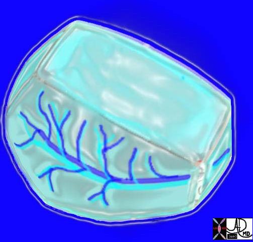
Here is a picture of the outside of the polyhedral pulmonary lobule from the side. It looked quite futuristic. Through the transparent side window we saw a couple similar to ourselves. From this vantage point the morphing did not look too different from what we had already been through – division after division – leaner and meaner. Ashley Davidoff MD. The Common Vein.net 42449b02
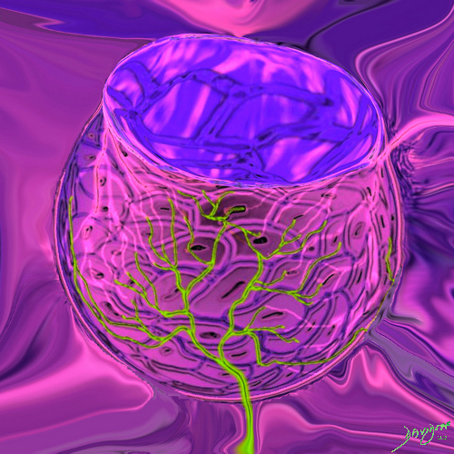
This drawing demonstrates the open mouth view of the alveolus, which is surrounded by its tree like capillary network. The lining single layer of squamous cells (pneumocytes) can be seen peaking through the vessels.
Ashley Davidoff MD. TheCommonVein.net lungs-0022
Ashley Davidoff
TheCommonVein.net lungs-0695

Ashley Davidoff
TheCommonVein.net
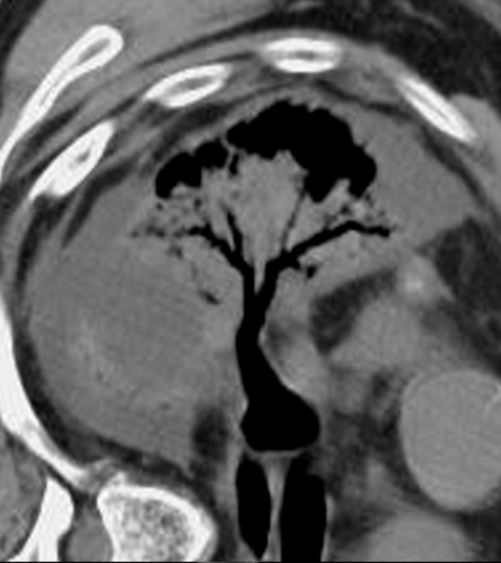
54-year-old female presents with a cough, fever and leukocytosis and multicentric lung abscesses. CT in the axial plane with the patient in decubitus position prior to diagnostic aspiration shows compressive atelectasis of the right upper and right lower lobes by a complex para-pneumonic effusion. Air bronchograms are noted. The residual air in the in the the airways of the compressed lungs is reminiscent of the tree in bud ? but in this case has different implications
Ashley Davidoff MD TheCommonVein.net 19599 art

Ashley Davidoff
TheCommonVein.net

Snow covered red berries ? the contrast between the cherry red and the snow white make them look delicious.
Ashley Davidoff MD
TheCommonVein.net
02160p