
Bronchiectasis (HCC-CMS)
Postinflammatory pulmonary fibrosis
Multiple pulmonary nodules
Seasonal allergic rhinitis
- 63 y.o. female with a
- history of
- HTN, HLD, spinal stenosis,
- LTBI s/p treatment,
- T2DM,
- breast CA s/p adjuvent XRT,
- history of inhalation injury
- inhalation injury about 15-20 years ago
- hot gas blast of unknown source
- walking down the street going to work and walked into this heat with a burning feeling.
- blistered skin and
- hair loss.
- hospitalized at Boston City
- smoking history
- 2-3 cigarettes per day x
- 25 years,
- quit >15 years ago
- PFT
- PFTs have shown significant reduction in DLCO. TTE with
- and abnormal CT,
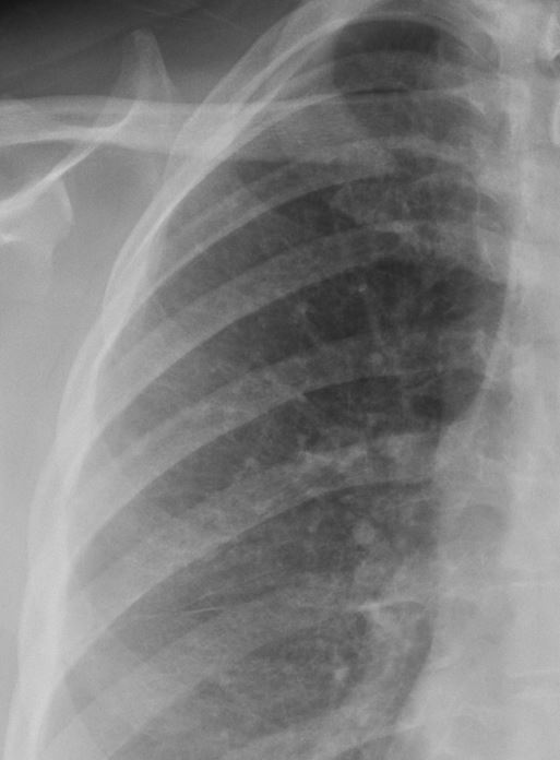
CXR 2yrs After Injury showing Nodules in the RUL
-
-
-

CXR 2yrs After Injury showing Nodules in the RUL
Subsequent Diagnosis likely Langerhans Cell histiocytosis (PLCH)
Ashley Davidoff TheCommonVein.net
CXR 2yrs After Injury showing Nodules in the RUL
Subsequent Diagnosis likely Langerhans Cell histiocytosis (PLCH)
Ashley Davidoff TheCommonVein.netCT scan of the 2yrs After Injury showing Mosaic Attenuation
-
-

Subsequent Diagnosis likely Langerhans Cell histiocytosis (PLCH)
Ashley Davidoff TheCommonVein.net
CXR 4yrs After Injury showing No Significant Change in the Nodules in the RUL

Subsequent Diagnosis likely Langerhans Cell histiocytosis (PLCH)
Ashley Davidoff TheCommonVein.net
Subsequent Diagnosis likely Langerhans Cell histiocytosis (PLCH)
Ashley Davidoff TheCommonVein.net
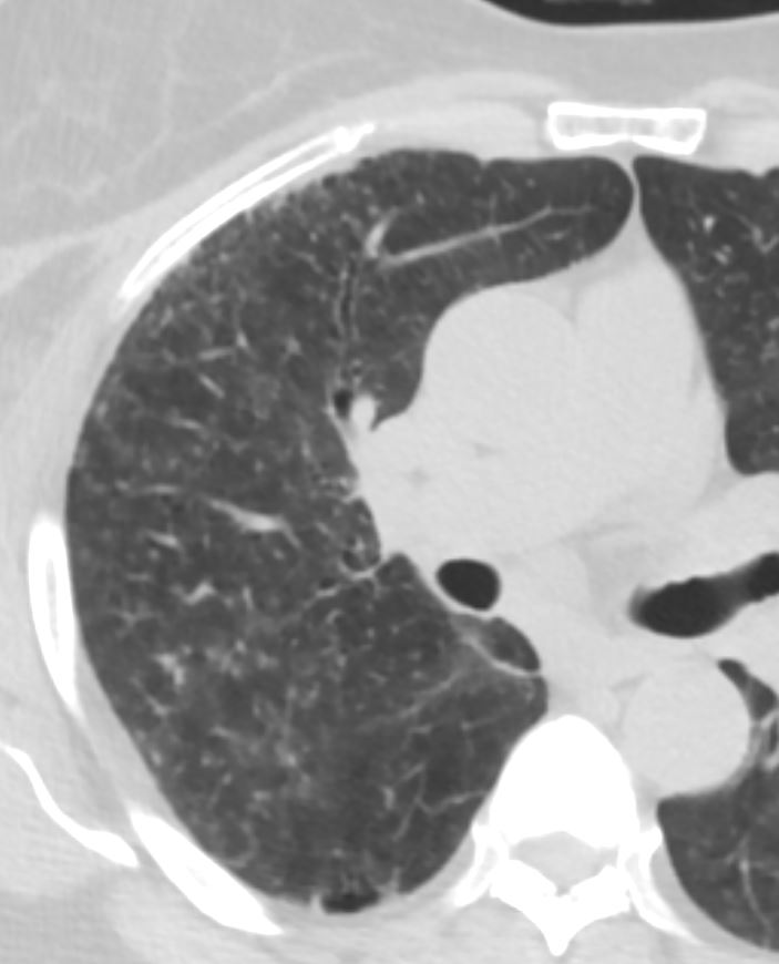
CT scan of the 5yrs After Injury showing Multiple Different Sized Cysts in the Upper Lobes and Diffuse Distribution of Micronodules Suggesting Small Airway Disease

Subsequent Diagnosis likely Langerhans Cell histiocytosis (PLCH)
Ashley Davidoff TheCommonVein.net
CT scan of the 5yrs After Injury showing Multiple Different Sized Cysts in the Upper Lobes and Micronodules Suggesting Small Airway Disease

CT scan of the 5yrs After Injury showing Multiple Different Sized Branching Cysts in the Upper Lobes and Micronodules Suggesting Small Airway Disease

Subsequent Diagnosis likely Langerhans Cell histiocytosis (PLCH)
Ashley Davidoff TheCommonVein.net

Subsequent Diagnosis likely Langerhans Cell histiocytosis (PLCH)
Ashley Davidoff TheCommonVein.net
CT scan of the 5yrs After Injury showing a Cyst Alongside a Pulmonary Arteriole Reflecting Bronchiolectasis

Subsequent Diagnosis likely Langerhans Cell histiocytosis (PLCH)
Ashley Davidoff TheCommonVein.net
CT scan of the 5yrs After Injury showing a Thick Walled Small Airway with Bronchiolectasis

Subsequent Diagnosis likely Langerhans Cell histiocytosis (PLCH)
Ashley Davidoff TheCommonVein.net
CXR 10 yrs After Injury showing No Significant Change in the Nodules in the RUL

Subsequent Diagnosis likely Langerhans Cell histiocytosis (PLCH)
Ashley Davidoff TheCommonVein.net
CT scan of the 11yrs After Injury showing Multiple Different Sized Cysts in the Upper Lobes Not Significantly Changed and Micronodules Suggesting Small Airway Disease

Subsequent Diagnosis likely Langerhans Cell histiocytosis (PLCH)
Ashley Davidoff TheCommonVein.net
CT scan of the 11yrs After Injury showing Multiple Different Sized Branching Cysts in the Upper Lobes and Micronodules Suggesting Small Airway Disease

Subsequent Diagnosis likely Langerhans Cell histiocytosis (PLCH)
Ashley Davidoff TheCommonVein.net
CT scan of the 11yrs After Injury showing Persistent Mosaic Attenuation in the Lower Lung Fields Suggesting Small Airway Disease

Subsequent Diagnosis likely Langerhans Cell histiocytosis (PLCH)
Ashley Davidoff TheCommonVein.net
CT scan of the 11yrs After Injury showing Persistent Mosaic Attenuation in the Lower Lung Fields and Micronodules Suggesting Small Airway Disease
Subsequent Diagnosis likely Langerhans Cell histiocytosis (PLCH)
Ashley Davidoff TheCommonVein.net
CT scan of the 12yrs After Injury showing mostly Stable Multiple Different Sized Cysts in the Upper Lobes and Micronodules Suggesting Small Airway Disease

Subsequent Diagnosis likely Langerhans Cell histiocytosis (PLCH)
Ashley Davidoff TheCommonVein.net

Subsequent Diagnosis likely Langerhans Cell histiocytosis (PLCH)
Ashley Davidoff TheCommonVein.net
CT scan of the 13yrs After Injury showing Slightly Progressive Reticulations and Stable Multiple Different Sized Cysts in the Upper Lobes and Micronodules Suggesting Small Airway Disease

Subsequent Diagnosis likely Langerhans Cell histiocytosis (PLCH)
Ashley Davidoff TheCommonVein.net
CXR 20 yrs After Injury showing Mild Increase in the Reticular Changes and No Significant Change in the in the Nodules in the RUL
