
Infection
Active Miliary TB
CT Miliary Tuberculosis
Nodules along the Fissures (b, Pink Arrowheads)

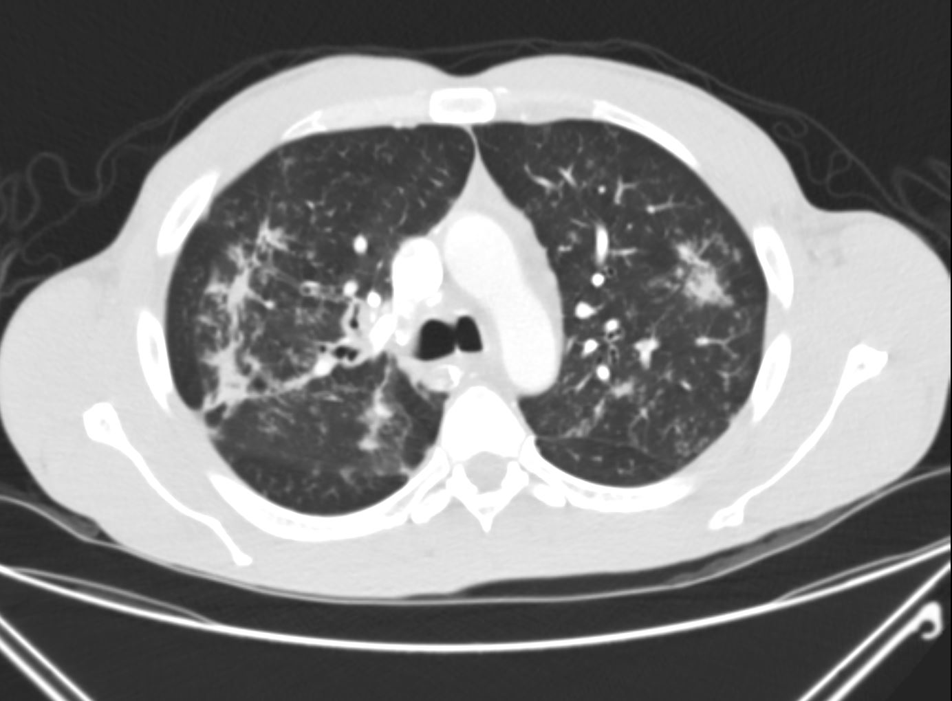
60-year-old immunocompromised female presents with a cough and weight loss. Axial CT shows miliary nodules throughout both lung fields. Some of these nodules are centrilobular or distributed along the bronchovascular bundles (c, maroon arrowheads) and others are fissural based (b, pink arrowheads) and along the pleura (yellow arrowheads suggesting at least a lymphatic distribution. There is bronchial wall thickening (b teal arrowhead). She responded well to treatment and final diagnosis was mycobacterium tuberculosis.
Ashley Davidoff MD TheCommonVein.net 265Lu 136202cL

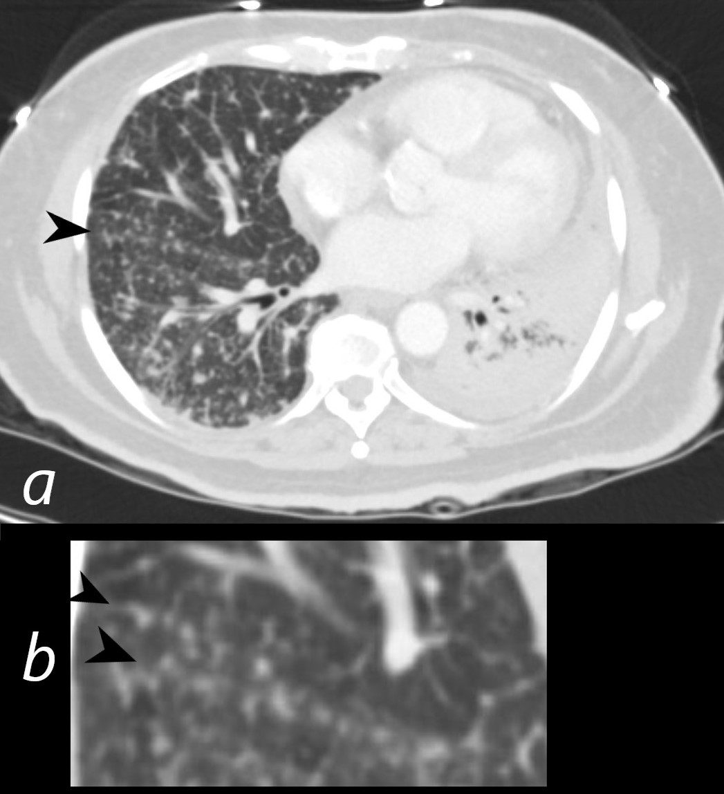
60-year-old immunocompromised female presents with a cough and weight loss. Axial CT shows miliary nodules throughout both lung fields. Some of these nodules are centrilobular or distributed along the bronchovascular bundles (b, c, maroon arrowheads) and others are fissural based (b and c pink arrowheads) , suggesting both bronchovascular and lymphatic distribution. She responded well to treatment and final diagnosis was mycobacterium tuberculosis. There is a healing right sided posterolateral rib fracture.
Ashley Davidoff MD TheCommonVein.net 265Lu 136202cL
Large Calcified Granuloma

CT scan of TB with Calcified Graulomata Centered around Major fissure and Bronchioles with Atelectasis and Bronchiolectasis
Ashley Davidoff MD The CommonVein.net granulomata-along-fissures-005
Inflammation
Sarcoidosis
The axial CTscan shows thickening and irregularity of the major fissure, a band of fibrosis in the right upper lobe, thickening of a segmental bronchus in the right upper lobe and bronchocentric fibrosis in the left upper lobe and multiple micronodules
Ashley Davidoff MD TheCommonvein.net lungs sarcoid 002

70-year-old female with micronodules along the fissures
Ashley Davidoff MD
TheCommonVein.netlenic involvement is noted
Ashley Davidoff MD

51-year-old male with history of sarcoidosis
The frontal CXR shows subtle nodular changes in the right upper peripheral lung field (red circles) and the lateral examination shows 3 regions of nodular changes (red arrowheads)
The CT examination scout film confirms 3 major regions of nodular change in the posterior and superior segment of the RUL along the confluence of the right major and minor fissure and in the posterior segment of the left upper lobe peripherally.
The axial images show a variety of characteristic changes including;
Ground glass opacity
Stellate or flame shaped nodules
Semisolid nodules
Fissural based nodules
Subpleural nodules
Micronodules along the
lymphovascular and
bronchovascular bundles of the secondary lobule
Calcified nodule some of which are surrounded by soft tissue of the granuloma
There are small calcified nodes in the mediastinum, but no significant pathological adenopathy
No obvious cardiac nor splenic involvement is noted
Ashley Davidoff MD
Malignancy
50-year-old female with primary adenocarcinoma of the left lung presenting with pneumonic consolidation of the left lower lobe, and diffuse reticulonodular changes bilaterally
The axial CT through the mid chest shows nodular changes along the major fissure (a and b, black arrowheads). These findings are consistent with the diagnosis of lymphangitis carcinomatosa.
The left lung shows consolidation in the posterior aspect of the left lower lobe
Ashley Davidoff MD TheCommonVein.net 158Lu 131029c01L

CT in the axial plane demonstrates a large, spiculated mass in the right upper lobe with surrounding halo likely reflecting hemorrhage or lymphatic edema around the mass. In addition, there is evidence of irregular interlobular septal thickening likely reflecting lymphatic invasion and indicating lymphangitis carcinomatosa. There is irregular thickening of the major fissure suggesting involvement.
Ashley Davidoff MD TheCommonVein.net 135865
Mechanical
Traction in Emphysema
Fissural Changes from Traction

51-year-old female smoker with a history of COPD asthma and pulmonary hypertension presents with progressive dyspnea. Axial CT through the upper lung fields at the level of the carina shows extensive changes of centrilobular emphysema and ground glasses changes in the anterior segments ? right more prominent than the left. In addition there is irregularity of the right major fissure (lower panel) seemingly as a result of the enlarged secondary lobule, and stress on the fissure by the interlobular septa. Path confirmed a diagnosis of DIP
Ashley Davidoff MD TheCommonVein.net 252Lu 135965c

51-year-old female smoker with a history of COPD asthma and pulmonary hypertension presents with progressive dyspnea. Axial CT through the upper lung fields at the level of the carina shows progression from extensive centrilobular changes to ground glass changes in the left anterior segment, and diffuse ground glass changes in the lower lobes. In addition, there is irregularity of the right major fissure (lower panel) seemingly as a result of the enlarged secondary lobule, and stress on the fissure by the interlobular septa. Path confirmed a diagnosis of DIP
Ashley Davidoff MD TheCommonVein.net 252Lu 135966c
/Atelectasis Trauma Metabolic
Circulatory CHF

CT
Ashley Davidoff MD
thecommonvein.net
Circulatory- Hemorrhage
75-year-old man on blood thinners s/p aortic valve replacement s/p trauma presents with hemoptysis. He was afebrile and without an elevated white count
Scout CT shows an elevated right hemidiaphragm and inferior displacement of the major fissure with a dense right upper lobe consolidation. The mass effect on the major fissure likely results from a hematoma, and the hemorrhage results in air bronchograms and groundglass changes.
Skin folds on the left results in a pseudo-pneumothorax. A loop recorder is noted overlying the left upper chest.
Ashley Davidoff MD TheCommonVein.net 165Lu 135850

75-year-old man on blood thinners s/p aortic valve replacement s/p trauma, presents with hemoptysis. He was afebrile and without an elevated white count
Axial CT at the level below the carina shows medial displacement of the major fissure by a dense right upper lobe consolidation. The mass effect on the major fissure likely results from a hematoma. Anterior to the consolidation there is a combination of ground glass opacity with thickened interlobular septa, and minor region of subsegmental consolidation with air bronchograms, likely resulting from hemorrhage.
There are bilateral effusions
Ashley Davidoff MD TheCommonVein.net 165Lu 135851

75-year-old man on blood thinners s/p aortic valve replacement, s/p trauma, presents with hemoptysis. He was afebrile and without an elevated white count
Coronal CT of the posterior lung fields shows inferior displacement of the major fissure by a dense right upper lobe consolidation. The mass effect on the major fissure likely results from a hematoma. Lateral to the consolidation there is a combination of ground glass opacity. There is elevation of the right hemidiaphragm. Left sided pleural effusion is present
Ashley Davidoff MD TheCommonVein.net 165Lu 135860
Immune
Infiltrative
Amyloidodis

Axial CT images through the chest shows a Fissural based amyloid nodule along the left major fissure (a,b) Images c and d show posterior peripheral centrilobular nodules. In image c the nodules are associated with mosaic attenuation. The ground glass nodule in d could reflect alveolar septal disease withground glass changes surrounding a centrilobular nodule
Ashley Davidoff Boston Medical Center TheCommonvein.net LV-014c
Idiopathic Iatrogenic Idiopathic