
lungs-00677-lo-res.jpg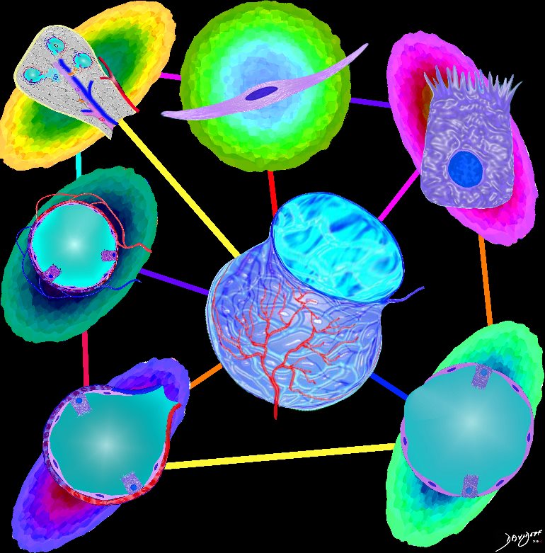
Parts and Bonds
Ashley Davidoff MD TheCommonVein.net lungs-0060
Airways are lined by a pseudostratified ciliated columnar epithelium interspersed with mucus secreting goblet cells
Ashley Davidoff
TheCommonVein.net lungs-00674b01-lo res
Ashley Davidoff TheCommonVein.net lungs-00675-lo-res
Ashley Davidoff
TheCommonVein.net lungs-00677-lo res
The Buck Ends Here
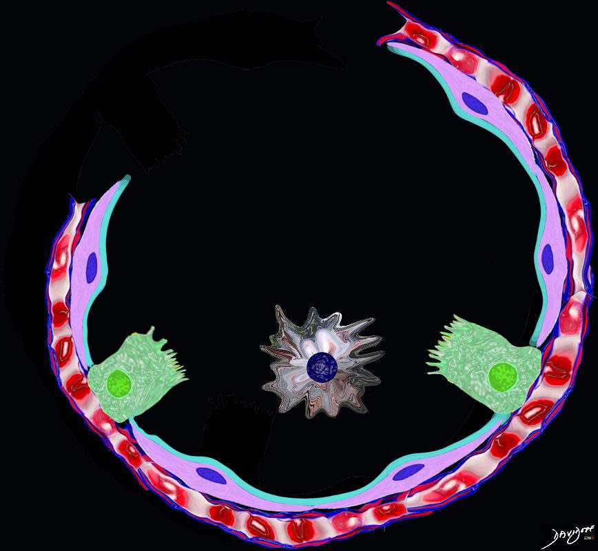
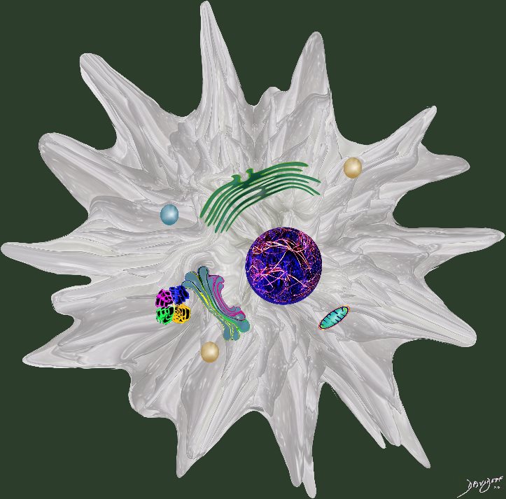
The alveolus is lined by a simple epithelium ? one cell layer thick. There are two types of lining cells; Type 1 pneumocytes are squamous cells that cover 90% of the surface of the inner lining of the lung , and type II cuboidal pneumocytes that are in fact much more numerous than Type I. They are involved in the production of surfactant . In the lumen there are resident macrophages which play a crucial role in the immune system. The mucosa is grounded by a basement membrane and a lamina propria, and connected to the lamina propria and basement membrane of the surrounding capillary. The alveolus is lined by a thin layer of surfactant. (teal blue)
Ashley Davidoff
TheCommonVein.net
Ashley Davidoff MD

It produces the phospholipid – part of the surfactant that reduces surface tension and allows the alveoli to remain open
#cells
Ashley Davidoff
TheCommonVein.net
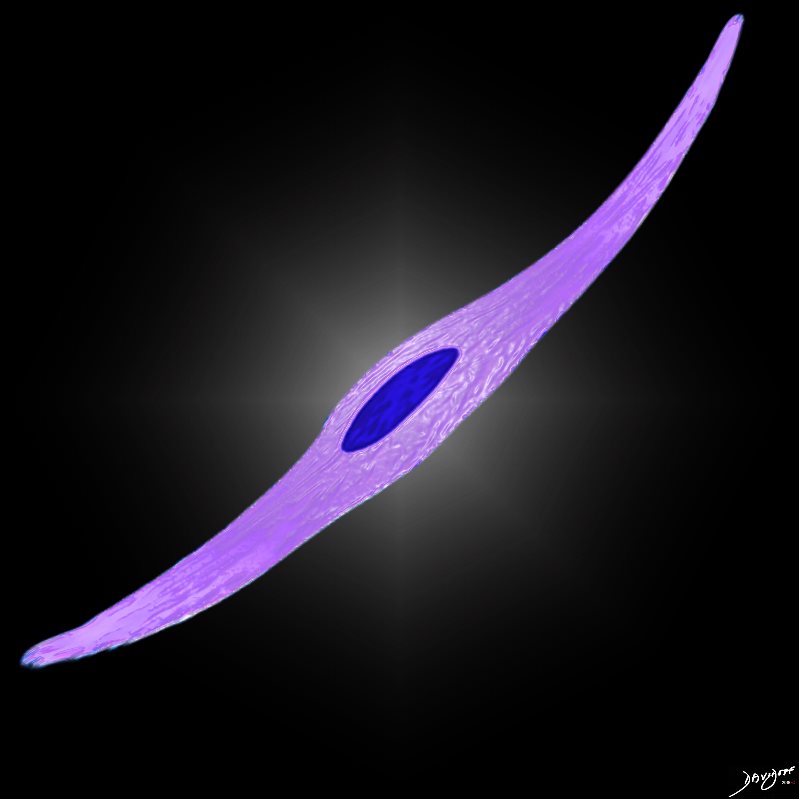
– flattened for gas exchange, forms a part of the Blood-Gas Barrier
nd sometimes vacuolated cytoplasm
It produces the phospholipid – part of the surfactant that reduces surface tension and allows the alveoli to remain open
#cells
Ashley Davidoff
TheCommonVein.net
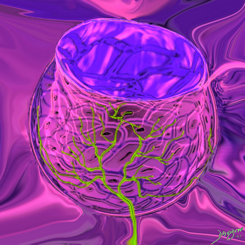
This drawing demonstrates the open mouth view of the alveolus, which is surrounded by its tree like capillary network. The lining single layer of squamous cells (pneumocytes) can be seen peaking through the vessels.
Ashley Davidoff MD. TheCommonVein.net lungs-0022
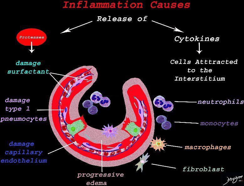
The cells of the immune system release cytokines, chemotactic agents and proteases. Immune cells , macrophages and fibroblasts are attracted to the interstitium. Some of proinflammatory agents are toxic to the cell lining causing damage to the surfactant, type 1 pneumocytes and the capillary endothelium. There is progressive edema.
Ashley Davidoff TheCommonVein.net lungs-0692-lo-res