
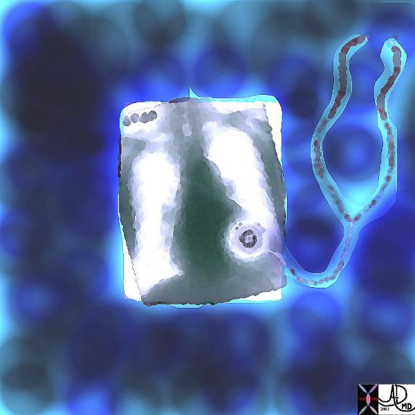
1-1-1-005-lo-res.jpg
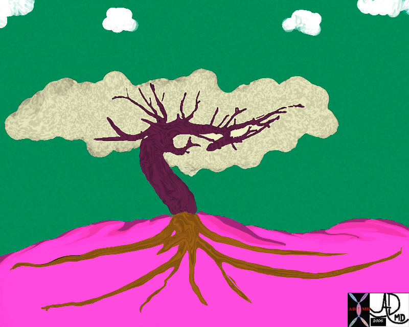
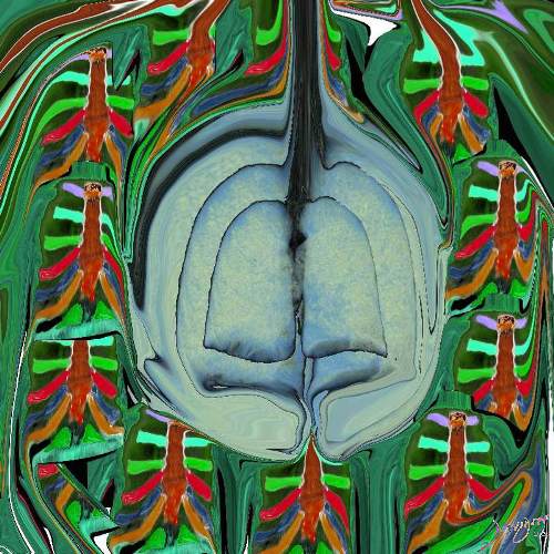
In biology, units bond with other units in a given environment, and create a new and miraculous unit, bigger than the parts and with greater functionality.
During life, the units are in a state of health (order) or disease (disorder), or somewhere in between. It is our function to identify disorder , and help bring disorder back to health.
Courtesy Ashley Davidoff MD TheCommonVein.net

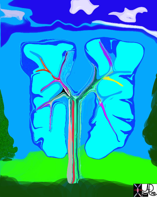
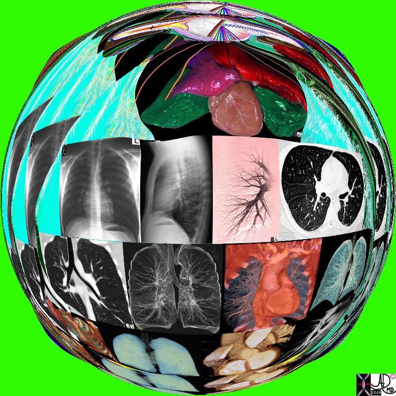
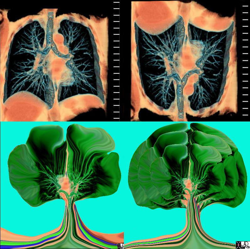
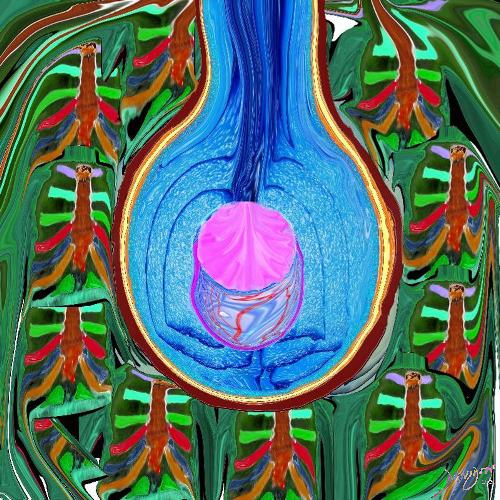
The image shows some of the major components of the lung that when bonded create a new and powerful unit – a vital organ. In the center is an example of the airways and parenchyma making up the 2 lungs. At 12 oclock the tracheo-bronchial tree with segmental and subsegmental airways. At 1 o?cloclock, is a cross section of the lungs showing some of the segments of the lung. At 5o?clock a cross section shows the arteries and veins of the lungs. At 7o?clock the drawing shows the pleura and pleural space of the lungs. At 9o?clock, a coronal reformat of the tracheobronchial tree shows the lymph node stations of the lungs. At 11 o?clock is the golden alveolus, the epicentral unit where gas exchange takes place
Ashley Davidoff MD TheCommonVein.net lungs-0696-lo res
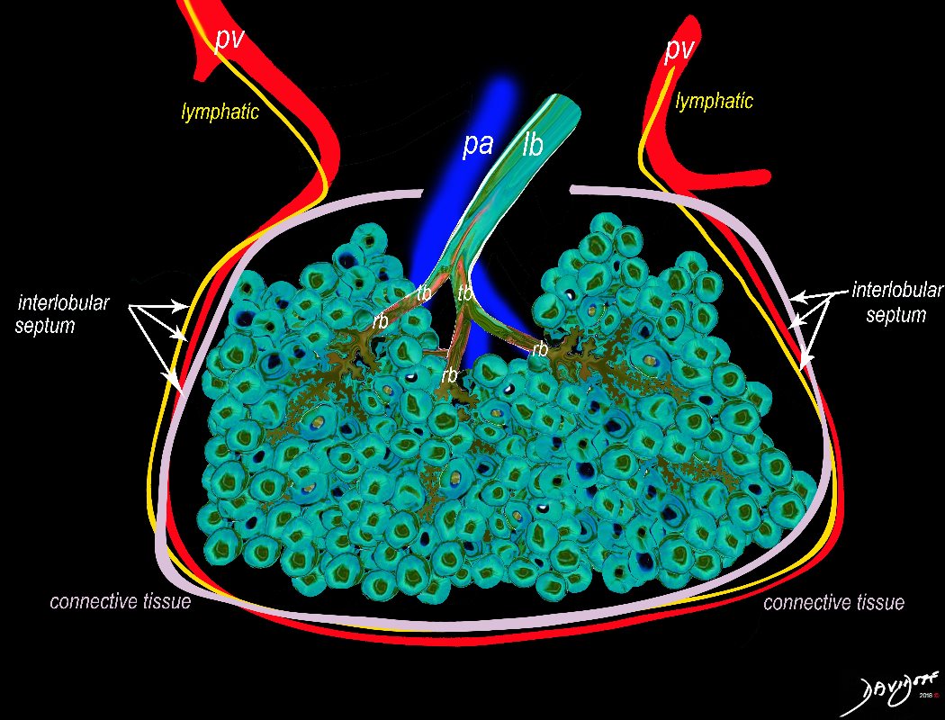
The secondary lobule is subtended by the lobular arteriole (a) and the lobular bronchiole (b) which which in turn branches into the respiratory bronchioles, alveolar ducts, and nd alveolar sacs (c) The acinus (d) consists of a respiratory bronchiole and its associated alveolar ducts, sacs, and alveoli and represents the functional unit of the lung.
The secondary lobule is drained by the pulmonary venule (e) which runs in the interlobular septum also containing the lymphatics (f). The whole unit is housed and surrounded by a connective tissue framework (g) . The latter 3 structures form the interlobular septum.
Ashley Davidoff MD TheCommonVein.net 0751 -lo resL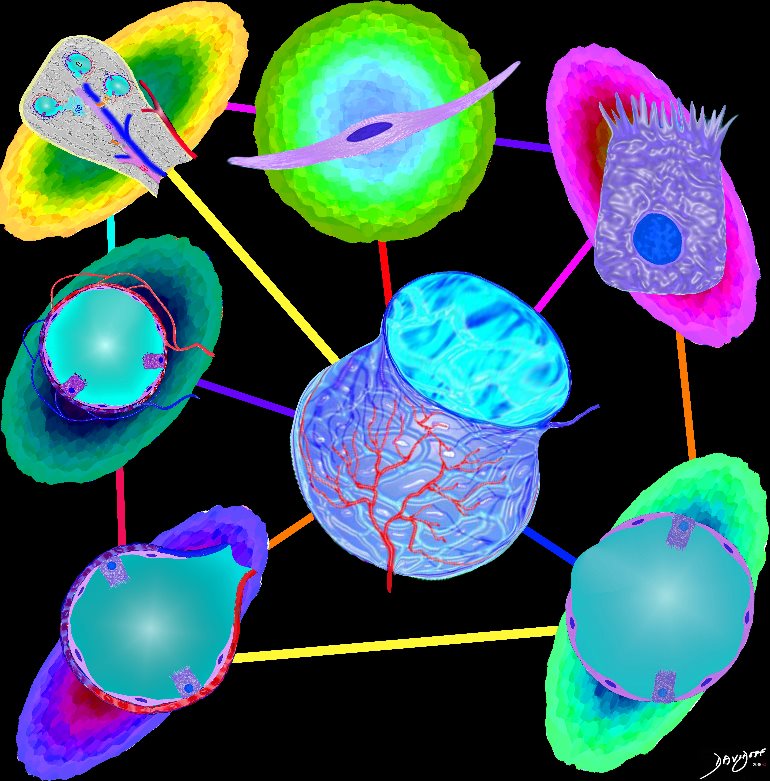
Parts and Bonds
Ashley Davidoff MD TheCommonVein.net lungs-0060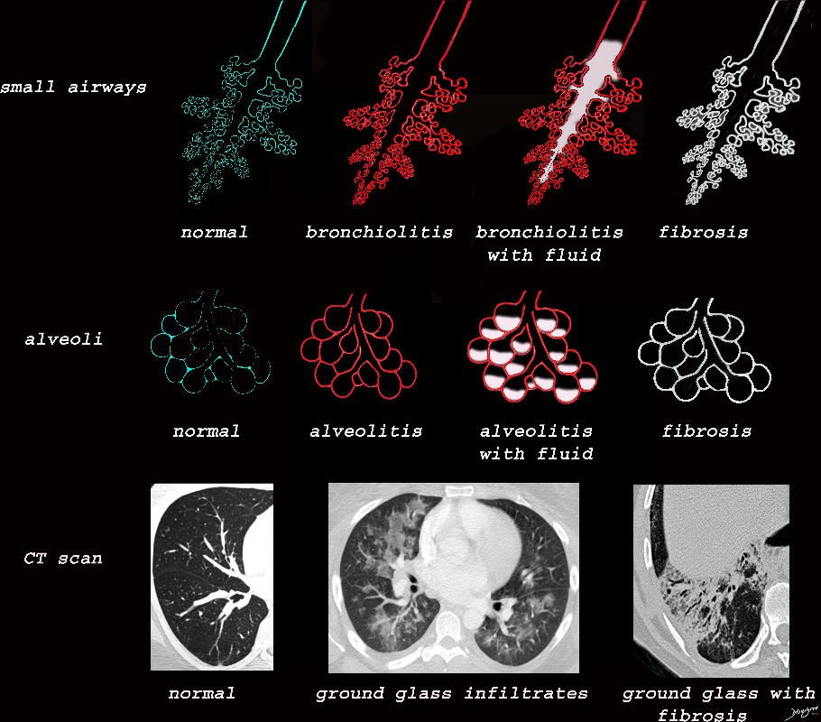
Ashley Davidoff MD TheCommonVein.net lungs-0733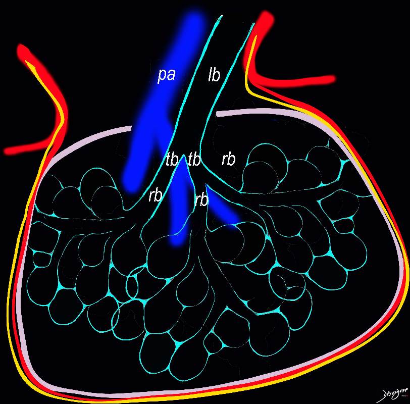
The secondary lobule is housed in a connective tissue framework in which run the lymphatic and venular tributaries . Together these 3 structures form the interlobular septum.
The lobar arteriole enters the framework, accompanied by the lobar bronchiole, and they all run together and form the interlobular septa. This structure measures between .5cms and 2cms and is visible on CT scan.
It is important in clinical radiology since many of the structures can be identified in health, and more particularly in disease, enabling the identification and characterization of many pathological processes.
Courtesy Ashley Davidoff MD The CommonVein.net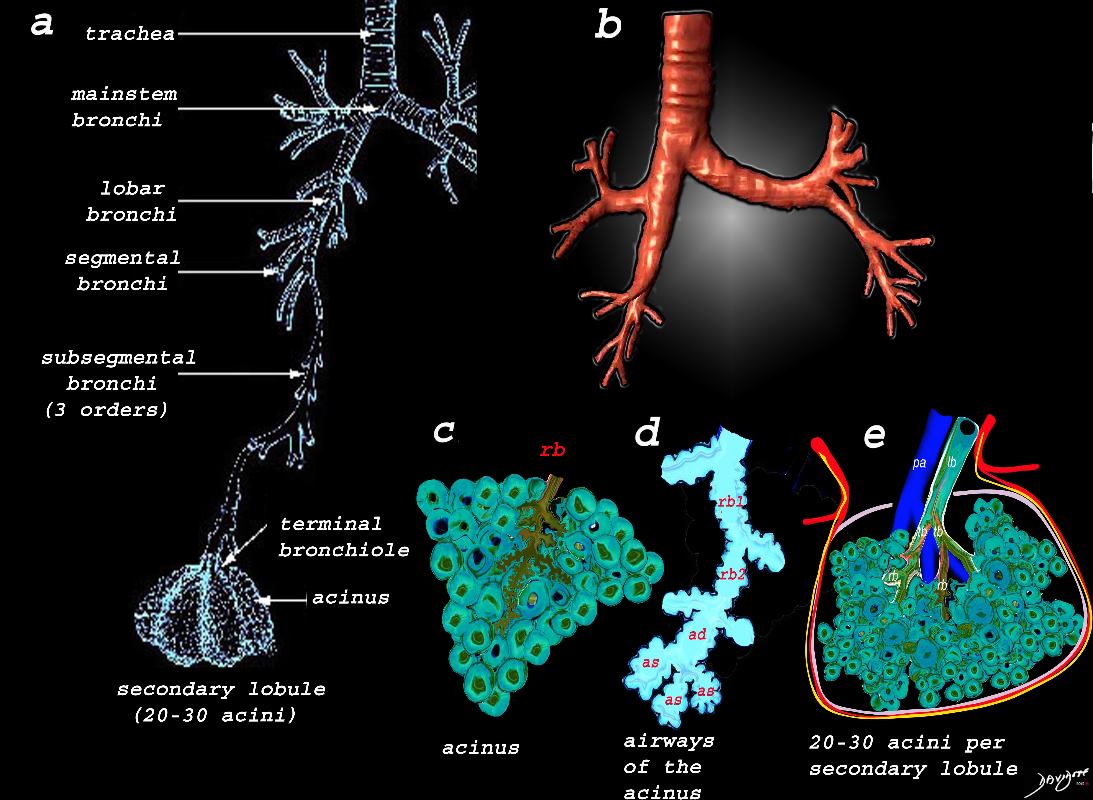
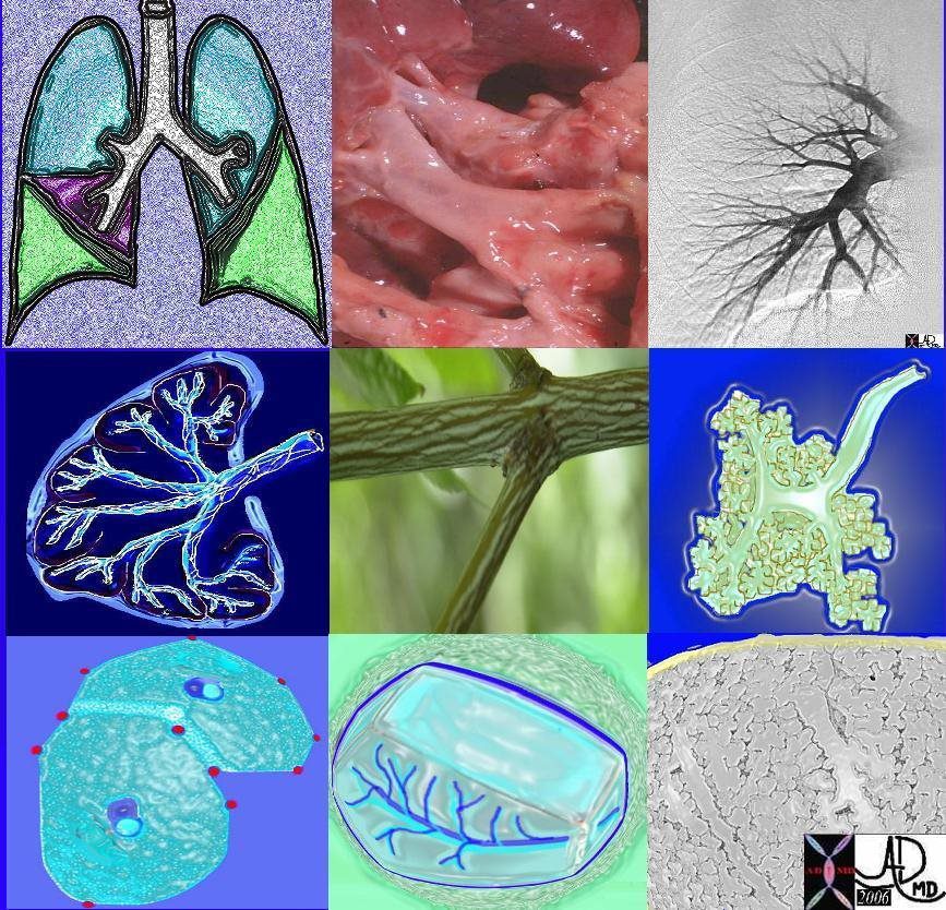
Image a shows the airways starting in the trachea and continuing to the mainstem bronchi, lobar bronchi, segmental bronchi, and subsegmental bronchi,. The subsegmental bronchi have 3 subsequent generations until the bronchiole is reached. The terminal bronchiole is the last of the transporting airways and is considered the most proximal small airway with a diameter of 2mm or less, and it gives rise to the respiratory bronchiole which is the feeding airway for the acinus . The acinus is the functional unit of the lung.
Image b is a 3D reconstruction of a CT scan showing the proximal airways from the trachea to the segmental airways.
Image c shows the structures that make up the acinus and the other parts of the small airways, starting with the respiratory bronchiole (rb) . The diagram in d, shows the detail of the small airways that participate in gas exchange, including the respiratory bronchiole, (rb) alveolar duct, (ad) and alveolar sac (as)
Image e shows the secondary lobule made from about 20-30 acini, arising from a single lobular bronchiole accompanied by a single pulmonary arteriole (pa).. Structure that surround and enclose the secondary lobule include the pulmonary venule, (red) lymphatics,(yellow) and a fibrous septum (pink).
Ashley Davidoff MD TheCommonVein.net
lungs-0739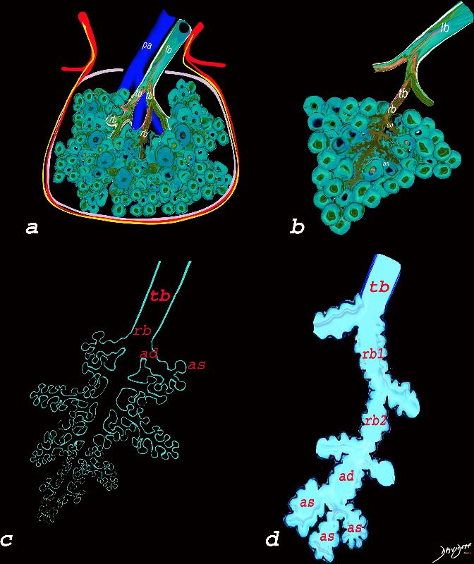
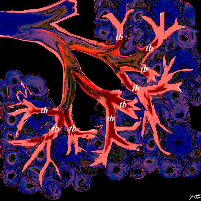
The diagram allows us to understand the the components and the position of the small airways starting in (a) which is a secondary lobule that is fed by a lobular bronchiole(lb) which enters into the secondary lobule and divides into terminal bronchioles (tb) which is the distal part of the conducting airways, and at a diameter of Ashley Davidoff MD TheCommonVein.net lungs-0744
The diagram allows us to understand the the components and the position of the small airways starting in (a) which is a secondary lobule that is fed by a lobular bronchiole(lb) which enters into the secondary lobule and divides into terminal bronchioles (tb) which is the distal part of the conducting airways, and at a diameter of 2mm or less . It divides into the respiratory bronchiole (rb) a transitional airway which then advances into the alveolar ducts(ad) and alveolar sacs (as) Diseases isolated to the small airways do not affect the alveoli and hence there is peripheral sparing Ashley Davidoff MD TheCommonVein.net
The club cell aka bronchiolar exocrine cells formerly known as the Clara cell is a low columnar cell with short microvilli and are most abundant in the bronchioles
Ashley Davidoff MD TheCommonVein.net lungs-0743
This upper diagram shows the ciliated columnar epithelium present throughout the 20- 25 generations of branching, until the airways start to transition their function from a transport system to a gas exchange system at the respiratory bronchiole level The ciliated columnar epithelium becomes a ciliated cuboidal epithelium. There are no goblet cells in the bronchioles In addition to the ciliated cells there are 2 other types of cells including the club cell (purple) and the neuroendocrine cell. The club cells Purple with dome shaped superior aspects – formerly Clara Cell) have many functions. The neuroendocrine cell (NE) (dark pink and round ) can be seen as a single cell (NE) and sometimes seen in a cluster, known as a neuroendocrine body (NEB) . The cells rest on a basement membrane, with prominent muscle layer (maroon) as well as elastic tissue (pink). There is no cartilage
Ashley Davidoff MD TheCommonVein.net
lungs-0741n
The secondary lobule is housed in a connective tissue framework in which run the lymphatic and venular tributaries . Together these 3 structures form the interlobular septum.
The lobar arteriole enters the framework, accompanied by the lobar bronchiole, and they all run together and form the interlobular septa. This structure measures between .5cms and 2cms and is visible on CT scan.
It is important in clinical radiology since many of the structures can be identified in health, and more particularly in disease, enabling the identification and characterization of many pathological processes.
Courtesy Ashley Davidoff MD The CommonVein.net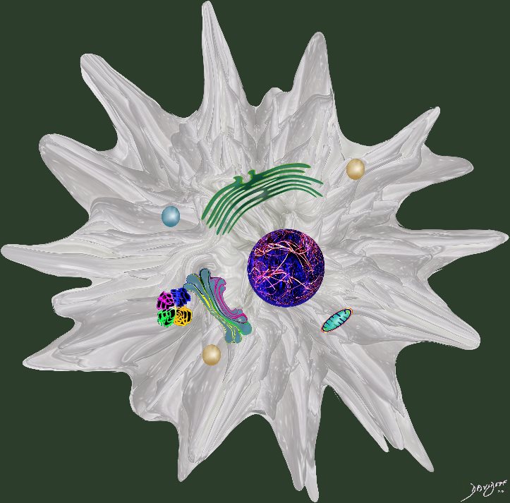
Ashley Davidoff MD
Light brown granules in the macrophage is characteristic of the smokers macrophage
Ashley Davidoff
TheCommonVein.net lungs-00686
It produces the phospholipid – part of the surfactant that reduces surface tension and allows the alveoli to remain open
#cells
Ashley Davidoff
TheCommonVein.net lungs-0062
– flattened for gas exchange, forms a part of the Blood-Gas Barrier
nd sometimes vacuolated cytoplasm
It produces the phospholipid – part of the surfactant that reduces surface tension and allows the alveoli to remain open
#cells
Ashley Davidoff
TheCommonVein.net lungs-0061
Ashley Davidoff MD
TheCommonVein.net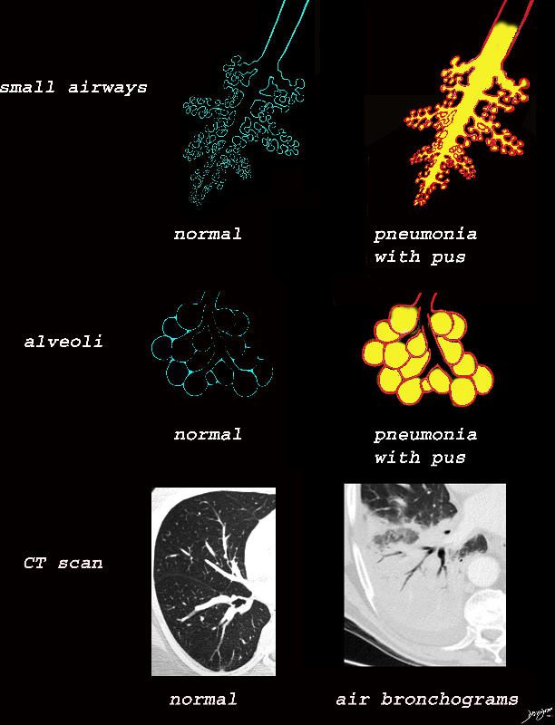
The collage provides a persepective of purulent accumulation in the small airways and the alveoli that results in consolidation. A process that increases the densityof the lungs to a net “white”regional density will result in a consolidation and in this case when the fluid is infected it is labelled “pneumonia” The net result on CT is aair bronchograms within the non aerated dense lung tissue.
Ashley Davidoff MD TheCommonVein.net lungs-0734
Ashley Davidoff MD TheCommonVein.net lungs-0733
Ashley Davidoff MD TheCommonvein.net lungs-0732b01

Ground glass opacification may be caused by partial filling of the alveolus with cellular material with partial replacement of air with solid material with the net density being gray rather than white if the alveolus were fully filled. The black of the airway nor the white of the vessels may blend with the gray density and hence they are not visualised in ground glass opacities. ore is usually lood vessels and airway walls The replacement may be due to cellular infiltration including inflammatory ,benign or malignant cells without or with fluid.
Ashley Davidoff MD TheCommonVein.net lungs-0707ad

Ashley Davidoff MD TheCommonVein.net lungs-0702d- lo res

Consolidation is the replacement of air with solid material resulting in obscuration of blood vessels and airway walls The replacement may be due to cellular infiltration including inflammatory ,benign or malignant cells without or with fluid.
Ashley Davidoff MD TheCommonVein.net
lungs-0707d- lo res

Ashley Davidoff MD TheCommonVein.net
lungs-0704d

The acute inflammatory process results in fluid exudation into the alveoli which can take the form of a serous transudate, and exudate or in the form of mucus, and when severe (eg ARDS) can result in tissue and vessel destruction and could be be blood tinged. Infected fluid could be mucoid or purulent. The extent of filling the alveoli results either in a ground glass appearance when partially filled or a consolidation when filled.
Ashley DAvidoff MD TheCommonVein.net
lungs-0701d- lo res
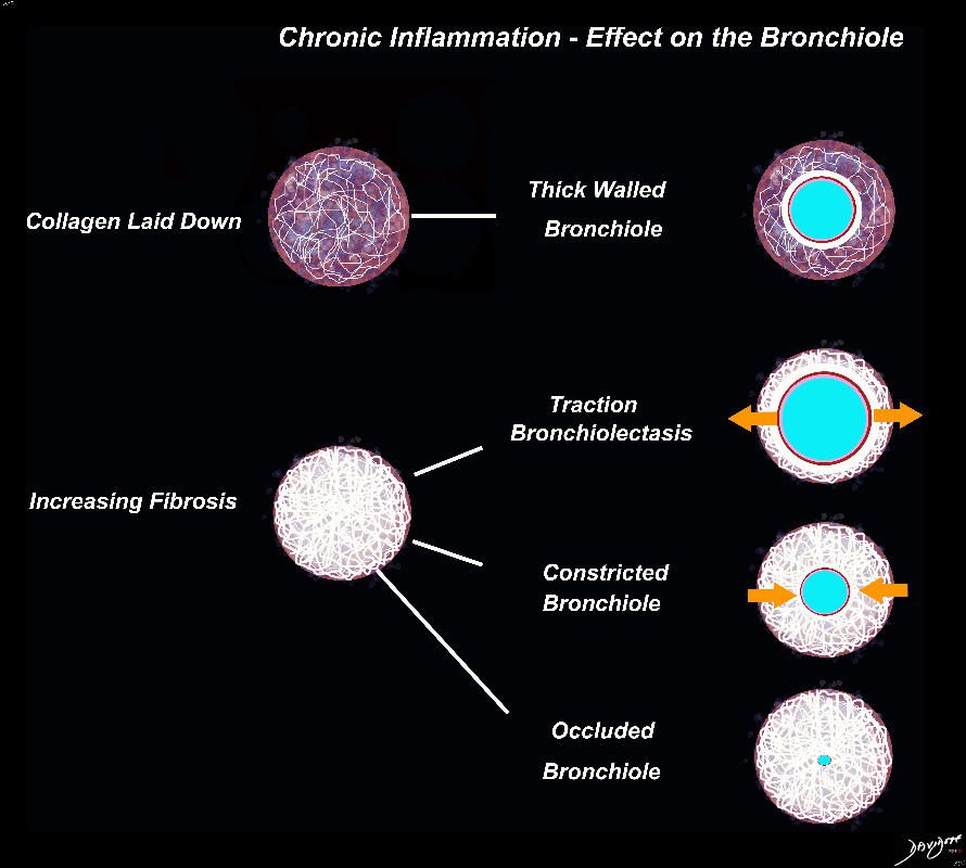
I the early phases collagen starts to get laid down resilting in a thick walled bronchiolesurrounde by a subacute inflanmmatory response of cells and resolving fluid. As the fibrotic process advances it gets denser resulting in traction bronchiectasis and bronchiolectasis. The ongoing may eventually constrict the aurway and subsequently occlude occlude the airway
Ashley Davidoff MD TheCommonVein.net

Ashley Davidoff MD TheCommonVein.net

Ashley Davidoff MD TheCommonVein.net lungs-0707

Black White and Gray Densities
The filling of alveoli with fluids or cells results in a density that is ?white? on X-ray and CT scan and is in distinct contrast to the black of the air filled airways. This contrast results in an air bronchogram. The smaller airways in a normal patient are not usually visualized because the ?black? of the of the airways and the black of the air filled alveoli does not create a contrast.
Ashley Davidoff MD TheCommonvein.net
lungs-0708

Ashley Davidoff MD TheCommonVein.net lungs-0725

Ashley Davidoff MD TheCommonVein.net lungs-0724

Ashley Davidoff MD TheCommonVein.net lungs-0723b
Ashley Davidoff thecommonvein.net

Ashley Davidoff
TheCommonVein.net

Ashley Davidoff
TheCommonVein.net
Ashley Davidoff
TheCommonVein.net
This artistic rendition of the small parts of the lung shows the beginning of the peripheral system just before it enters the acinus. This duct is called the terminal duct and it is the last part of the ductal system that has no ability for gas exchange. After its first division, the bronchioles become the respiratory bronchioles, and they are the first in the system to have an ability to both transport the gases as well as enable gas exchange.
Ashley Davidoff
TheCommonVein.net
32645b04b05.8s

This diagram shows the basic division of the tracheobronchial tree into lobes. The right lung is divided into right upper (RUL) (teal) right middle, (RML pink) and right lower lobe (RLL green). The left lung is divided into left upper (LUL teal), which includes the lingula(dark blue), and left lower lobe (LLL= green). Note that the two mainstem bronchi are of unequal length and size. The right mainstem is short and fat while the left is long and thin. This irregular dichotomous branching pattern is characteristic of the branching pattern of all the conducting systems within the lungs.
Ashley Davidoff
TheCommonVein.net
32686b05
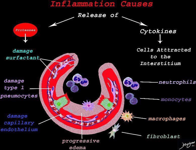
The cells of the immune system release cytokines, chemotactic agents and proteases. Immune cells , macrophages and fibroblasts are attracted to the interstitium. Some of proinflammatory agents are toxic to the cell lining causing damage to the surfactant, type 1 pneumocytes and the capillary endothelium. There is progressive edema.
Ashley Davidoff
TheCommonVein.net
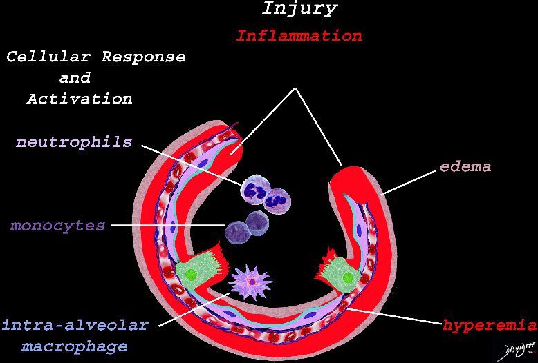
The initial injury results in an acute severe inflammatory response consisting hyperemia , edema with migration initially of neutrophils in the first 6-24 hours followed by monocytes (24-48hours). The intra -alveolar macrophages are activated.
Ashley Davidoff
TheCommonVein.net
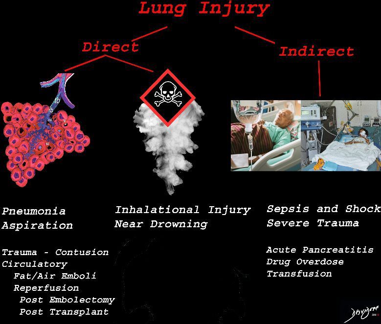
The lung is injured either by direst causes most commonly pneumonia, aspiration or from inhalation of toxic substances. Severe systemic illnesses, most commonly sepsis with shock, and severe trauma are considered indirect causes.
Ashley Davidoff
TheCommonVein.net

The diagram shows the lining of the normal alveolus composed of type 1 pneumocyte squamous in nature and the cuboidal cell (type pneumocyte) which rest on a lamina propria, and basement membrane (not shown) shared with the inner endothelial layer of the capillary. Intra-alveolar macrophage lies within the alveolar lumen
Ashley Davidoff
TheCommonVein.net
42474b18.800 lung trachea bronchi tracheobronchial tree
Ashley Davidoff MD
TheCommonVein.net

Snow covered red berries ? the contrast between the cherry red and the snow white make them look delicious.
Ashley Davidoff MD
TheCommonVein.net
02160p

This is a drawing of a cluster of alveoli surrounded by the capillary network, fed by an arteriole in blue, and drained by a venule in red. The second image shows the exchange of life giving oxygen for the by product of metabolic activity ? carbon dioxide
Ashley Davidoff MD
TheCommonVein.net
32165c

Tree in bud nociceptors free nerve endings trees in the body
Ashley Davidoff MD
TheCommonVein.net87559pb04b07b.8s
Ashley Davidoff MD
TheCommonVein.net.
32697
82738p chest lung connective tissue pulmonary artery pulmonary vein axial interstitial tissue secondary lobule lobes segments trachea bronchi interlobular septa polygonal Ashley Davidoff MD
TheCommonVein.net

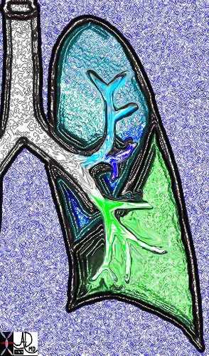
This diagram illustrates the branching pattern of the tracheobronchial tree that extends from the bronchi to the terminal bronchioles transitioning into the alveoli via the alveolar sacs.
32645b04b04 lung D
Ashley Davidoff MD
TheCommonVein.net 32645a10.800
32647 Davidoff
Ashley Davidoff MD
TheCommonVein.net
42444b18.8 lungs anatomy X-ray
Ashley Davidoff MD
TheCommonVein.net
46649c01.800 lung pulmonary artery pulmonary trunk
Ashley Davidoff MD
TheCommonVein.net

32368 Ashley Davidoff MD
TheCommonVein.net
46649b04b.800 lung pulmonary artery pulmonary trunk Ashley Davidoff MD
TheCommonVein.net
46649b11.800b01 lung pulmonary Ashley Davidoff MD
TheCommonVein.net
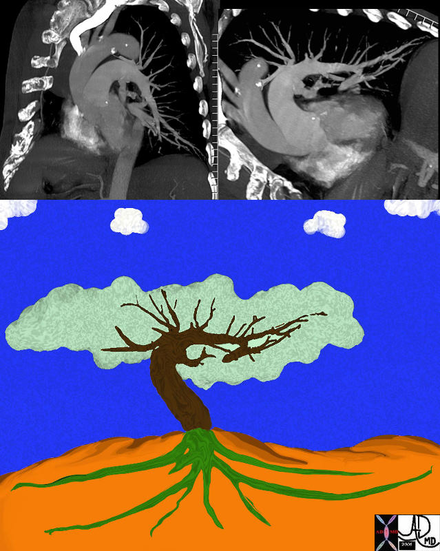
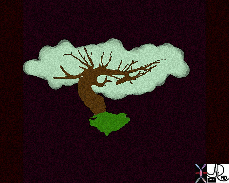
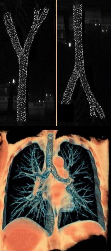
The tracheobronchial tree turned upside down shows it?s similarity to the branching pattern of a tree.
keywords lung bronchus tracheobronchial tree airway tree the common vein applied biology
Ashley Davidoff MD TheCommonVein.net
32620c02.800

Tracheobronchial Tree
Tree, flower, tracheobronchial tree, trachea bronchi lung
Ashley Davidoff Art 32620b14.800b02p
Ashley Davidoff MD
TheCommonVein.net
lungs-00687-lo-res.jpg
Smoking and the Alveolus –The effect of the proteases and and elastases cause destruction of the alveoli and loss of elasticity, and therefore overall function. The destruction leads to bullous disease
The accumulation of smokers macrophage, and in the case of Langerhans cell histiocytosis leads to space occupation of the alveoli also reducing function
Ashley Davidoff
TheCommonVein.net
lungs-00687-lo res

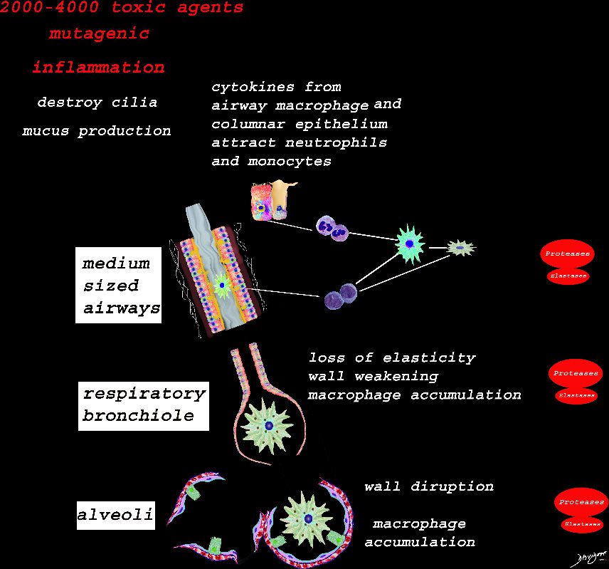
Inhaled smoke contains between 2000-4000 toxic substances. The cancer producing properties are well known. The smoke causes inflammation of the medium sized airways resulting in activation of the resident macrophage in the lumen of the airway, hyperemia of the mucosa, activation of the neutrophils, monocytes, nonresident macrophages, and fibroblasts. Cytokines are released which also induce the secretion of proteases and elastases among many others. Structurally there is destruction of the cilia, hypersecretion of mucus, hypertrophy of the muscularis and mild fibrosis. These findings are the hallmark of chronic bronchitis
Ashley Davidoff
TheCommonVein.net
Ashley Davidoff
TheCommonVein.net
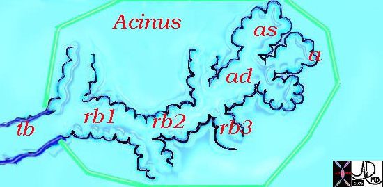
This diagram illustrates the acinus which consists of the respiratory bronchioles (rb 1, 2, 3) the alveolar duct (ad) the alveolar sac (as) and the alveoli. (a)
Courtesy Ashley Davidoff MD 42446b12
TheCommonVein.net
Parts and Bonds
Ashley Davidoff MD
lungs-0023catalogue-signed-small.jpg

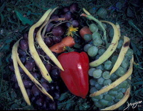
Photograph of the Heart and Lungs created with a red pepper (the heart, grapes (alveoli) carrots (pulmonary arteries), dandelion(mediastinum) and banana peels (ribs)
Ashley Davidoff MD TheCommonVein.net


Ashley Davidoff TheCommonVein.net . 22071b01.800
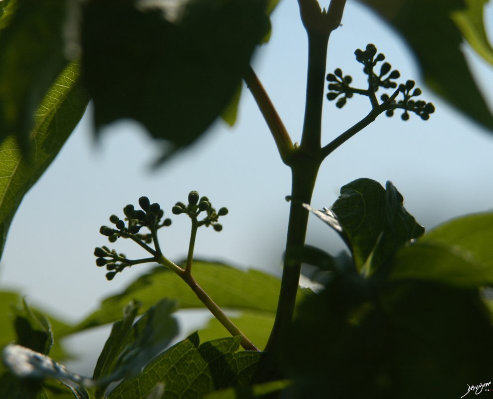
Two leaves of the coleus plant, with a pyramidal or conical shape that reminded the photographer of a set of lungs. The branching system originates from the hilum of the leaf almost at its center, but unlike the tracheobronchial tree it is not irregularly dichotomous.
Ashley Davidoff TheCommonVein.net . 42643
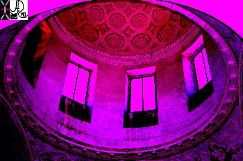
This cupola or dome was photographed in the church of the Villa Melzi gardens in Bellagio, Italy. If you imagine yourself in the chest cavity and you look up towards the neck, this is what you will see ? the dome shaped structure of the apex of the lung and pleura.
Ashley Davidoff TheCommonVein.net 78115pb01
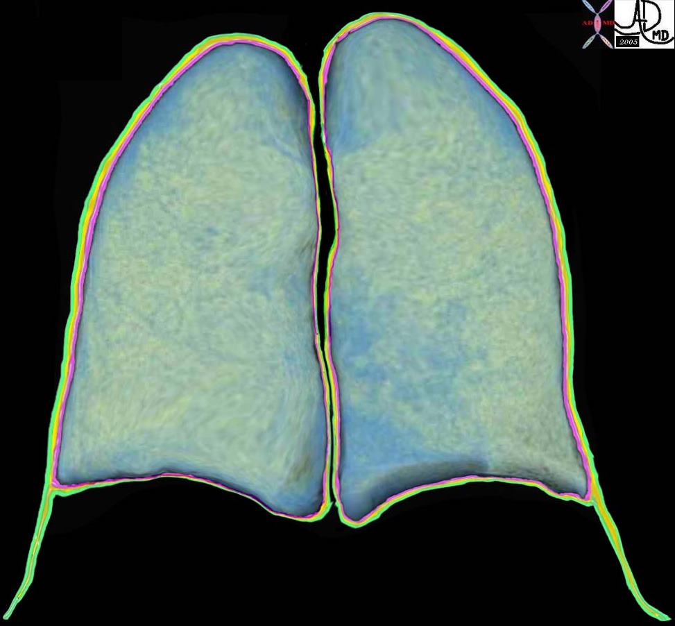
The coronally reformatted image of the lung parenchyma has been outlined with the visceral pleura, (pink) the pleural fluid in the pleural space, (orange) and the parietal pleura. (green) Note how at end expiration the parietal pleura in the costophrenic sulcus extends beyond the lung margin so that the visceral pleura is absent in the costophrenic sulcus and there are two layers of parietal pleura facing each other. During inspiration the lung expands into this space. 32634b10
Key Words lung pleura pulmonary
Ashley Davidoff MD TheCommonVein.net
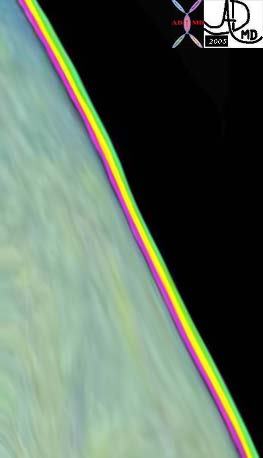

The inferior aspect of the left lung base is magnified in this image s. It shows the costophrenic sulcus where the visceral pleura is absent and two layers of parietal pleura face each other. Ashley Davidoff MD TheCommonVein.net 32634b11b04b

The white arrow points to the potential path of a needle to a lesion in the liver. To the observer it would seem low and out of range of the pleura and lung. Since the costophrenic sulcus would not be visible to ultrasound nor CT interrogation it is difficult to know exactly where it lies. One just has to knowthat it may lie quite low, and that on inspiration the lung may in fact fill that space as well. Hence the potential complications include a pneumothorax from this approach, and if long term catheter drainage is contemplated transgression of the pleural space may cause an effusion or an infection resulting in an empyema.
Ashley Davidoff MD TheCommonVein.net 32634b15
The Segments
The secondary lobules, connect, and unite, linked through the airways, blood vessels, lymphatics and nerves, to form segments in the lungs
There are ten bronchopulmonary segments in the right lung: three in the upper lobe, two in the middle lobe, and five in the lowerlobe. Some of the segments may fuse in the left lung to form usually eight to nine segments (four to five in the upper lobe and four to five in the lower lobe.
Ashley Davidoff MD
TheCommonVein.net lungs-0015catalogue-signed-small
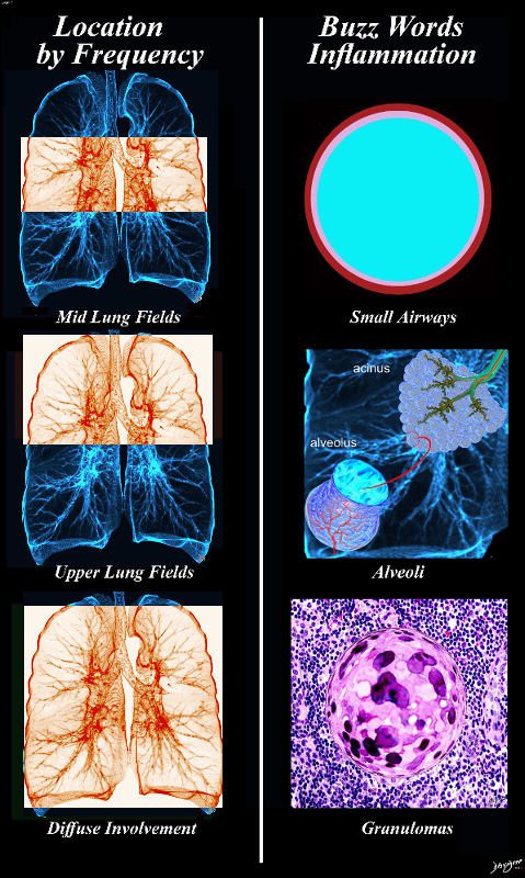
The Airways
Ashley Davidoff MD TheCommonvein.net
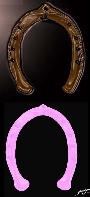
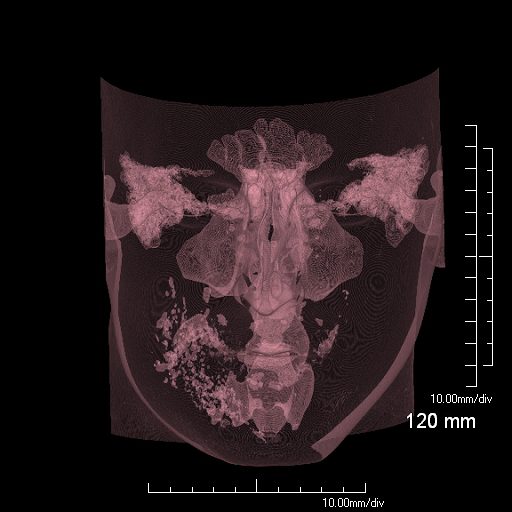
Normal chest CT of the upper lobes of both lungs. The trachea is horse shoe shaped.
Ashley Davidoff TheCommonVein.net 32158 42636c01
The classical branching pattern of many trees
Ashley Davidoff MD

This diagram shows the segmental branches of the right bronchial system. The RUL has three branches, the apical, posterior and anterior segments. (teal overlay) The middle lobe has two segmental branches called lateral and medial segments. (pink) The right lower lobe has five: the superior, anterior basal, lateral basal, posterior basal and medial basal segments. Ashley Davidoff MD. TheCommonVein.net 32686b03
This is a drawing is of a left lung in coronal section. Note there are only two lobes separated by the major fissure
Courtesy Ashley Davidoff MD The CommonVein.net 32686b05L01


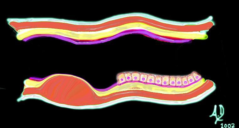
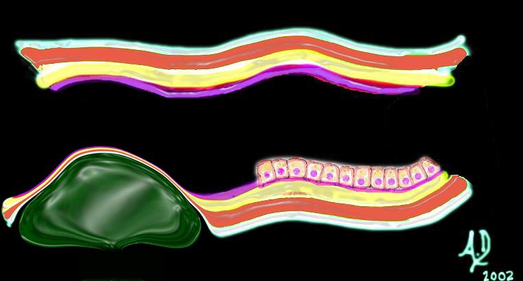
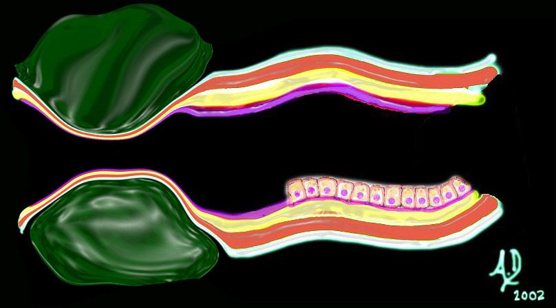
Airways are lined by a pseudostratified ciliated columnar epithelium interspersed with mucus secreting goblet cells
Ashley Davidoff
TheCommonVein.net

Ashley Davidoff
TheCommonVein.net
Ashley Davidoff
TheCommonVein.net
lungs-00677-lo-res.jpg
At the level of the mebranous airways (respiratory bronchiole, alveolar duct, alveolar sac and alveoli, the mucosa becomes mostly a simple squamous epitheliumAshley Davidoff
TheCommonVein.net
The Secondary Lobule
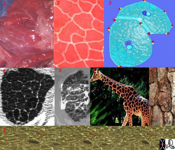
This is a series of images demonstrating the shape of the secondary lobule. The first image (1) is a post mortem specimen with congested lungs showing the interlobular septa, while the next (2), is an overlay of the septa in white showing their polygonal shape. The next drawing reveals side-by-side secondary lobules with central bronchovascular bundles and peripheral lympho-vascular bundles. Image 4 is a CT image through the apex of the lung showing thickened secondary lobules in a patient with mild emphysema, and 5 shows marked thickening of the interlobular septa in a patient with end stage sarcoidosis. 6,7,8 show the shape of the secondary lobules in the skin of a giraffe, the bark of a pine, and the ripples of the water respectively.
Ashley Davidoff MD TheCommonVein.net 31866collage
The secondary lobule is housed in a connective tissue framework in which run the lymphatic and venular tributaries . Together these 3 structures form the interlobular septum.
The lobar arteriole enters the framework, accompanied by the lobar bronchiole, and they all run together and form the interlobular septa. This structure measures between .5cms and 2cms and is visible on CT scan.
It is important in clinical radiology since many of the structures can be identified in health, and more particularly in disease, enabling the identification and characterization of many pathological processes.
Courtesy Ashley Davidoff MD The CommonVein.net
lungs-0036-low res
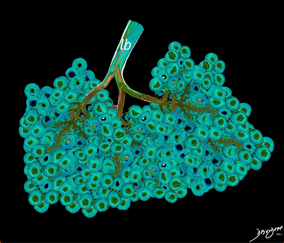
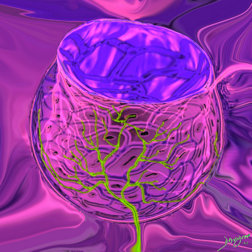
Between 10 and 30 acini combine to form a secondary lobule which is between .5- 2 cms in diameter. It is subtended by a single lobar bronchiole (lb), and is accompanied by arterioles, venules, lymphatics and connective tissue. It is important in clinical radiology since many of the structures can be identified in health, and more particularly in disease, enabling the identification and characterization of many disease processes.
Courtesy Ashley Davidoff MD
lungs-0035-low res
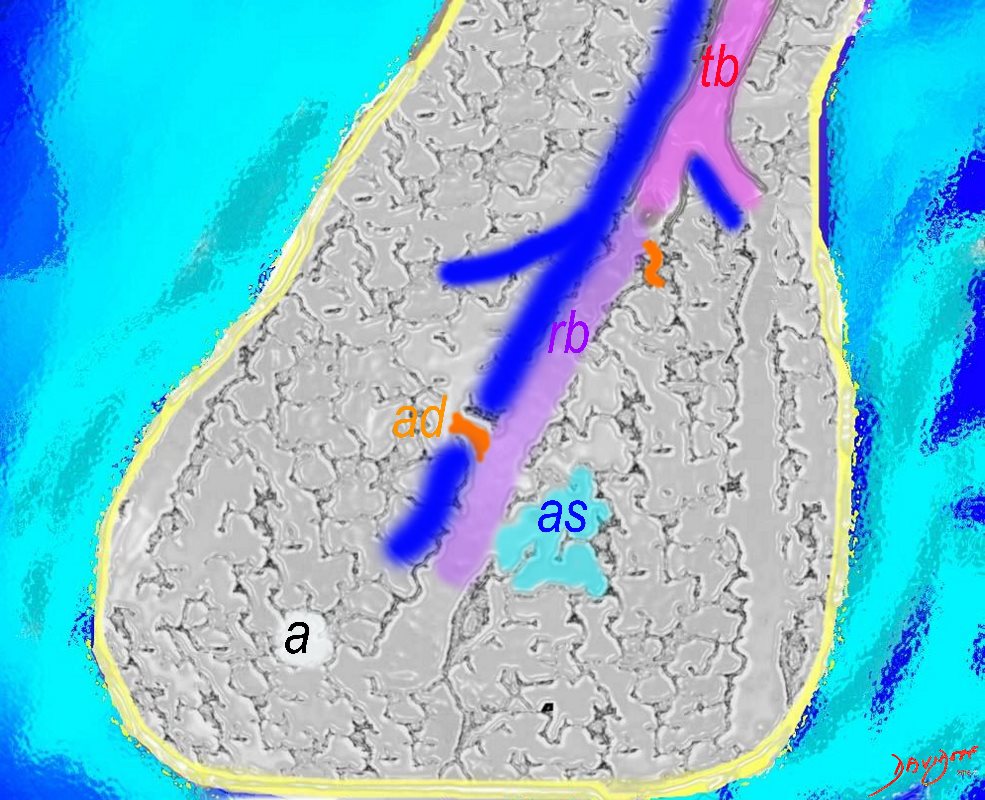
The Acinus

The subsegmental medium sized airways give rise to the terminal bronchiole (tb) which gives rise to the membranous airways. These include in order, the respiratory bronchiole (rb), alveolar duct (ad) and alveolar sac (as)
Ashley Davidoff TheCommonvein.net
The Duct, and the Artery
The pulmonary arteriole accompanies the airway as it carries oxygen from the trachea to the alveoli. They part ways at the alveoli where the pulmonary venule then takes the oxygenated blood from capillary network around the alveoli back to the left atrium.
The intimate relationship of the airways and the pulmonary artery and their close approximation in size, is helpful in radiology, firstly to identify theese structures and secondly to define disease such as heart failure and bronchiectasis.
The acinus as shown in this image is defined as a unit of lung consisting of a single first order respiratory bronchiole that subtending a cluster of alveoli reminiscent of a bunch of grapes or berries (acinus in Latin means berry) . The lobular bronchiole (lb) branches into the terminal bronchiole (tb), which then branches into the first order respiratory bronchiole (rb). Subsequent branching after the respiratory bronchiole, includes in order, the alveolar duct (ad), alveolar sac (as), and then finally the berry like alveoli.
Courtesy Ashley Davidoff 2019
lungs-0033-low res

Ashley Davidoff MD TheCommonVein.net
Here is a picture of the outside of the polyhedral pulmonary lobule from the side. It looked quite futuristic. Through the transparent side window we saw a couple similar to ourselves. From this vantage point the morphing did not look too different from what we had already been through – division after division – leaner and meaner. Ashley Davidoff MD. The Common Vein.net 42449b02
This picture was taken just before the real drama started. The image gives a sense of what was to come. You can see here in the house of the lobule that we were all dividing into smaller parts and were getting smaller and the picture was quite colorful and rosy. I fully expected to have intimate contact with the arteriole? but it did not happen as I expected…… Ashley Davidoff MD. The Common Vein.net 42447b05b02
This picture shows us on the left with a white ring around us (we were the tallest) and the other couples who looked so much like us (also ringed). We called our tribe the “bronchovascular bundle” with the one part of the bundle being the progeny of the bronchus and the other the progeny of the pulmonary artery. In the distance at the periphery we could see the pairs from the other friendly tribe – the red pulmonary vein with its smaller yellow buddy the lymphatic. Behind them we could see the transparent window membrane through which we had peaked earlier. Oh my goodness!!! Look what has happened to my body!!!!!!!…… Ashley Davidoff MD. The Common Vein.net 42447b03b01
There are many references to the shape bell in biology architecture and statistics. The thoracic cage in this instance is bell shaped.
Ashley Davidoff MD TheCommonVein.net . 61829
Between 10 and 30 acini combine to form a secondary lobule which is between .5- 2 cms in diameter. It is subtended by a single lobar bronchiole (lb), and is accompanied by arterioles, venules, lymphatics and connective tissue. It is important in clinical radiology since many of the structures can be identified in health, and more particularly in disease, enabling the identification and characterization of many disease processes.
Courtesy Ashley Davidoff MD
lungs-0035-low res

Courtesy Ashley Davidoff MD
lungs-0028-low res
The Alveolus

key words
key words RS lung alveolus respiratory bronchiole artery vein pulmonary capillary normal anatomy histology drawing
Ashley Davidoff MD TheCommonVein.net 32164

This drawing demonstrates the open mouth view of the alveolus, which is surrounded by its capillary network. The lining single layer of squamous cells (pneumocytes) can be seen peaking through the vessels.
Ashley Davidoff MD.
TheCommonVein.net
32166

Ashley Davidoff MD TheCommonVein.net
The Cells
The Buck Ends Here
The alveolus is lined by a simple epithelium ? one cell layer thick. There are two types of lining cells; Type 1 pneumocytes are squamous cells that cover 90% of the surface of the inner lining of the lung , and type II cuboidal pneumocytes that are in fact much more numerous than Type I. They are involved in the production of surfactant . In the lumen there are resident macrophages which play a crucial role in the immune system. The mucosa is grounded by a basement membrane and a lamina propria, and connected to the lamina propria and basement membrane of the surrounding capillary. The alveolus is lined by a thin layer of surfactant. (teal blue)
Ashley Davidoff
TheCommonVein.net
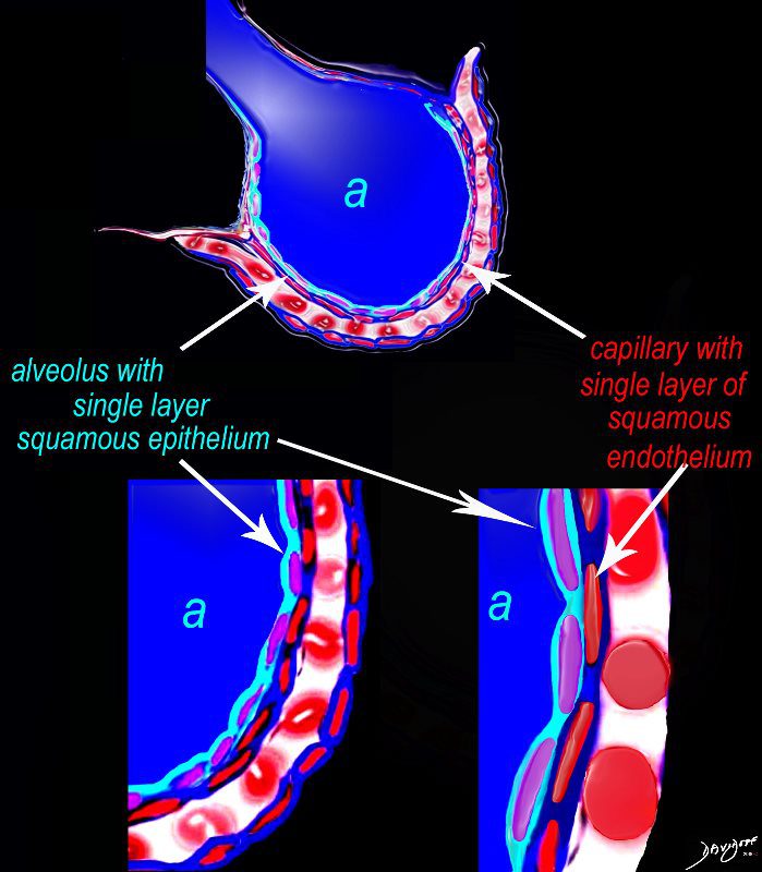
The diagram shows an alveolus (a) above, lined by a single layer of squamous cells, surrounded by a capillary with red cells which is also lined by a single layer of squamous endothelial cells . The images below show progressive magnification of the alveolar wall demonstrating the two thin layer of the alveolar membrane .
Courtesy Ashley Davidoff 2019
lungs-0028-low res
First line of defense against infections of the lung.
Reside in alveolar walls, lymphatic channels and lymph node.
Originate in the bone marrow and are part of the Mononuclear Phagocytic System.
Ashley Davidoff
TheCommonVein.net
It produces the phospholipid – part of the surfactant that reduces surface tension and allows the alveoli to remain open
#cells
Ashley Davidoff
TheCommonVein.net
– flattened for gas exchange, forms a part of the Blood-Gas Barrier
nd sometimes vacuolated cytoplasm
It produces the phospholipid – part of the surfactant that reduces surface tension and allows the alveoli to remain open
#cells
Ashley Davidoff
TheCommonVein.net
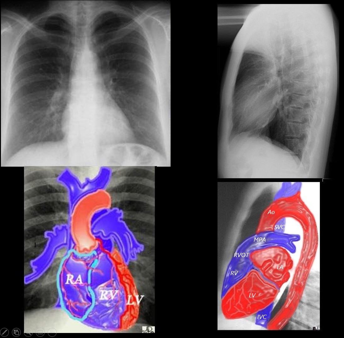
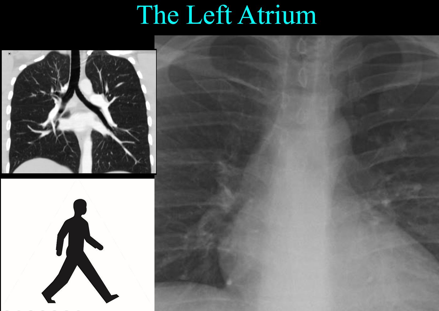
Blood Supply and Venous Drainage
Normal 3D reconstruction of a CT scan of the Heart Showing the Left Atrium and Pulmonary Veins
Ashley Davidoff MD TheCommonVein.net
77612b.3kb07.8s
Normal 3D reconstruction of a CT scan of the Heart Showing the Left Atrium and Pulmonary Veins
Ashley Davidoff MD TheCommonVein.net 77612b.3kb08.8s
In this coronally oriented CTA the RPA can be seen traveling horizontally above the LA and the LPA origin hides behind the MPA as it courses posteriorly. Courtesy Ashley Davidoff MD TheCommonVein.net 32807b03.8s
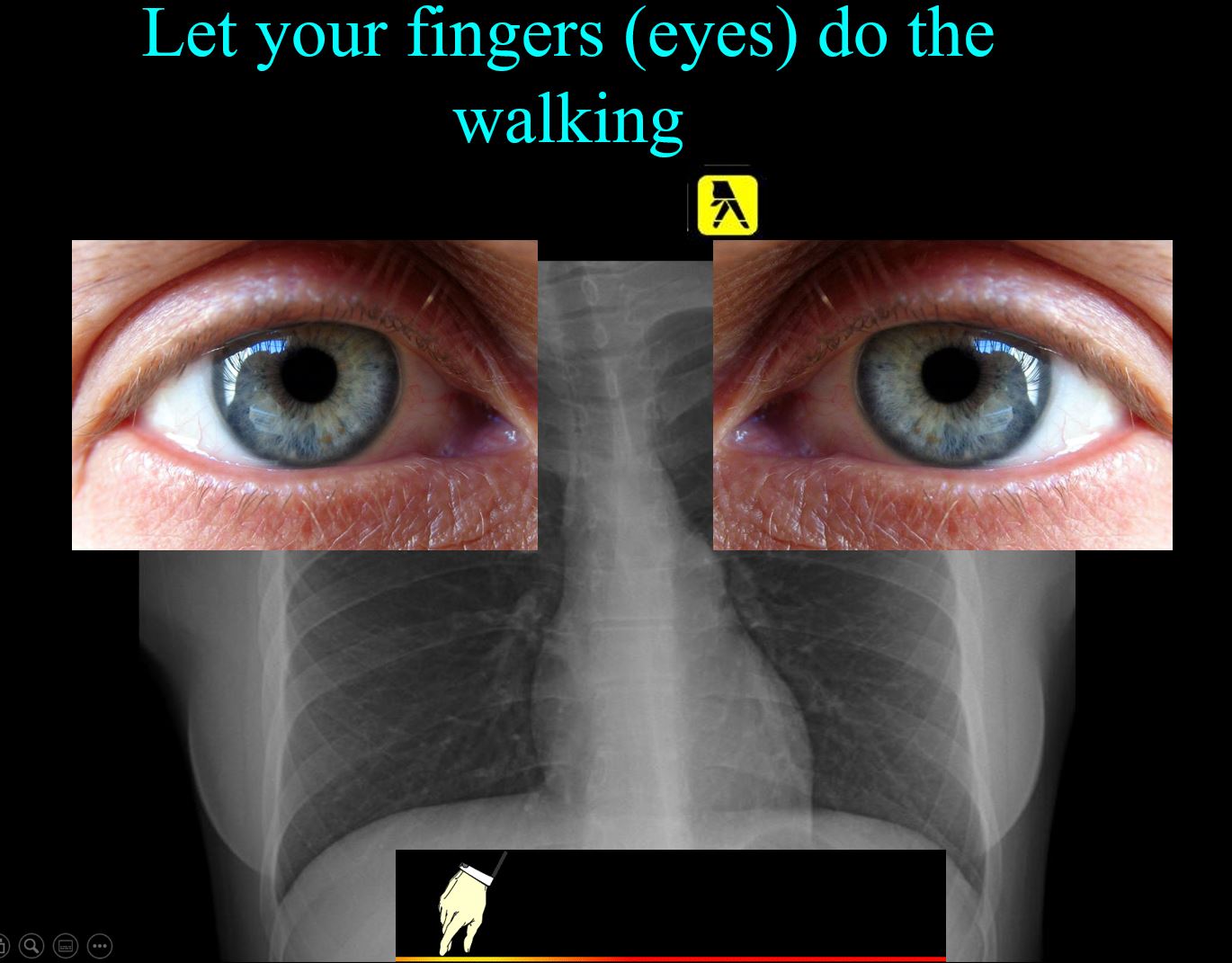
Imaging
Learning to Read
Ashley Davidoff thecommonvein.net
Ashley Davidoff thecommonvein.net

Ashley Davidoff
thecommonvein.net
Ashley Davidoff thecommonvein.net
Disease
Principles

32347d01 key words mucosa submucosa muscularis adventitia serosa mucosal mass polyp neoplasm carcinoma acute angles with the lumen histopathology imaging diagnosis Ashley Davidoff TheCommonVein.net
32347d02 key words mucosa submucosa muscularis adventitia serosa submucosal mass edema hemorrhage obtuse angles or right angle 90 degree ninety degree angle with the lumen histopathology imaging diagnosis
Ashley Davidoff TheCommonVein.net
32347d03 key words mucosa submucosa muscularis adventitia serosa submucosal mass edema hemorrhage obtuse angles or right with the lumen histopathology imaging diagnosis Ashley Davidoff TheCommonVein.net
32347d04 key words mucosa submucosa muscularis adventitia serosa submucosal mass edema hemorrhage neoplasm malignancy benign obtuse angles with the lumen histopathology imaging diagnosis
Ashley Davidoff TheCommonVein.net
32347d06
key words mucosa submucosa muscularis adventitia serosa submucosal mass edema hemorrhage neoplasm malignancy benign obtuse angles with the lumen circumferential narrowing constriction obstruction histopathology imaging diagnosis Davidoff art Davidoff MD
lungs-0021catalogue-signed-smallb.jpg
Inflamed AlveoliAshley Davidoff MD TheCommonvein.net
Links and References
- Maps of Art and Culture in the