
Infection Inflammation Malignancy Mechanical/Atelectasis Trauma Metabolic Circulatory- Hemorrhage Immune Infiltrative Idiopathic Iatrogenic Idiopathic
Speckled Calcification

Ashley Davidoff MD TheCommonVein.net hamartoma 003c
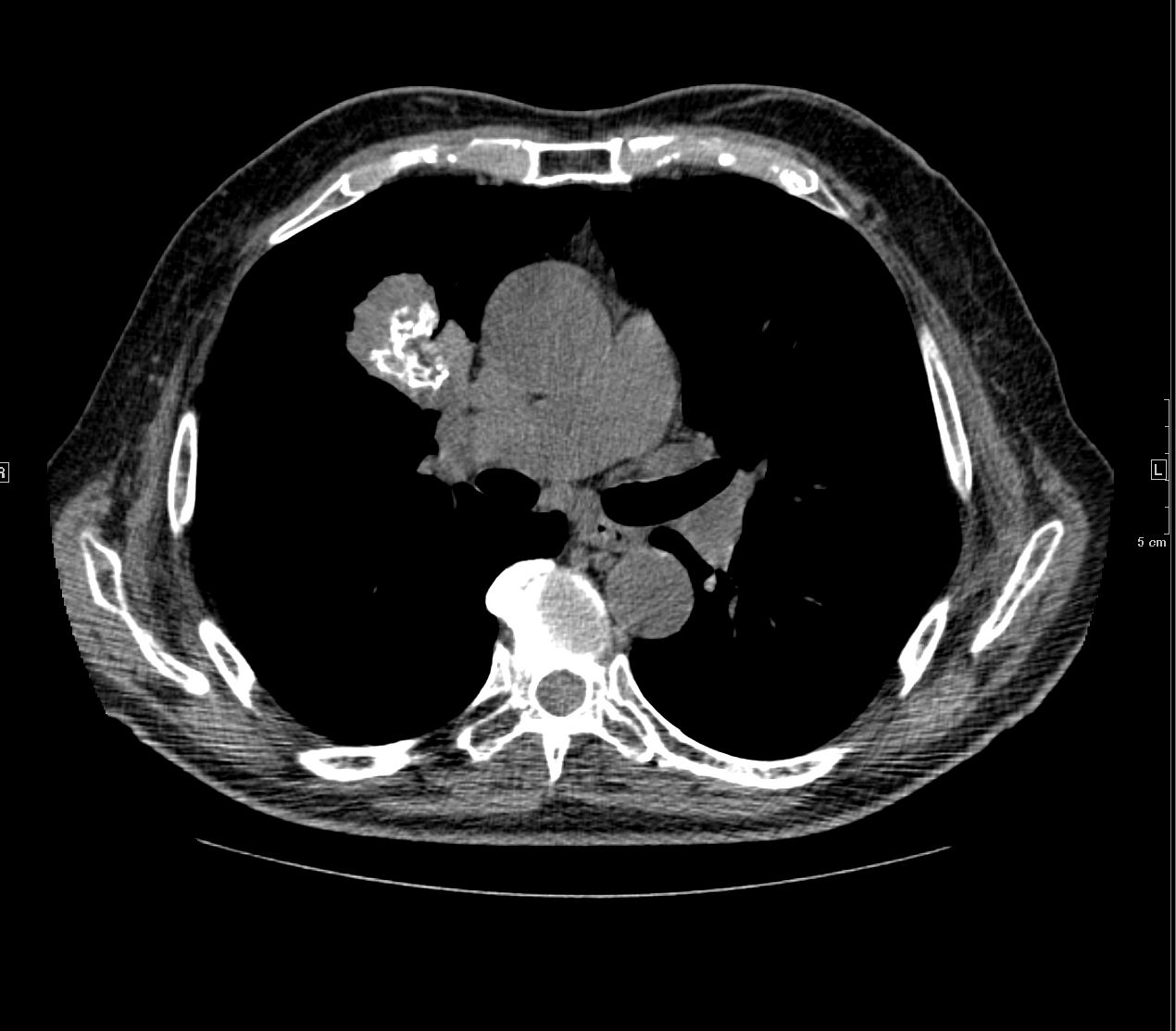
Popcorn Calcifications

Ashley Davidoff TheCommonvein.net
hamartoma 0001c01 86f
Heavy Homogeneous Calcification – Hamartoma

and amyloidoma
Ashley Davidoff TheCommonvein.net
hamartoma calcifications 004c stable
Amyloid
83 y.o. male with biopsy proven nodular lung AL amyloidosis diagnosed by lung biopsy 5 years ago . His underlying amylogenic protein was typed by liquid chromatography mass spectrometry as kappa.
Ashley Davidoff
TheCommonVein.net
Metastases
31491c.jpg
65 year old male with rectal mucinous adenocarcinomaCT scan of the chest shows a metastatic nodule with psammomatous calcifications
Ashley Davidoff
TheCommonVein.net
31491c
Calcified Metastases in the Lung from Leiomyosarcoma of the Uterus

CT scan through right lower lobe of the lung shows a calcified mass representing a metastasis from a primary uterine leiomyosarcoma.
Ashley Davidoff MD TheCommonVein.net 135680