
Centrilobular Emphysema
Emphysema
A drawing showing the normal acinus in teal and the abnormal emphysematous acinus in green characterised by destruction of the septal walls, enlargement of the alveoli, and loss of elasticity. The absence of involvement of the respiratory bronchiole makes the pathological diagnosis of centrilobular emphysema. Ashley Davidoff MD
TheCommonVein.net 32645

The red arrows point to the soft tissues of the centrilobular emphysema consisting of the arterioles and bronchiolar walls (not usually visible.
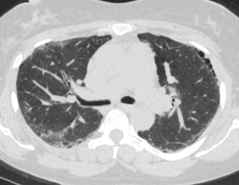
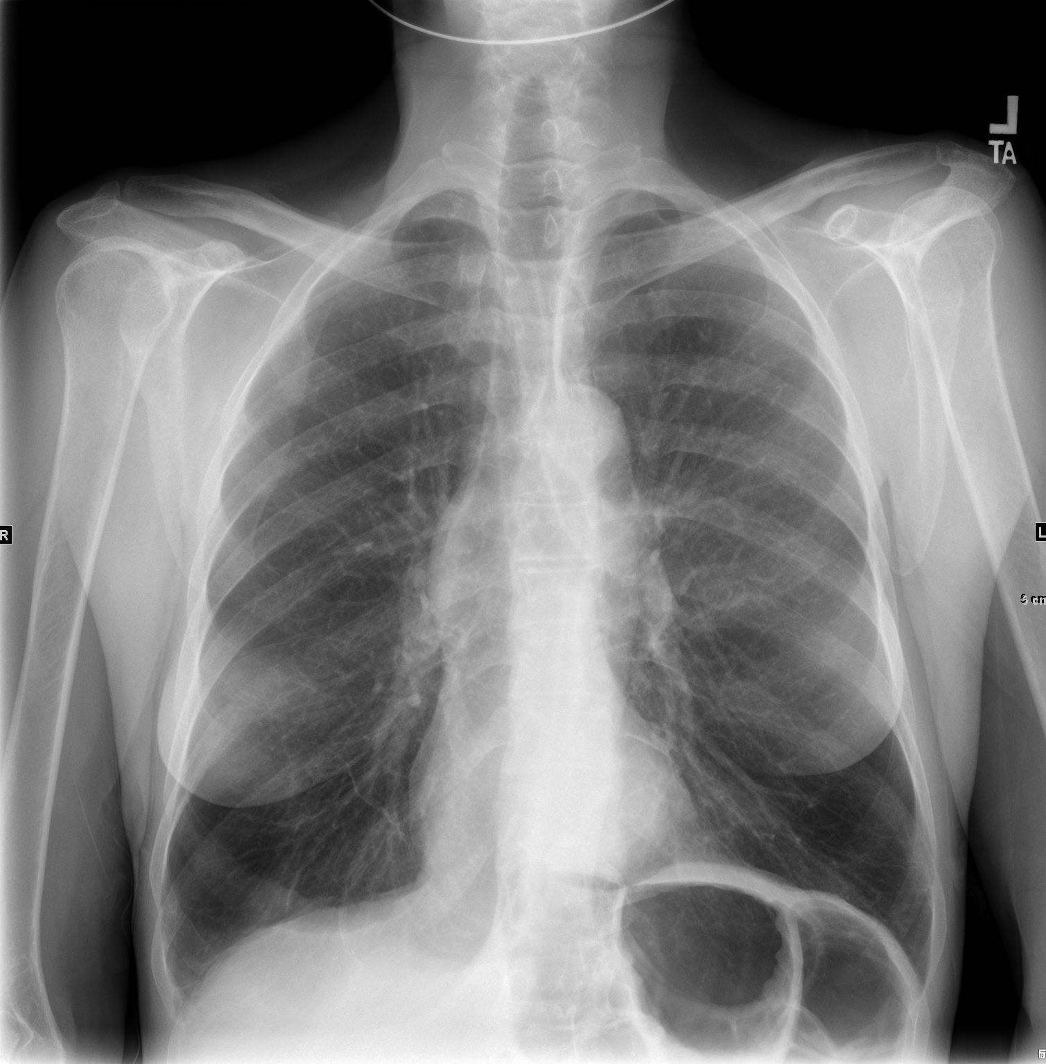
71-year-old female presents with history emphysema
Chest X-ray shows hyperinflated lungs with flattened hemidiaphragms and increase in the retrosternal space and right ventricular enlargement based on the decrease in the retrosternal air space
CT scan confirms the presence of centrilobular emphysema, predominantly in the upper lobes with associated right atrial, right ventricular and pulmonary arterial enlargement. The LA and LV are normal
These findings are consistent with cor pulmonale and pulmonary hypertension, secondary to emphysema.
Ashley Davidoff MD

Ashley Davidoff MD TheCommonvein.net
Moderate CHF with Interstitial Edema in a
Background of Emphysema

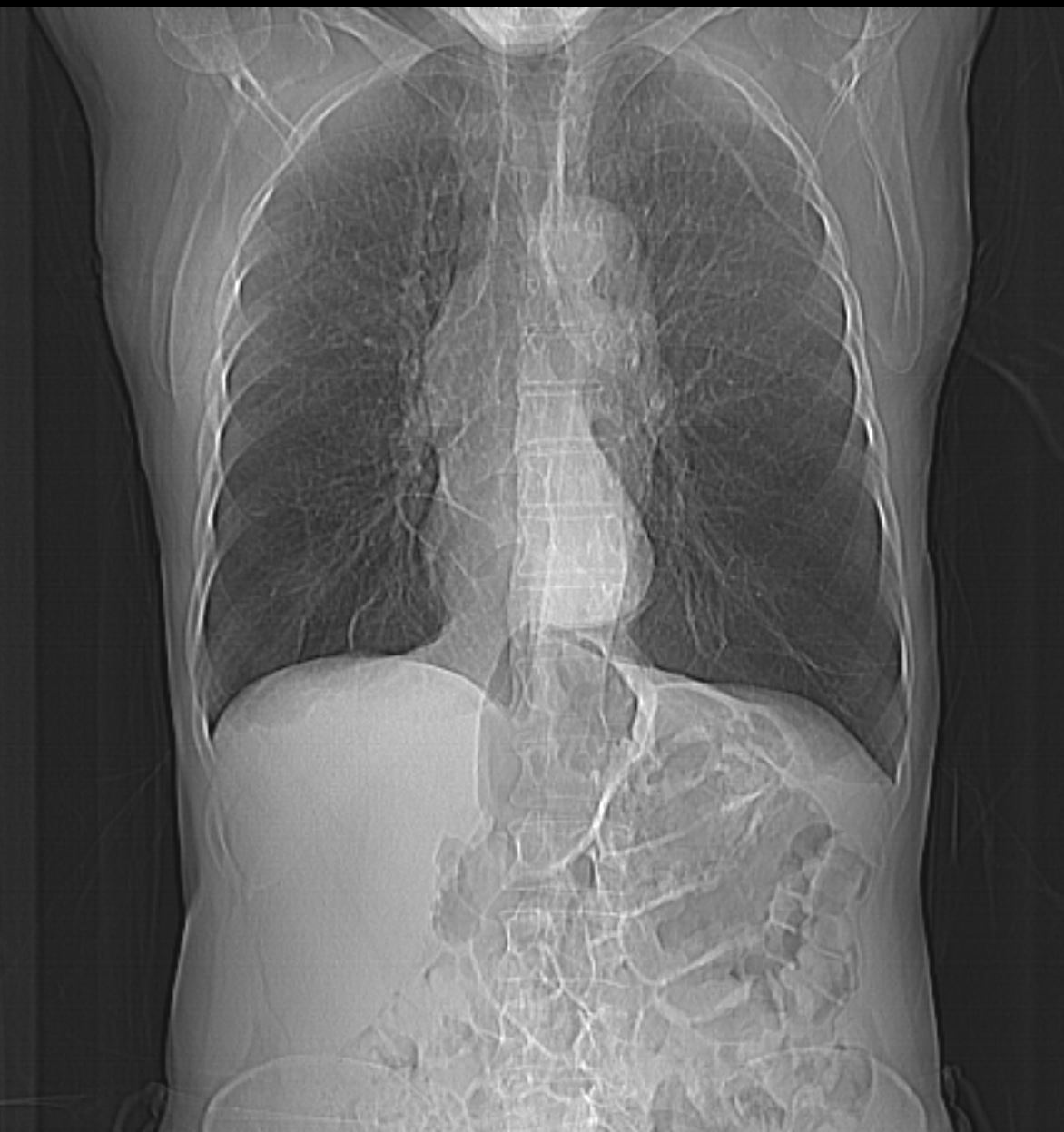
68 year old male with a history of emphysema presents with increasing dyspnea, Frontal Chest X-ray shows an enlarged left atrium and diffuse interstitial thickening most prominent in the upper lobes.
There is global cardiomegaly, an enlarged main pulmonary artery indicating pulmonary hypertension and a single lead pacemaker.
Ashley Davidoff MD TheCommonVein.net 136556 288Lu

68 year old male with a history of emphysema presents with increasing dyspnea. CT in the axial plane shows predominant upper lobe centrilobular emphysema with diffuse thickening of the interlobular septa .
Ashley Davidoff MD TheCommonVein.net 288Lu 136558

68 year old male with a history of emphysema presents with increasing dyspnea. CT in the coronal plane y shows predominant upper lobe centrilobular emphysema with diffuse thickening of the interlobular septa (b white circle) with evidence of Kerley B lines (c red arrowheads) best exemplified in the left upper lung field, and thickening of the minor fissure (b, pink arrowhead). The left atrium (LA) is enlarged. These findings are consistent with moderate CHF with interstitial edema
Ashley Davidoff MD TheCommonVein.net 288Lu 136557cL
Paraseptal Emphysema


Panlobular Emphysema Alpha-1-Antitrypsin Deficiency
CXR shows hyperinflation with basilar prominence consistent with panlobular emphysema secondary to Alpha-1 antitrypsin deficiency
Ashley Davidoff TheCommonVein.net 216Lu
Scout for CT scan shows hyperinflation with basilar prominence consistent with panlobular emphysema secondary to Alpha-1 antitrypsin deficiency
Ashley Davidoff TheCommonVein.net 216Lu

Axial CT scan shows lower lobe panlobular emphysema and bronchitis
Ashley Davidoff TheCommonVein.net 216Lu

Axial CT scan shows lower lobe panlobular emphysema and bronchitis
Ashley Davidoff TheCommonVein.net 216Lu