
Infection
Inflammation Mechanical Small Airway Disease
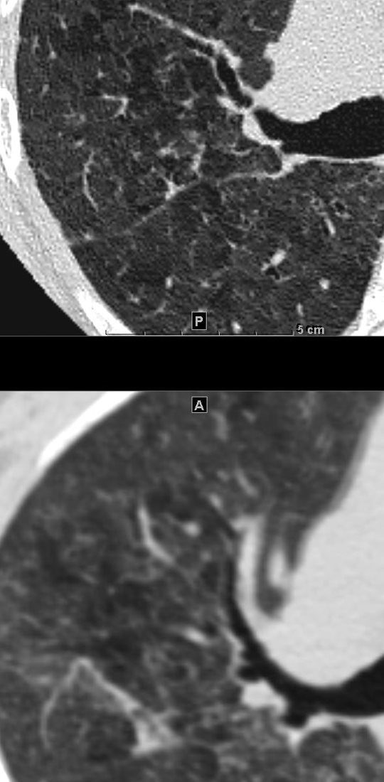
Desquamative Interstitial Pneumonia (DIP) Patchy Ground Glass Changes and Mosaicism and Air Trapping – Inspiration Expiration Views

51-year-old female smoker with a history of COPD asthma and pulmonary hypertension presents with progressive dyspnea. Axial CT through the right posterior recesses at end inspiration (upper panel) and end expiration (lower panel) confirms the presence of air trapping indicating the presence of mild small airway disease
Pathology confirmed a diagnosis of DIP
Ashley Davidoff MD TheCommonVein.net 252Lu 135987
Air Trapping ?
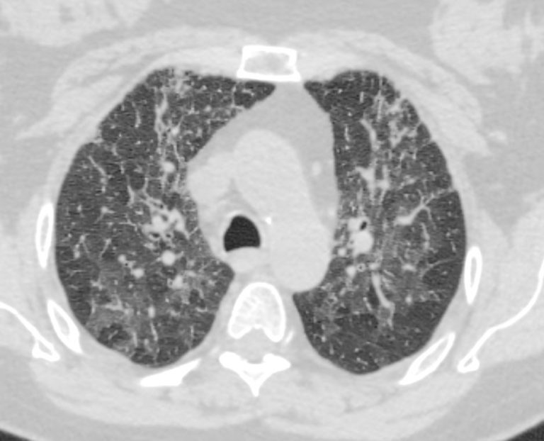
Hypersensitivity Pneumonitis Bird Fancier?s Disease

55-year-old female with shortness of breath. She keeps birds for pets Axial CT through the mid lung fields on inspiration, shows diffuse ground glass changes with a combination of mosaic attenuation and normal lung giving the appearance of the head cheese sign.
On the expiration phase the mosaic attenuation remains indicating air trapping and inferring small airway disease
Ashley Davidoff MD TheCommonVein.net 242Lu 13551aL

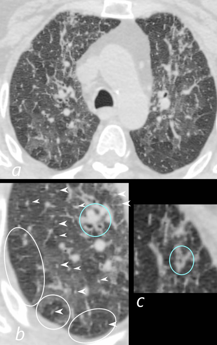
70-year-old female presents with dyspnea
CT performed in expiration shows multicentric foci of differing densities that include ground glass (seen on inspiration images) normal, and mosaic attenuation with air trapping and prominent centrilobular nodules (teal blue arrowheads). These findings confirm small airway disease and in the appropriate clinical context, are consistent with hypersensitivity pneumonitis (HP)
Ashley Davidoff MD TheCommonvein.net 135792cL 144Lu

55-year-old female with shortness of breath. She keeps birds for pets Axial CT through the mid lung fields on inspiration (upper image) shows diffuse ground glass changes with low attenuation regions (mosaic attenuation) Expiration images (below) shows persistent regions of mosaic attenuation indicating air trapping
Ashley Davidoff MD TheCommonVein.net 13557

55-year-old female with shortness of breath. She keeps birds for pets Axial CT through the mid lung fields on inspiration (upper image) shows diffuse ground glass changes with low attenuation regions (mosaic attenuation) Expiration images (below) shows persistent regions of mosaic attenuation indicating air trapping
Ashley Davidoff MD TheCommonVein.net 13556

55-year-old female with shortness of breath. She keeps birds for pets Axial CT through the mid lung fields on inspiration (upper image) shows diffuse ground glass changes with low attenuation regions (mosaic attenuation) Expiration images (below) shows persistent regions of mosaic attenuation indicating air trapping
Ashley Davidoff MD TheCommonVein.net 13555
55-year-old female with shortness of breath. She keeps birds for pets Axial CT through the mid lung fields on inspiration (upper image) shows diffuse ground glass changes with low attenuation regions Expiration images (below)shows increase in the density of this region indicating that this region is normal lung
Ashley Davidoff MD TheCommonVein.net 13554

55-year-old female with shortness of breath. She keeps birds for pets Axial CT through the mid lung fields on inspiration (upper image) shows diffuse ground glass changes with mosaic attenuation Expiration images (below) shows some regions of regions of air trapping (left lower lobe) and some of normalizing (right lower lobe )
Ashley Davidoff MD TheCommonVein.net 13553
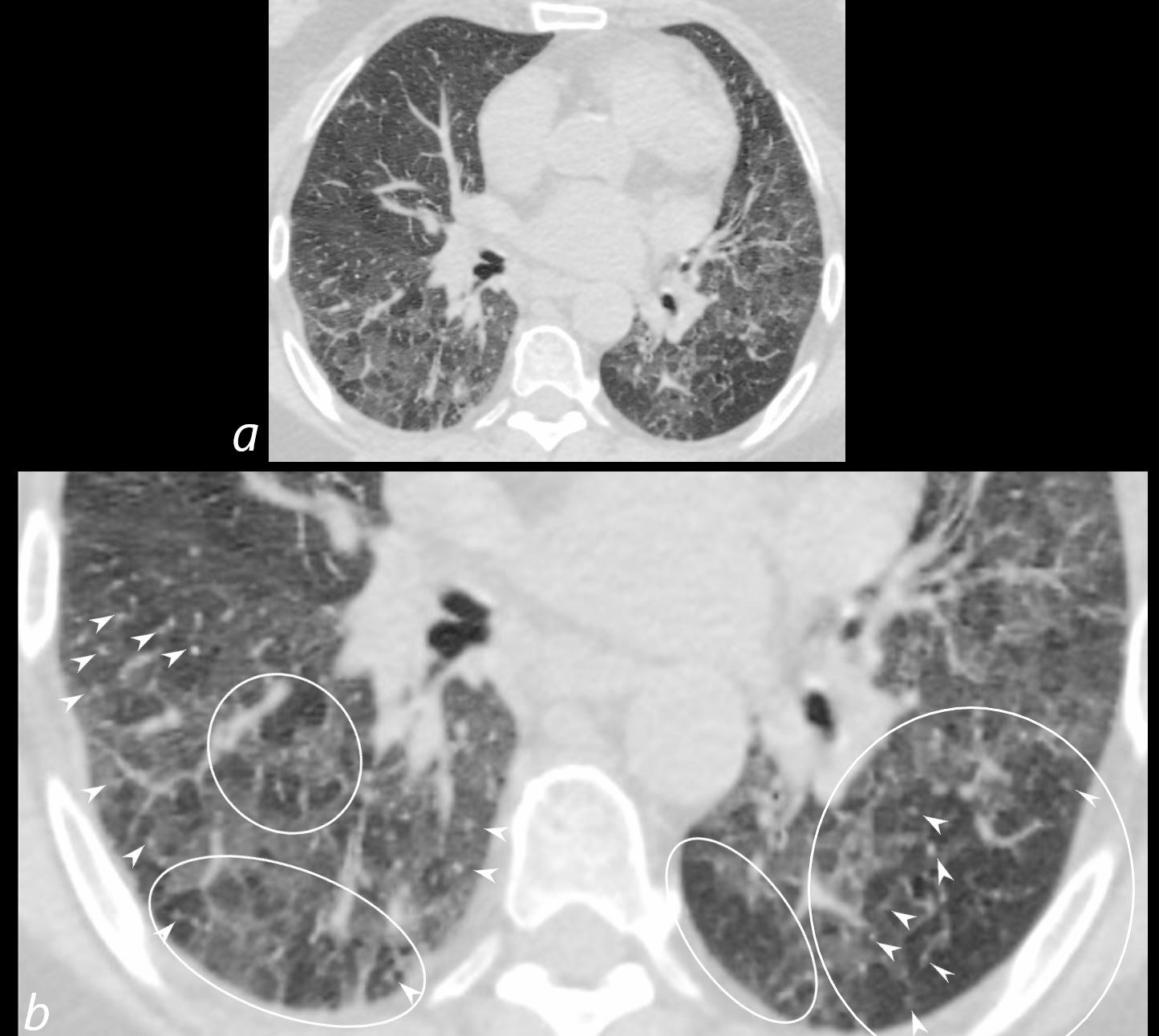
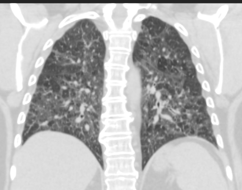
Follicular Bronchiolitis and RA
70-year-old female former smoker with long standing history of RA presents with chronic dyspnea.
Axial CT of the chest at the level of the aortic arch reveals centrilobular nodules, ground-glass opacities, and mosaic attenuation (likely due to air trapping in this context) and bronchial wall thickening. In the context of a patient with rheumatoid arthritis a diagnosis of follicular bronchiolitis is likely. However radiologically fibrotic hypersensitivity pneumonitis (HP) is included in the differential diagnosis
Ashley Davidoff MD TheCommonVein.net 132Lu 136652
70-year-old female former smoker with long standing history of RA presents with chronic dyspnea.
Axial CT of the chest at the level of the aortic arch reveals centrilobular nodules (b, white arrowheads) , ground-glass opacities, and mosaic attenuation (b, white rings) likely due to air trapping in this context, and bronchial wall thickening (b, c teal rings). There is some irregular thickening of the interlobular septa. In the context of a patient with rheumatoid arthritis a diagnosis of follicular bronchiolitis is likely. However radiologically fibrotic hypersensitivity pneumonitis (HP) is included in the differential diagnosis
Ashley Davidoff MD TheCommonVein.net 132Lu 136652cL
70-year-old female former smoker with long standing history of RA presents with chronic dyspnea.
Axial CT of the chest at the level of the lower lung fields reveals centrilobular nodules (b white arrowheads), ground-glass opacities, and mosaic attenuation (b, white rings) likely due to air trapping in this context.
In the context of a patient with rheumatoid arthritis a diagnosis of follicular bronchiolitis is likely. However radiologically fibrotic hypersensitivity pneumonitis (HP) is included in the differential diagnosis
Ashley Davidoff MD TheCommonVein.net 132Lu 136657cL
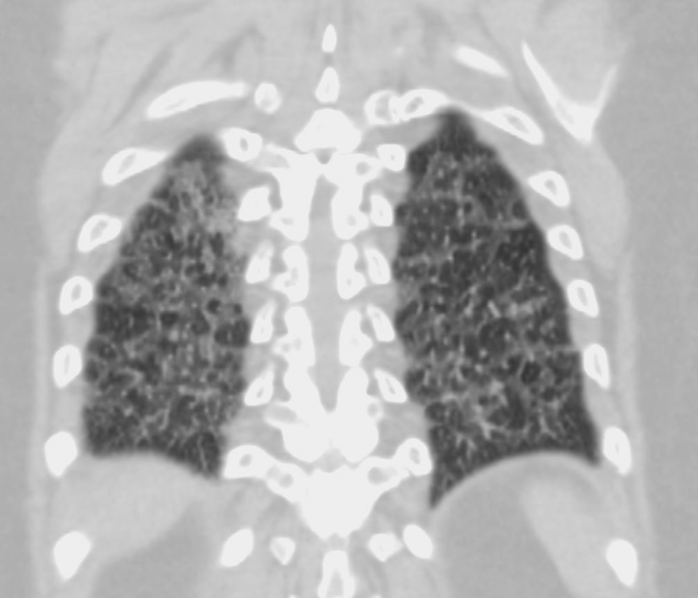
70-year-old female former smoker with long standing history of RA presents with chronic dyspnea.
CT in the coronal plane of the chest at the level of the spine reveals bilateral diffuse changes in the lungs characterized by centrilobular nodules, ground-glass opacities, mosaic attenuation (likely due to air trapping in this context) and irregular thickening of the interlobular septa.
In the context of a patient with rheumatoid arthritis a diagnosis of follicular bronchiolitis is likely. However radiologically fibrotic hypersensitivity pneumonitis (HP) is included in the differential diagnosis
Ashley Davidoff MD TheCommonVein.net 132Lu 136663
70-year-old female former smoker with long standing history of RA presents with chronic dyspnea.
CT in the coronal plane of the chest at the level of the spine reveals bilateral diffuse changes in the lungs characterized by centrilobular nodules, ground-glass opacities, mosaic attenuation (likely due to air trapping in this context) and irregular thickening of the interlobular septa.
In the context of a patient with rheumatoid arthritis a diagnosis of follicular bronchiolitis is likely. However radiologically fibrotic hypersensitivity pneumonitis (HP) is included in the differential diagnosis
Ashley Davidoff MD TheCommonVein.net 132Lu 136664
Aspiration
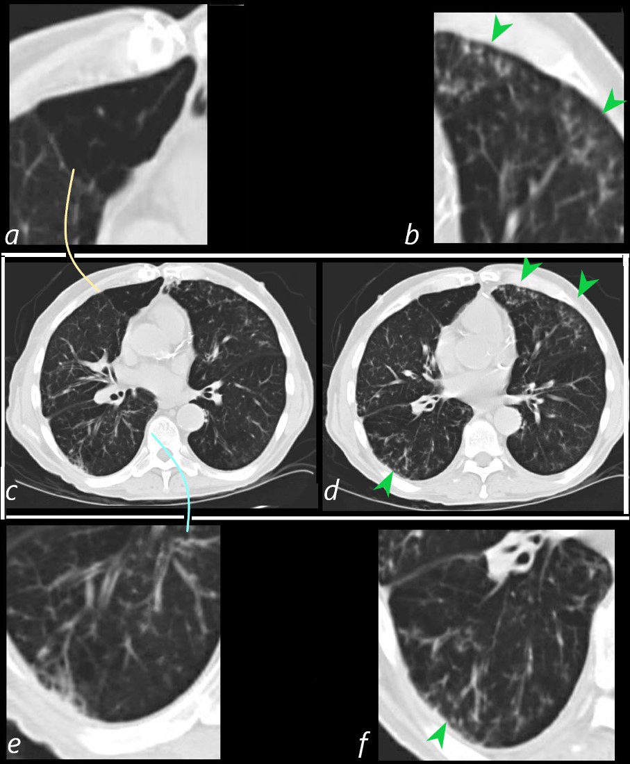
86year old patient with known emphysema and chronic bronchitis presents with a fever and an acute on chronic productive cough The CT scan of the chest through the mid lung fields shows a subsegmental region of air trapping in the medial segment of the middle lobe (c and magnified in a ? orange arc). There is evidence of segmental and subsegmental airway thickening (c magnified in e ? teal arc) with evidence of small airway disease characterised by tree in bud changes (green arrowhead d magnified in b and f). These findings are non-specific, but in the current context represent manifestations of aspiration.
Ashley Davidoff MD TheCommonVein.net 30602bL
Malignancy
Mechanical
Metastatic Mediastinal Adenopathy Encasement of the Airways with Ball Valve Effect on the Left Upper Bronchus
Carcinoma of the Cervix

33-year-old female with known primary carcinoma of the cervix presents with infiltrating and encasing mediastinal adenopathy with severe stenosis of both the left and right main stem bronchi. The upper panels reveal hyperinflation of the left upper lobe (a,b blue arrrowheads) likely secondary to air trapping. The lower panel reveals the severe stenosis of the left main stem bronchus (c white arrowhead) with subtotal occlusion appreciated in d, (white arrowhead). This severe subtotal occlusion probably accounts for the hyperinflation secondary to ball valve mechanism
Ashley Davidoff MD TheCommonVein.net 254Lu 136050b02L
Atelectasis Trauma Metabolic Circulatory- Hemorrhage Immune Infiltrative Idiopathic Iatrogenic Idiopathic