
74 year old male with a cough.
CT shows split pleura sign with thickened visceral and parietal pleura with regions of early spiraling of an atelectatic process in the right lower lobe consistent with early rounded atelectasis
Ashley Davidoff MD TheCommonVein.net
31563c
CT shows focal region of pleural thickening with calcification
Also note hyperlucent right lower lobe
Ashley Davidoff MD TheCommonVein.net
CT shows focal region of pleural thickening with calcification
Also note hyperlucent right lower lobe
Ashley Davidoff MD TheCommonVein.net
CT shows focal region of pleural thickening with calcification
Also note hyperlucent right lower lobe
Ashley Davidoff MD TheCommonVein.net
CT shows focal region of pleural thickening with calcification
Also note hyperlucent right lower lobe
Ashley Davidoff MD TheCommonVein.net
CT shows focal region of pleural thickening with calcification
Also note hyperlucent right lower lobe
Ashley Davidoff MD TheCommonVein.net
Courtesy Ashley Davidoff MD.
TheCommonVein.net
32426_03cl keywords
lung bronchus lyphatic infiltrate mass obstruction atelectasis thickening interlobular septa neoplasm malignant primary malignancy small cell carcinoma imaging radiology CTscan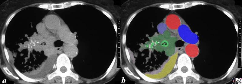
This CT scan is from a middle aged female with poorly differentiated small cell carcinoma, with extensive mediastinal and hilar involvement (dark green in b) extending into and almost occluding the right main stem bronchus light green with black air in the centre) and occluding the smaller airways (light green surrounded by white bronchial cartilage). A complex right effusion (yellow) and atelectasis is seen (light pink). The relationship to the SVC (light blue ) is noted without mass effect at this time, with aorta in red and left pulmonary artery (royal blue). Parenchymal disease is suspected based on the increased soft tissue noted in the mediastinal windows in the right lower lobe.
Courtesy Ashley Davidoff MD
TheCommonVein.net
32429bc03.8s
Source
Signs in Thoracic Imaging
Journal of Thoracic Imaging 21(1):76-90, March 2006.
