
Infection

TheCommonVein.net
ABPA
Early

ABPA 3 years prior CT Scan RUL
72 year old female with asthma presented 3 years prior with acute dyspnea.
CT in the axial plane shows thickening of the segmental and subsegmental airways of the posterior segment of the right upper lobe with mucoid impaction
Ashley Davidoff MD TheCommonVein.net 294Lu 135116b.0001c
ABPA Advanced

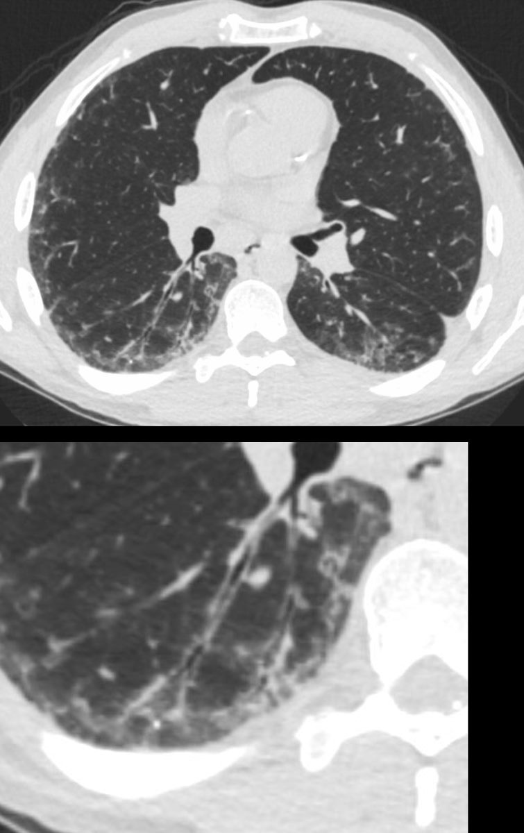
60 year old male with history of asthma, allergic bronchopulmonary aspergillosis (ABPA)
CT scan shows bibasilar bronchiectasis and soft tissue/fluid impaction of the bronchovascular bundles
Ashley Davidoff TheCommonVein.net
CT Allergic Bronchopulmonary Aspergillosis (ABPA)
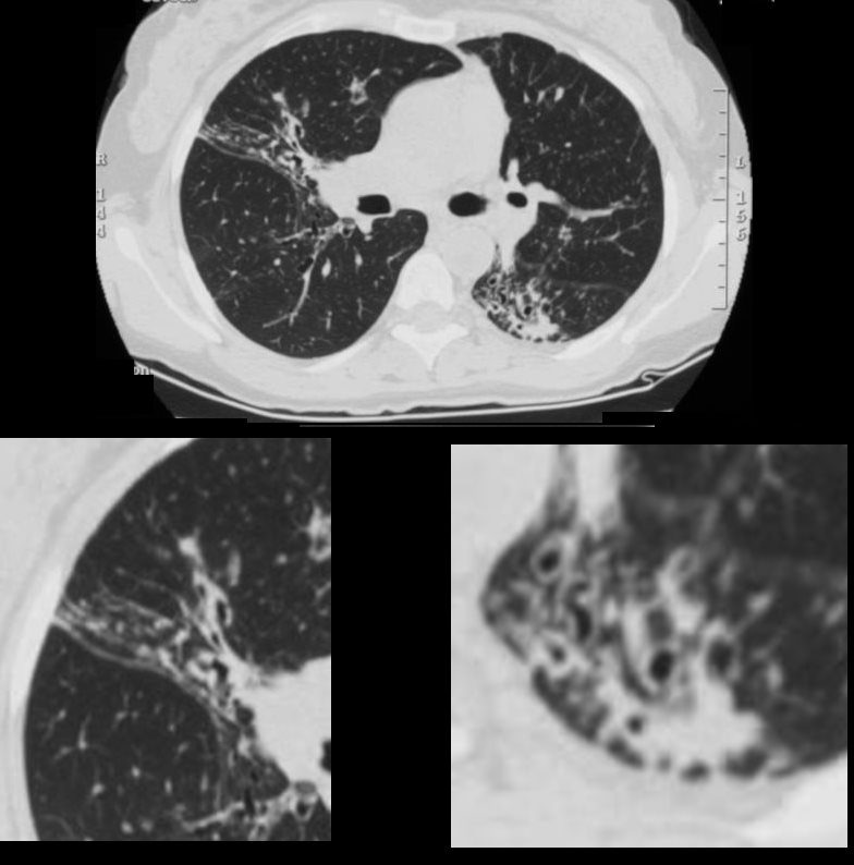
48 year old female with a history of asthma presents with productive cough. CT scan 18 months prior shows multicentric foci of bronchial wall thickening , in the segmental and subsegmental airways, in the middle lobe with crowding of the airways in the RML indicating atelectasis. (upper panel magnified lower left). In the LLL there is bronchiolectasis with thickened airways and a focal subsegmental consolidation (lower panel right) . Flexible bronchoscopy revealed mucus plugs and aspergillus was isolated.
Ashley Davidoff MD TheCommonVein.net
Inflammation
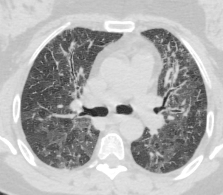
70-year-old female former smoker with long standing history of RA presents with chronic dyspnea.
Axial CT of the chest at the level of the carina reveals centrilobular nodules, ground-glass opacities, and mosaic attenuation (likely due to air trapping in this context) and bronchial wall thickening . In the context of a patient with rheumatoid arthritis a diagnosis of follicular bronchiolitis is likely. However radiologically fibrotic hypersensitivity pneumonitis (HP) is included in the differential diagnosis
Ashley Davidoff MD TheCommonVein.net 132Lu 136654
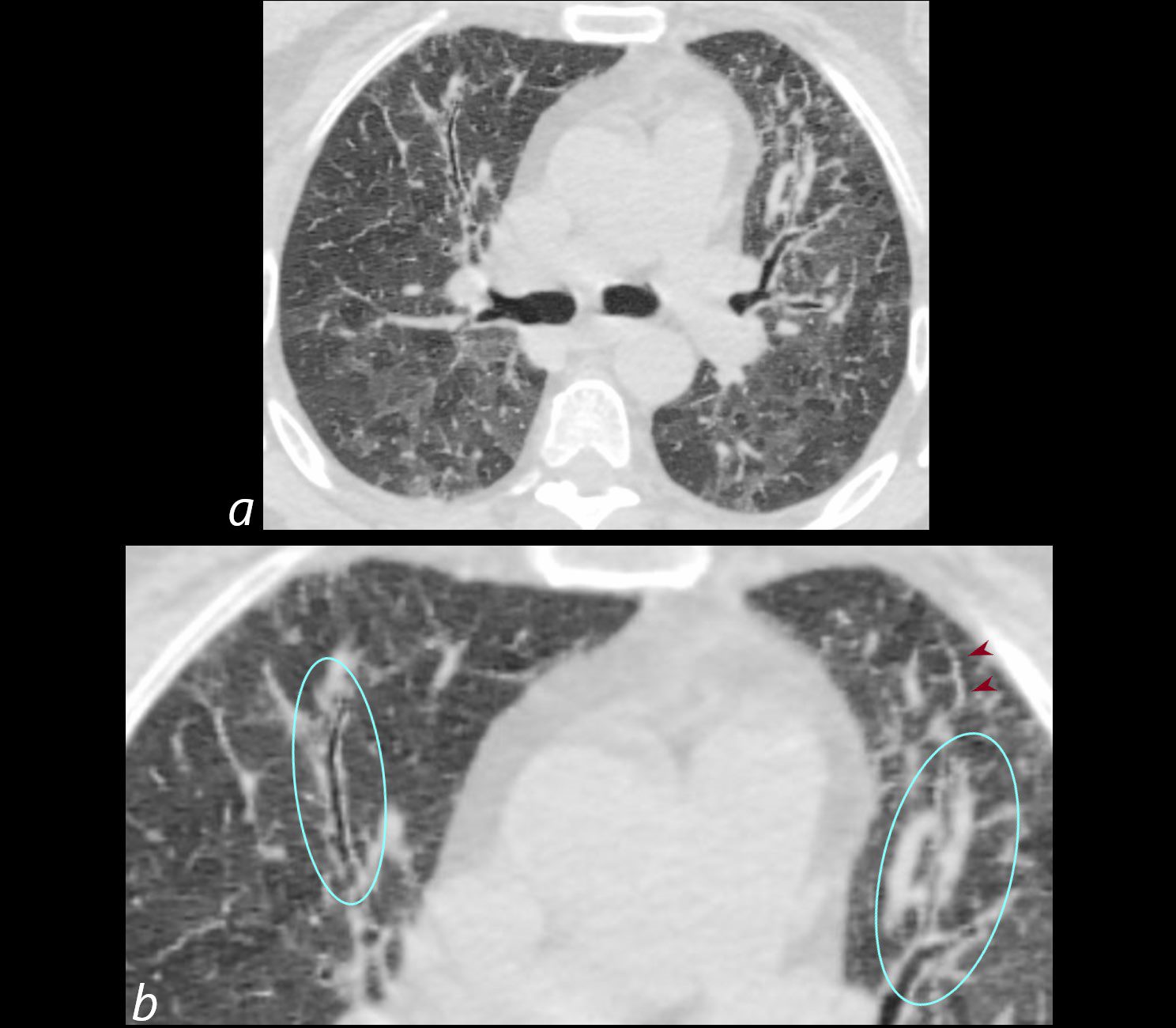
70-year-old female former smoker with long standing history of RA presents with chronic dyspnea.
Axial CT of the chest at the level of the carina reveals centrilobular nodules, ground-glass opacities, and mosaic attenuation (likely due to air trapping in this context) and bronchial wall thickening . Bronchial wall thickening (b, maroon arrowheads) and irregular septal thickening (b maroon arrowheads) are noted.
In the context of a patient with rheumatoid arthritis a diagnosis of follicular bronchiolitis is likely. However radiologically fibrotic hypersensitivity pneumonitis (HP) is included in the differential diagnosis
Ashley Davidoff MD TheCommonVein.net 132Lu 136654cL
70-year-old female former smoker with long standing history of RA presents with chronic dyspnea.
Axial CT of the chest at the level of the superior lingula bronchus shows prominent segmental and segmental bronchial wall thickening together with mosaic attenuation and ground glass opacities (GGO’s)
Ashley Davidoff MD TheCommonVein.net 132Lu 136656
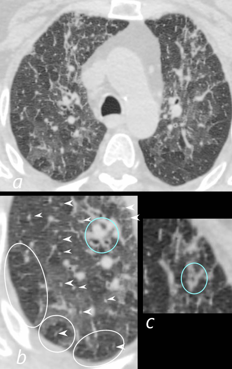
70-year-old female former smoker with long standing history of RA presents with chronic dyspnea.
Axial CT of the chest at the level of the aortic arch reveals centrilobular nodules (b, white arrowheads) , ground-glass opacities, and mosaic attenuation (b, white rings) likely due to air trapping in this context, and bronchial wall thickening (b, c teal rings). There is some irregular thickening of the interlobular septa. In the context of a patient with rheumatoid arthritis a diagnosis of follicular bronchiolitis is likely. However radiologically fibrotic hypersensitivity pneumonitis (HP) is included in the differential diagnosis
Ashley Davidoff MD TheCommonVein.net 132Lu 136652cL
Constrictive Bronchiolitis
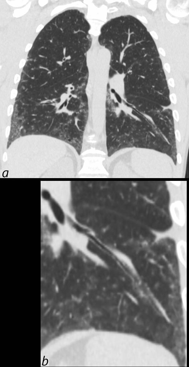
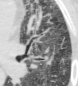
39-year-old-male with a history of scleroderma associated with ILD and digital vasculopathy with ulcers.
Coronal CT shows thickening of the segmental and subsegmental airway supplying the lateral basal segment of the left lower lobe. In addition there is a background of peripheral ground glass changes poorly defined ground glass centrilobular nodules and mild reticulation.
In this clinical setting obliterative bronchiolitis (aka bronchiolitis obliterans aka constrictive bronchiolitis) is suggested. Cellular NSIP is also a radiological consideration.
Ashley Davidoff MD TheCommonVein.net 132Lu 136669c
39-year-old-male with a history of scleroderma associated with ILD and digital vasculopathy with ulcers.
Axial CT shows thickening of the segmental, subsegmental and small airways supplying the posterior basal segment of the right lower lobe. In addition there is a background poorly defined ground glass changes and mild reticulation.
In this clinical setting obliterative bronchiolitis (aka bronchiolitis obliterans aka constrictive bronchiolitis) is suggested. Cellular NSIP is also a radiological consideration.
Ashley Davidoff MD TheCommonVein.net 132Lu 136669c
Aspiration
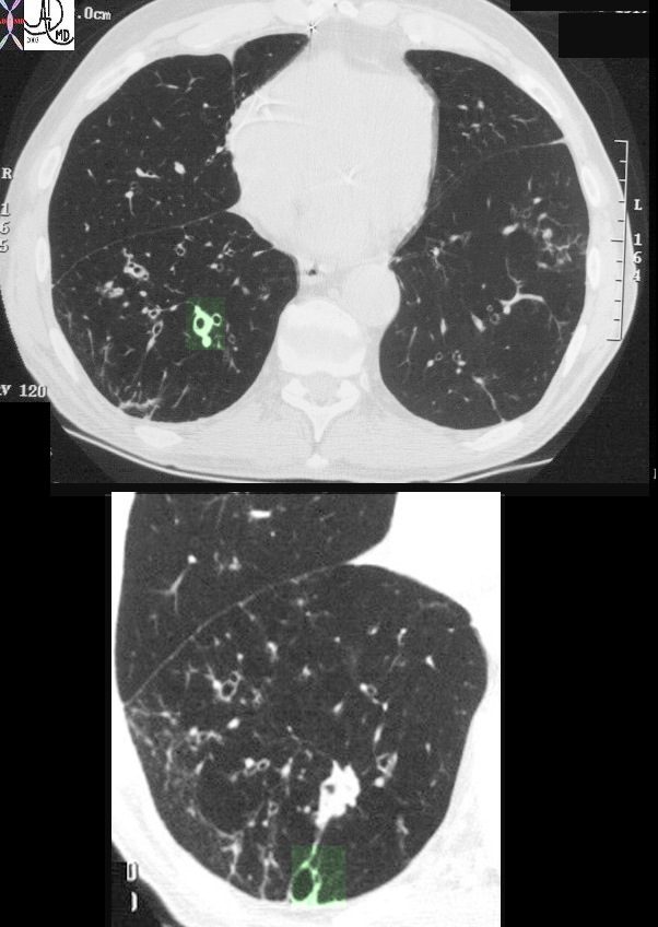
This image represents the CT scan of the chest with a focus on the right lower lobe in an 86year old patient who presented with long standing history of a productive cough. The segmental airways in the upper image are thickened (green overlay) as are the subsegmental airways in the lower panel. Additionally an small airways is abnormally visualised within 1cms of the pleura, indicating traction bronchiolectasis. Barium swallow revealed aspiration as a cause of the bronchial wall thickening.
Ashley Davidoff MD TheCommonVein.net 30602c 245Lu
Immune Diseases
60 year old male with history of asthma, allergic bronchopulmonary aspergillosis (ABPA)
CT scan shows bibasilar bronchiectasis and soft tissue/fluid impaction of the bronchovascular bundles
Ashley Davidoff TheCommonVein.net
Infiltrative Disorders
Amyloidosis
Mainstem

44-year-old female with immunoglobulin light chain (AL) amyloidosis of the mainstem bronchi and left upper lobe bronchi. The axial CT shows circumferential thickening of the mainstem bronchi with minor calcification in the walls and without obstruction. The wall of the right upper lobe bronchus is calcified.
Ashley Davidoff MD TheCommonVein.net 135868 249Lu
Amyloidosis
Segmental Bronchi

44-year-old female with immunoglobulin light chain (AL) amyloidosis of segmental bronchi. The axial CT shows circumferential thickening of the segmental bronchi with punctate calcifications in the walls and without obstruction.
Ashley Davidoff MD TheCommonVein.net 135869b 249Lu
Malignancy

50-year-old female with primary adenocarcinoma of the left lung presenting with pneumonic consolidation, and diffuse reticulonodular changes bilaterally
Images from the CT in a coronal plane (a, and magnified b-f)
Image b shows peribronchial thickening (yellow arrowheads) likely reflecting lymphovascular spread..
Image c shows centrilobular nodular changes (teal arrowheads)
Image d shows peribronchial thickening (yellow arrowhead) likely reflecting lymphovascular spread..
Image e shows centrilobular nodules (teal arrowheads) and diffuse nodular thickening of the interlobular septa representing lymphatic spread along the lymphovascular bundles
Image f shows the primary carcinoma presenting as a pneumonic consolidation.(green oval)
Ashley Davidoff MD TheCommonVein.net 158Lu 131025cL
Mechanical/Atelectasis Trauma Metabolic
Circulatory-
CT Acute CHF Peribronchial Cuffing
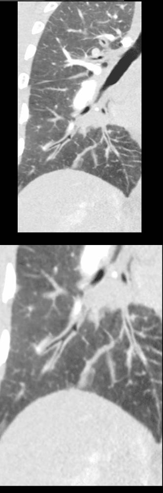
50 year old female with diabetes, chronic renal failure with congestive heart failure. CT in the coronal plane shows diffuse ground glass changes, with prominent peribronchial cuffing in the right upper lobe and and magnified in the lower image in the right lower lobe Prominent Kerley B lines are noted medially – a few laterally and there is with thickening of the right transverse fissure
Ashley Davidoff MD TheCommonvein.net 135777c 193Lu
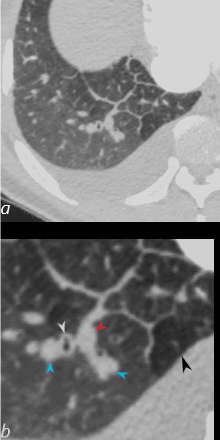
50-year-old female with diabetes, chronic renal failure and congestive heart failure. CT in the axial plane through the right posterior recess, shows thickened interlobular septa at the right base, congested arterioles (light blue arrowheads, b), alongside the bronchioles, peribronchial cuffing (white arrowheads, b), a congested pulmonary venule in the interlobular septum (red arrowhead arrowheads, b), ground glass changes and a secondary lobule demonstrating mosaic attenuation (black arrowhead arrowheads, b). The IVC is dilated and a small complex effusion is present.
Ashley Davidoff MD TheCommonvein.net 135783cL 193Lu
Circulatory Hemorrhage Immune
Infiltrative

44-year-old female with immunoglobulin light chain (AL) amyloidosis of segmental bronchi. The axial CT shows circumferential thickening of the segmental bronchi with punctate calcifications in the walls and without obstruction.
Ashley Davidoff MD TheCommonVein.net 135870 249Lu
44-year-old female with immunoglobulin light chain (AL) amyloidosis of segmental bronchi. The axial CT shows circumferential thickening of the segmental bronchi with punctate calcifications in the walls and without obstruction.
Ashley Davidoff MD TheCommonVein.net 135869b 249Lu
Idiopathic Iatrogenic Idiopathic