
- 61 y.o. male,
- never smoker, fro
- bilateral severe bronchiectasis i
- pulmonary TB in 13 years ago
- s/p 6 month course of therapy
- (unclear if LTBI vs active infection),
- MAI i10 years ago
- s/p 6 months of azithro, rifampin, and ethambutol,
- MAC 2 years ago (untreated), and now
- M. abscessus infection
- treatment (IV amikacin, imipenem, azithromycin and linezolid,
LAbs- ANA positive 1:320 diffuse pattern
- CF genotype testing negative
- Etiology likely secondary to Mounier Kuhn Syndrome compounded by recurrent infections
- Imaging
- 61-year-old male with a history of tracheomegaly (suggestive of Mounier Kuhn syndrome) and varicoid bronchiectasis dominantly involving the middle
lobe and lingula with most recent CT 6 months prior with noted multiple lung nodules, presenting for follow-up. In addition he has been treated
for latent tuberculosis, and pulmonary MAC - CT without contrast reveals the following
Unchanged appearance of multiple pulmonary nodules as above.
Unchanged tracheobronchomegaly and mid-lower lung predominant varicose
bronchiectasis.
- 61-year-old male with a history of tracheomegaly (suggestive of Mounier Kuhn syndrome) and varicoid bronchiectasis dominantly involving the middle
- never smoker, fro
61 year old male with a
History of treated mycobacterial infections
and chronic cough
CXR Frontal View Bronchiectasis Shaggy Heart Border
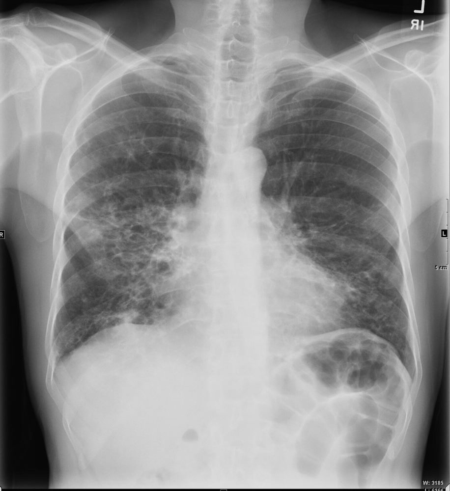
61 year old male with a history of treated mycobacterial infections and chronic cough
Frontal view shows shaggy heart borders with bibasilar cystic changes consistent with bronchiectasis in the middle lobe and lingula
Ashley Davidoff MD TheCommonVein.net 250Lu 135871
Enlarged Trachea and Bronchiectasis

61 year old male with a history of treated mycobacterial infections and chronic cough
Lateral view shows an enlarged trachea and thick walled cystic changes overlying the heart consistent with known bronchiectasis. There is evidence of hyperinflation
Ashley Davidoff MD TheCommonVein.net 250Lu 135872a

61 year old male with a history of treated mycobacterial infections and chronic cough
Lateral view shows an enlarged trachea and thick walled cystic changes overlying the heart consistent with known bronchiectasis. There is evidence of hyperinflation
Lateral view (a magnified in b, and shows an enlarged trachea (white arrowheads) and thick walled cystic changes overlying the heart consistent with known bronchiectasis
Ashley Davidoff MD TheCommonVein.net 250Lu 135872ac01L
Tracheomegaly and Mucus

61 year old male with a history of treated mycobacterial infections and chronic cough
Axial CT at the level of the brachiocephalic vessels shows an enlarged trachea with a strand of mucus straddling the lateral walls. The trachea measures up to 3cms which is abnormally enlarged. There are thin walled cystic changes of the airways along the subsegmental arteries in the upper lobes likely reflecting bronchiectasis
Ashley Davidoff MD TheCommonVein.net 250Lu 135873a
Mounier Kuhn Tracheomegaly and Bronchiectasis

61 year old male with a history of treated mycobacterial infections and chronic cough
Axial CT at the level of the brachiocephalic vessels shows an enlarged trachea that measures 3cms which is abnormally enlarged. There are thin-walled cystic changes of the airways along the subsegmental arteries in the upper lobes likely reflecting bronchiectasis
Ashley Davidoff MD TheCommonVein.net 250Lu 135874ac

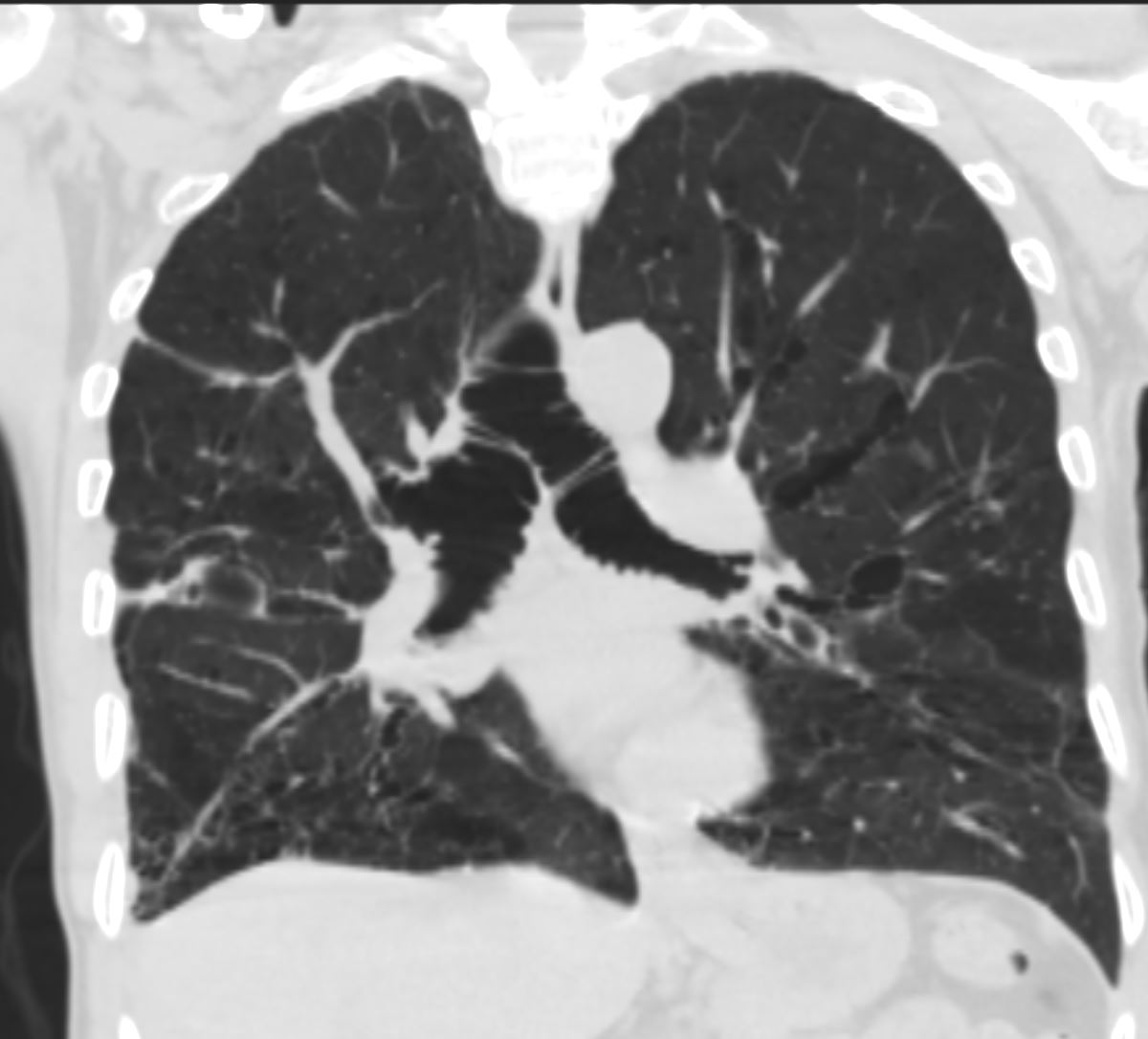
61 year old male with a history of treated mycobacterial infections and chronic cough
Coronal CT at the level of the trachea shows an enlarged trachea that measures 3cms which is abnormally enlarged. There are both thin-walled and mildly thickened cystic changes of the airways along the subsegmental bronchovascular bundle in the upper lobes and lower lobes reflecting bronchiectasis
Ashley Davidoff MD TheCommonVein.net 250Lu 135880

61 year old male with a history of treated mycobacterial infections and chronic cough
Sagittal CT at the level of the trachea shows an abnormally enlarged trachea. Mild thin walled bronchiectasis is also noted Flattened hemidiaphragm indicates hyperinflation
Ashley Davidoff MD TheCommonVein.net 250Lu 135884
Enlarged Mainstem Bronchi and Bronchiectasis

61 year old male with a history of treated mycobacterial infections and chronic cough
Axial CT at the level of the carina shows bilaterally enlarged mainstem bronchi that measure 1.9cms. each which are abnormally enlarged. There are both thin-walled cystic changes of the airways along the subsegmental arteries in the upper lobes likely reflecting bronchiectasis . Some of these cystic changes in the right upper lobe (upper panel) have thicker walls
Ashley Davidoff MD TheCommonVein.net 250Lu 135875a
61-year-old male with a history of treated mycobacterial infections and chronic cough
Coronal CT at the level of the carina shows bilaterally enlarged mainstem bronchi that measure 1.9cms. each which are abnormally enlarged. There are both thin-walled cystic changes of the airways along the subsegmental bronchovascular bundles in the upper lobes reflecting bronchiectasis .
Ashley Davidoff MD TheCommonVein.net 250Lu 135881
Lady Windermere Syndrome
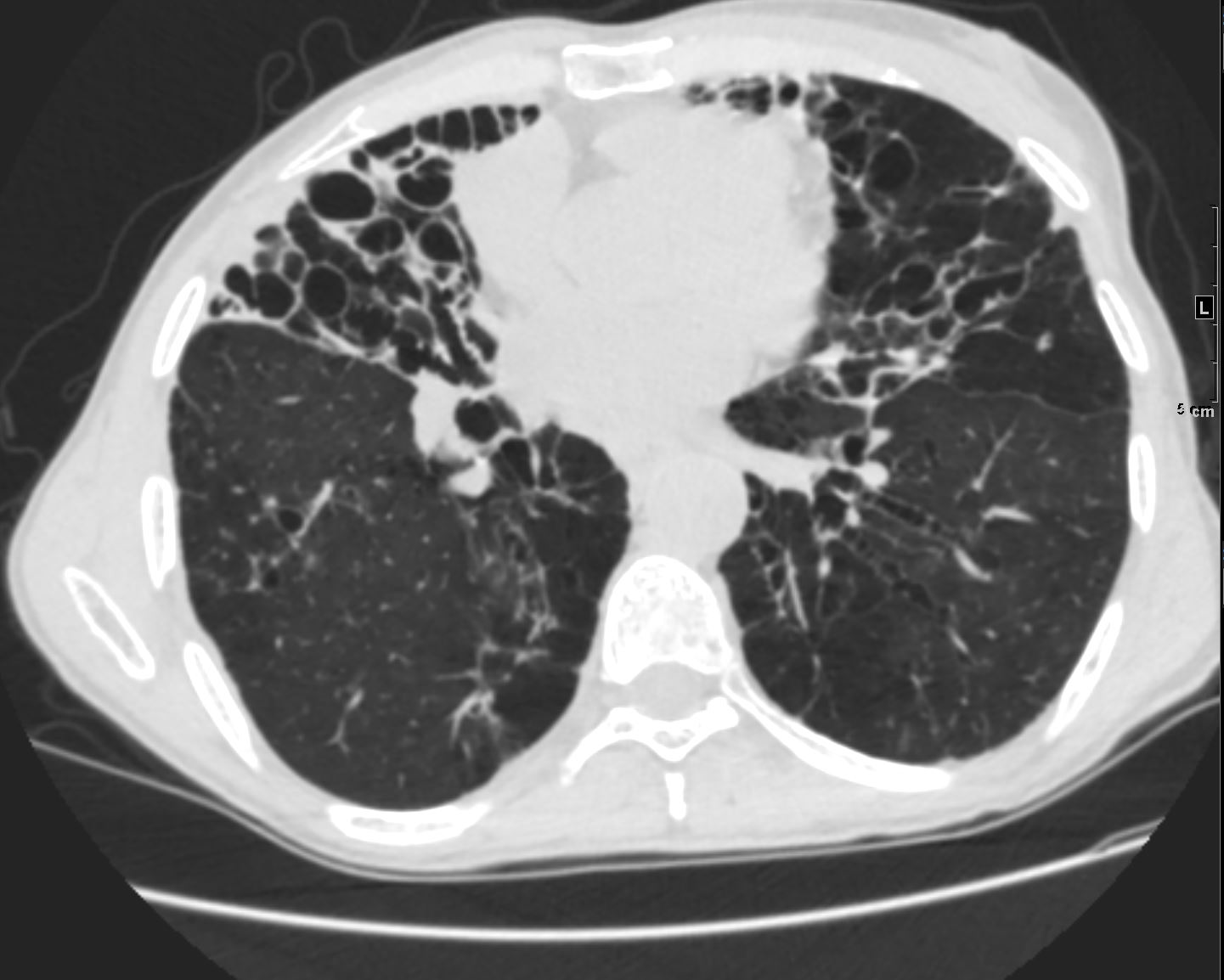
61-year-old male with a history of treated mycobacterial infections including MAC and chronic cough.
Axial CT at the level of the mid to lower chest shows mildly ectatic segmental airways to the lower, and middle lobe bronchi but significant bronchiectasis to the middle lobe and lingula involving the subsegmental airways. There is a relative paucity of mucus in the ectatic airways. The history of MAC and the distribution of the bronchiectasis in the middle lobe and lingula are reminiscent of the diagnosis of Lady Windermere syndrome
Ashley Davidoff MD TheCommonVein.net 250Lu 135876

61-year-old male with a history of treated mycobacterial infections including MAC and chronic cough.
Axial CT at the level of the mid to lower chest shows mildly ectatic segmental airways to the lower, and middle lobe bronchi but significant bronchiectasis to the middle lobe and lingula involving the subsegmental airways. There is a relative paucity of mucus in the ectatic airways. The history of MAC and the distribution of the bronchiectasis in the middle lobe and lingula are reminiscent of the diagnosis of Lady Windermere syndrome
Ashley Davidoff MD TheCommonVein.net 250Lu 135877

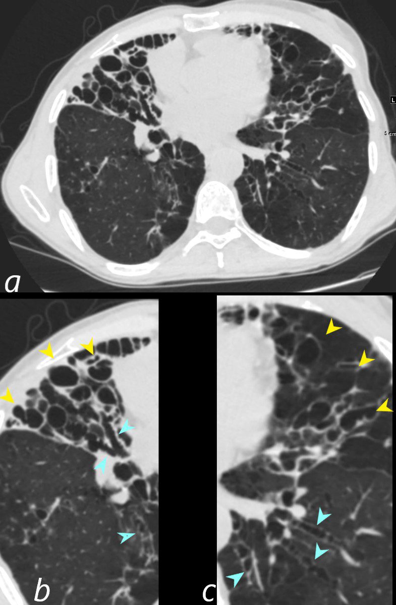
61-year-old male with a history of treated mycobacterial infections including MAC and chronic cough.
Axial CT at the level of the mid to lower chest shows mildly ectatic segmental airways to the lower, and middle lobe bronchi (teal arrowheads (b and c) but significant bronchiectasis to the middle lobe and lingula involving the subsegmental airways (yellow arrowheads b and c). There is a relative paucity of mucus in the ectatic airways. The history of MAC and the distribution of the bronchiectasis in the middle lobe and lingula are reminiscent of the diagnosis of Lady Windermere syndrome
Ashley Davidoff MD TheCommonVein.net 250Lu 135877cL

61-year-old male with a history of treated mycobacterial infections including MAC and chronic cough.
Coronal CT at the level of the heart shows significant bronchiectasis to the middle lobe and lingula and as a result abut the right and left heart border accounting for the CXR findings of a ?shaggy heart border?. There is a relative paucity of mucus in the ectatic airways. The history of MAC and the distribution of the bronchiectasis in the middle lobe and lingula are reminiscent of the diagnosis of Lady Windermere syndrome
Ashley Davidoff MD TheCommonVein.net 250Lu 135879

Barrel Chest Lady Windermere Syndrome
61-year-old male with a history of treated mycobacterial infections including MAC and chronic cough.
Right sagittal CT shows mildly ectatic segmental airways to the upper, middle and lower lobe airways, but significant bronchiectasis to the middle lobe subsegmental airways. There is a relative paucity of mucus in the ectatic airways. The history of MAC and the distribution of the bronchiectasis in the middle lobe and lingula are reminiscent of the diagnosis of Lady Windermere syndrome. The barrel chest reflects hyperinflation and the obstructive nature of the Mounier Kuhn Syndrome
Ashley Davidoff MD TheCommonVein.net 250Lu 135883

61-year-old male with a history of treated mycobacterial infections and chronic cough
Coronal CT at the level of the spinal column shows bilateral mildly ectatic mostly thin-walled cystic changes of the airways along the subsegmental bronchovascular bundles in the upper and lower lobes reflecting bronchiectasis. There is a paucity of mucus accumulation.
Ashley Davidoff MD TheCommonVein.net 250Lu 135882
Links and References
- TCV
- Refrence 11024