
26-year-old female with scleroderma with dyspnea presents for chest evaluation.
The absence of significant fibrotic component and lack of volume loss in the lower lobes makes cellular NSIP more likely
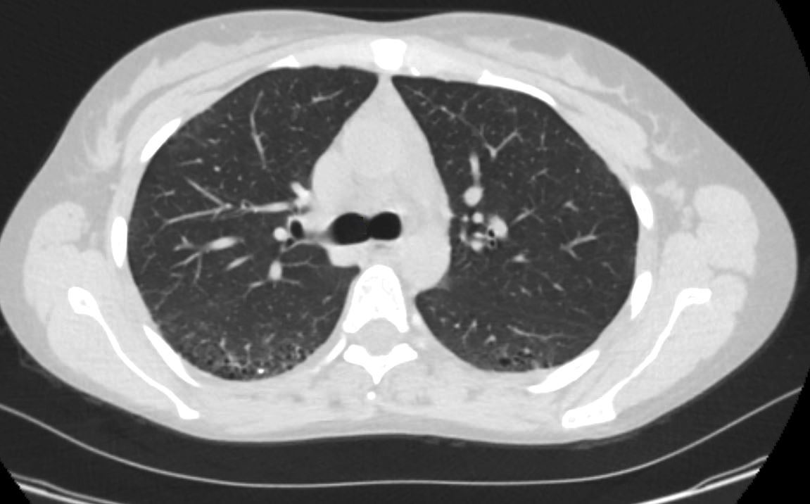
Anterolateral Reticulations in the Lung Apices

26-year-old female with scleroderma with dyspnea presents for evaluation. Axial CT through the lung apices shows mild reticulation in the anterolateral aspects of both lungs. There is an air-fluid level in the esophagus indicating reflux
Ashley Davidoff MD TheCommonVein.net 272Lu 136242
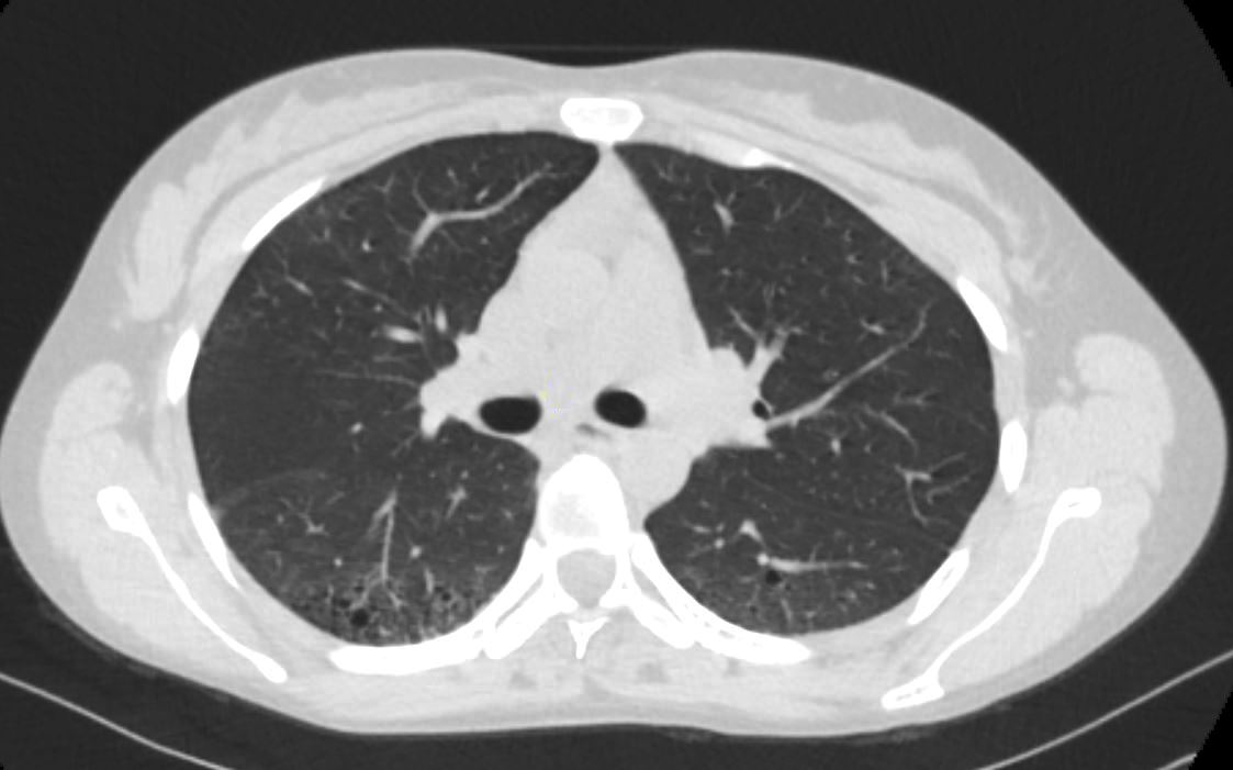
Ground Glass Changes and Mild Reticulations in the Periphery of the Apical Segments of the Lower Lobes

26-year-old female with scleroderma with dyspnea presents for evaluation. Axial CT through the lungs at the level of the carina shows peripherally located, ground glass changes, mild reticulation and bronchiolectasis in the peripheral aspects of the apical segments of the lower lobes with lesser findings in the anterolateral aspects predominantly in the right upper lobe.
Ashley Davidoff MD TheCommonVein.net 272Lu 136243
Ground Glass Changes Bronchiolectasis Mild Reticulations and Dystrophic Calcification in the Periphery of the Apical Segments of the Lower Lobes
26-year-old female with scleroderma with dyspnea presents for evaluation. Axial CT through the lungs at the level of the carina shows peripherally located, ground glass changes, mild reticulation and bronchiolectasis in the peripheral aspects of the apical segments of the lower lobes and a focal dystrophic calcification posteriorly in the right lower lobe. There is an air-fluid level in the esophagus indicating reflux.
Ashley Davidoff MD TheCommonVein.net 272Lu 136244
Ground Glass Changes Bronchiolectasis Mild Reticulations in the Periphery of the Apical Segments of the Lower Lobes
No Significant Volume Loss
26-year-old female with scleroderma with dyspnea presents for evaluation. Axial CT through the lungs at the level of the carina shows peripherally located, ground glass changes, mild reticulation and bronchiolectasis along the bronchovascular bundle in the RLL. The changes are most prominent in the peripheral aspects of the apical segments of the lower lobes with lesser findings in the anterolateral aspects predominantly in the right upper lobe. There is an air-fluid level in the esophagus indicating reflux.
Ashley Davidoff MD TheCommonVein.net 272Lu 136245
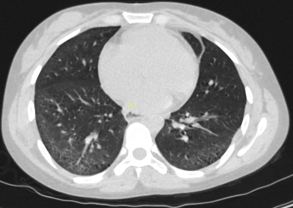
Minimal Bronchial Wall Thickening

26-year-old female with scleroderma with dyspnea presents for evaluation. Axial CT through the lungs at the level of the MPA shows peripherally located, ground glass changes, mild reticulation, bronchiolectasis and a thickened subsegmental airway in the right lower lobe. The fissures are normally placed with no obvious loss of volume of the lower lobes.
Ashley Davidoff MD TheCommonVein.net 272Lu 136246
Ground Glass Changes Bronchiolectasis Mild Reticulations and Subpleural Sparing in the Periphery of the Lower Lobes
No Significant Volume Loss
Note Air-Fluid level in the Esophagus
26-year-old female with scleroderma with dyspnea presents for evaluation. Axial CT through the lungs through the lower lungs shows peripherally located, ground glass changes, mild reticulation bronchiolectasis and subpleural sparing. The fissures are normally placed with no obvious loss of volume of the lower lobes. There is no honeycombing
Ashley Davidoff MD TheCommonVein.net 272Lu 136247
Ground Glass Changes Bronchiolectasis Mild Reticulations and Subpleural Sparing in the Periphery of the Lower Lobes
Note Air-Fluid level in the Esophagus
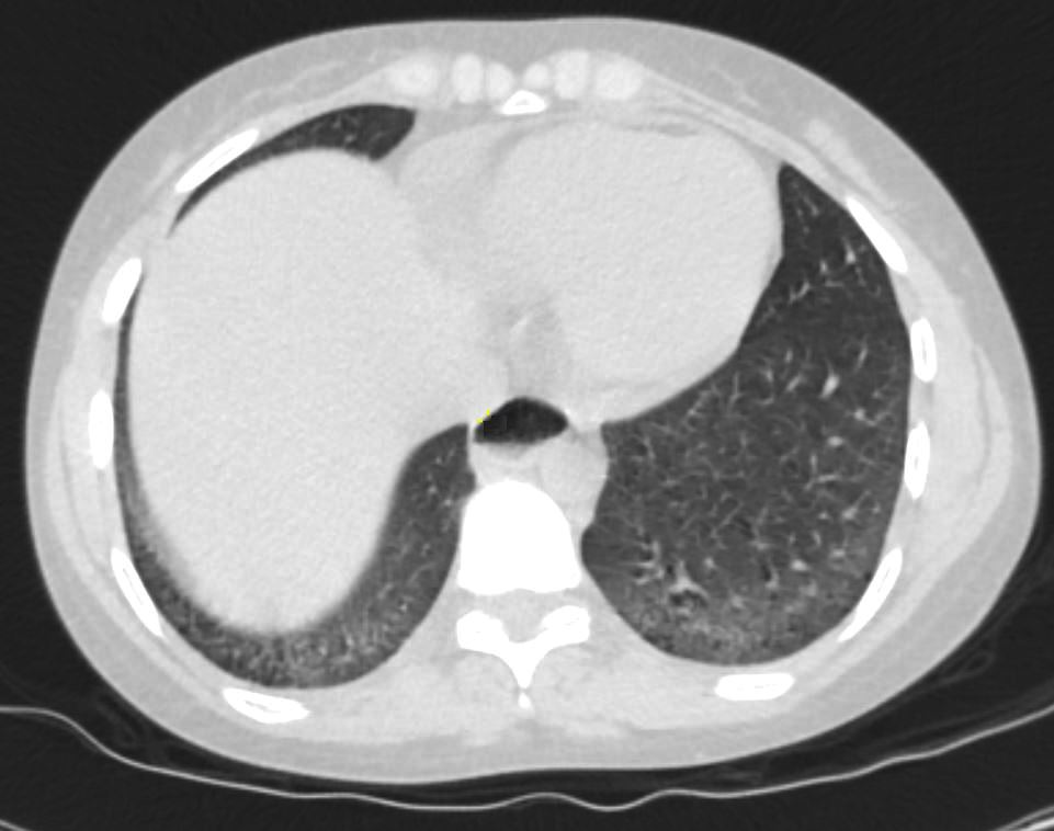
26-year-old female with scleroderma with dyspnea presents for evaluation. Axial CT through the lungs bases shows peripherally located, ground glass changes, bronchiolectasis and subpleural sparing. There is no honeycombing
Ashley Davidoff MD TheCommonVein.net 272Lu 136248
Scleroderma Acro osteolysis and Soft Tissue Calcification-Ossification

26-year-old female with scleroderma. X-Ray through the hands in A-P view shows acro-osteolysis of the distal phalanges of 5 digits of the right hand associated with curling of the nails. The changes of acro-osteolysis are magnified and ringed in b, and soft tissue calcification and ossification (c, white arrowhead).
Ashley Davidoff MD TheCommonVein.net 272Lu 136249c

26-year-old female with scleroderma. X-Ray through the hands in A-P, oblique and lateral views show acro-osteolysis of the distal phalanges of 5 digits of the right hand associated with curling of the nails. Note the soft tissue calcification- ossification near the PIP of the 3rd digit.
Ashley Davidoff MD TheCommonVein.net 272Lu 136250cL