
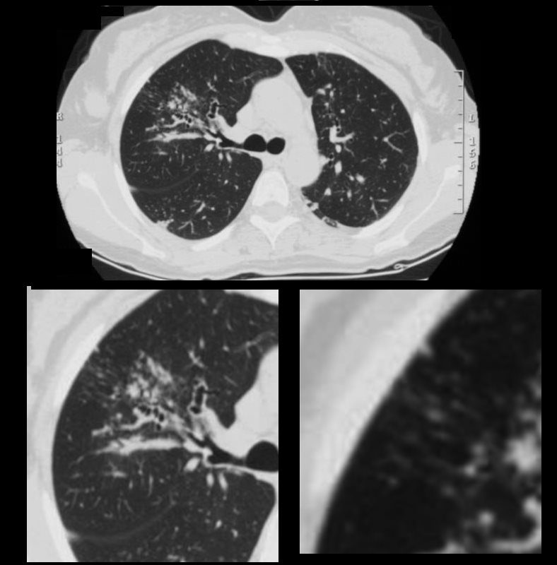
Mucoid Impaction and Bronchiectasis Micronodules RUL
CT Allergic Bronchopulmonary Aspergillosis (ABPA)

CT Allergic Bronchopulmonary Aspergillosis (ABPA)
48 year old female with a history of asthma presents with productive cough. CT scan 18 months prior shows multicentric foci of bronchial wall thickening , in the segmental and subsegmental airways, bronchiectasis and mucoid impaction in the RUL with suggestion of ill defined micronodules indicating small airways disease affecting the upper lobes of the lung. Flexible bronchoscopy revealed mucus plugs and aspergillus was isolated.
Ashley Davidoff MD TheCommonVein.net
Mucoid Impaction Bronchiectasis Micronodules in RML
CT Allergic Bronchopulmonary Aspergillosis (ABPA)
CT Allergic Bronchopulmonary Aspergillosis (ABPA)
48 year old female with a history of asthma presents with productive cough. CT scan 18 months prior shows multicentric foci of bronchial wall thickening , in the segmental and subsegmental airways, bronchiectasis and mucoid impaction in the middle lobe Upper panel magnified lower left) with suggestion of ill defined micronodules (lower panel right) indicating small airways disease affecting the middle lobe of the lung. Flexible bronchoscopy revealed mucus plugs and aspergillus was isolated.
Ashley Davidoff MD TheCommonVein.net
Bronchial Wall Thickening, Micronodules in RML and an Infiltrate in the LLL
CT Allergic Bronchopulmonary Aspergillosis (ABPA)

CT Allergic Bronchopulmonary Aspergillosis (ABPA)
48 year old female with a history of asthma presents with productive cough. CT scan 18 months prior shows multicentric foci of bronchial wall thickening , in the segmental and subsegmental airways in the middle lobe (upper panel magnified lower left) with suggestion of ill defined micronodules (lower panel right) indicating small airways disease . Flexible bronchoscopy revealed mucus plugs and aspergillus was isolated.
Ashley Davidoff MD TheCommonVein.net
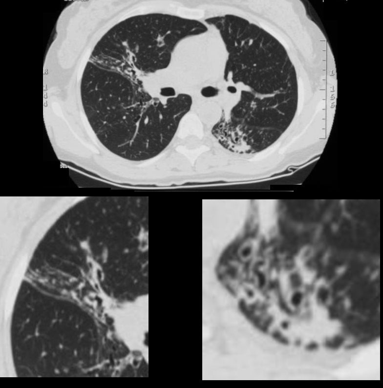
Bronchial Wall Thickening, Bronchiolectasis Atelectasis Micronodules in RML and an Infiltrate in the LLL
CT Allergic Bronchopulmonary Aspergillosis (ABPA)
CT Allergic Bronchopulmonary Aspergillosis (ABPA)
48 year old female with a history of asthma presents with productive cough. CT scan 18 months prior shows multicentric foci of bronchial wall thickening , in the segmental and subsegmental airways, in the middle lobe with crowding of the airways in the RML indicating atelectasis. (upper panel magnified lower left). In the LLL there is bronchiolectasis with thickened airways and a focal subsegmental consolidation (lower panel right) . Flexible bronchoscopy revealed mucus plugs and aspergillus was isolated.
Ashley Davidoff MD TheCommonVein.net
Atelectasis, Mild Bronchial Wall Thickening, and Bronchiolectasis in the RML, Lingula and Bilateral Lower Lobes

CT Allergic Bronchopulmonary Aspergillosis (ABPA)
48 year old female with a history of asthma presents with productive cough. CT scan 18 months prior confirms atelectasis in the middle lobe (upper panel and right lower panel) . There is diffuse mild multicentric foci of bronchial wall thickening in the segmental and subsegmental airways of the middle lobe, lingula and the lower lobes bilaterally (upper panel magnified in lower 3 panels).
Ashley Davidoff MD TheCommonVein.net
Atelectasis Bronchial Wall Thickening, Bronchiolectasis and Micronodules in RML, Lingula and Bilateral Lower Lobes

CT Allergic Bronchopulmonary Aspergillosis (ABPA)
48 year old female with a history of asthma presents with productive cough. CT scan 18 months prior confirms atelectasis in the middle lobe (upper panel and right lower panel)with anterior bulging of the fissure . There is diffuse mild multicentric foci of bronchial wall thickening in the segmental and subsegmental airways of the the lower lobes bilaterally (upper panel magnified in lower 3 panels).
Ashley Davidoff MD TheCommonVein.net
18 Months Later Presents with RUL Atelectasis
CXR Allergic Bronchopulmonary Aspergillosis (ABPA) with Right Upper Lobe Atelectasis
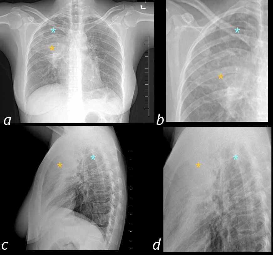
50 year old female with a history of asthma represents with productive cough. CXR in the PA projection (a magnified in b) shows an ill defined density in the right upper lobe of the lung (orange asterisk) and a relatively lucent right apex (blue asterisk) with only minimal elevation of the right hemidiaphragm and minimal rightward mediastinal deviation . The lateral CXR shows a poorly defined density of the atelectatic RUL (orange asterisks c and D) filling in the retrosternal air space, with the hyperinflated right lower lobe reaching the right apex (orange asterisks c and d) . The significantly hyperinflated right lower lobe likely reduces the overall volume loss and hence the subtle compensatory changes of the elevated right hemidiaphragm and the mediastinal shift.
Ashley Davidoff MD TheCommonVein.net
Explaining the CXR based on the CT Findings
The Lucent Apex, Ill Defined Consolidation/Atelectasis, and the Relatively Lucent periphery of the Consolidation

The Lucent Apex, Ill Defined Consolidation/Atelectasis, and the Relatively Lucent periphery of the Consolidation
50 year old female with a history of asthma represents with productive cough.
The ill defined atelectasis in the RUL.
The CXR in the PA projection (a ) shows an ill defined density in the right upper lobe of the lung, with a relatively lucent right apex (blue asterisk in c ) . This image aims to explain the radiological findings integrating the appearance on the CXR and the appearance on CT
Why a lucent apex on CXR?
There is almost complete right upper lobe collapse seen on subsequent CT imaging and the lucent apex (blue asterisk on the magnified view of the CXR, c) correlates with the lucent hyperinflated RLL which reaches to the apex (blue asterisk on the magnified view of the CT, d also see image135097)
Why a lucent periphery of the consolidation?
In the periphery of the atelectatic right upper lobe there are aerated bronchiectatic airways circumscribed with green circle in the magnified view of the chest (c) and confirmed on the CT (green circle d). The overall lucency of this region reflects a relatively low density rather than the expected soft tissue density of the atelectasis
The soft tissue density medially?
The medial aspect of the atelectasis is consolidated and hence displays the characteristic soft tissue density in the magnified view of the CXR (orange asterisk c) and similarly on the CT (orange asterisk d)Ashley Davidoff MD TheCommonVein.net
CT Atelectasis Hyperinflation and Bronchiectasis
ABPA

ABPA
The coronal image is relatively anterior and hence presents as a dense consolidation of atelectasis (orange asterisk, a) In the axial images the hyperinflated RLL is seen posteriorly (teal asterisks in b and c) The region of varicose bronchiectasis is noted posteriorly (lime green arrow, c) When the net density of these 3 findings (consolidation, hyperinflated RLL and bronchiectasis in the LUL) are superimposed on CXR they present with a difficult interpretation since it is the overall net density that gets reflected. The CT scan helps us understand the findings
Ashley Davidoff MD TheCommonVein.net
CT Varicose Bronchiectasis ABPA

The axial images of the RUL are focused on the air filled varicoid bronchiectasis within the RUL atelectasis (lime green arrow in d) The shape of the posterior subsegmental airways has varicoid shape, and hence the terminology. The axial cuts on the left at 2 different levels, are magnified on the right images. The consolidation (yellow asterisk is noted in all 4 images – solid anteriorly, with the bronchiectatic posterior subsegmental airways posteriorly. There is a small focus of mucoid accumulation in the proximal segmental airway (yellow arrowhead d) The hyperinflated RLL is marked with a teal asterisk in all 4 images .
Ashley Davidoff MD TheCommonVein.net
The interesting part of this case is the interpretation of the CXR and how combining it with the CT findings an understanding of the CXR becomes clearer Also aspergillosis presenting with atelectasis is not common