
Aspergillomas
Ashley Davidoff TheCommonVein.net
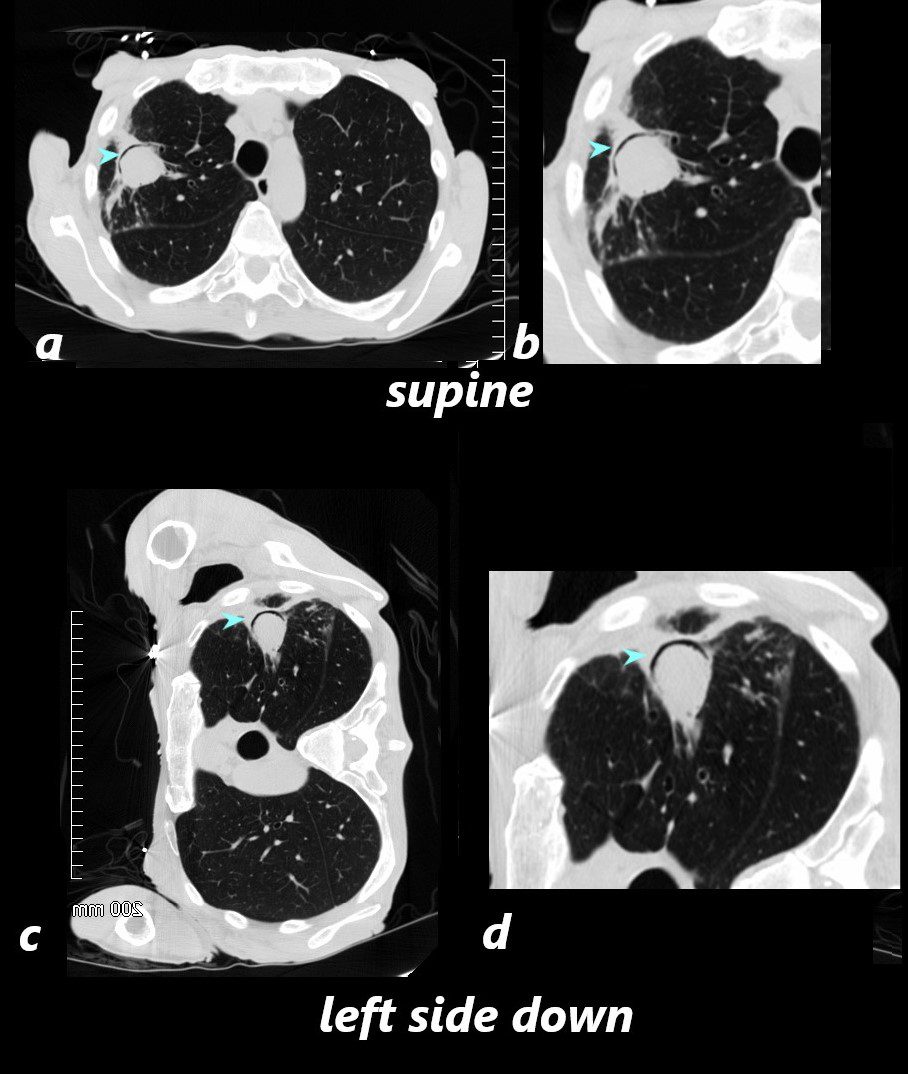
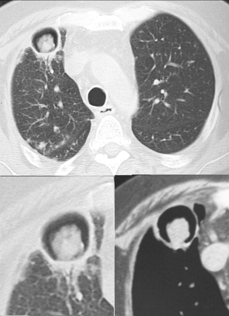
CT scan in the axial projection with the patient in the supine position shows a soft tissue mass in the right upper lung field with an anterior crescent shaped rim of air (blue arrowheads) anterior to the aspergillus fungus ball (a, magnified in b)
Since the fungus ball is movable, when the patient is placed in the the left decubitus position c and d), the fungus ball “sinks” to the dependent position and the air moves to the most superior position (blue arrowheads)
Ashley Davidoff TheCommonVein.net 78612a L

Ashley Davidoff TheCommonVein.net

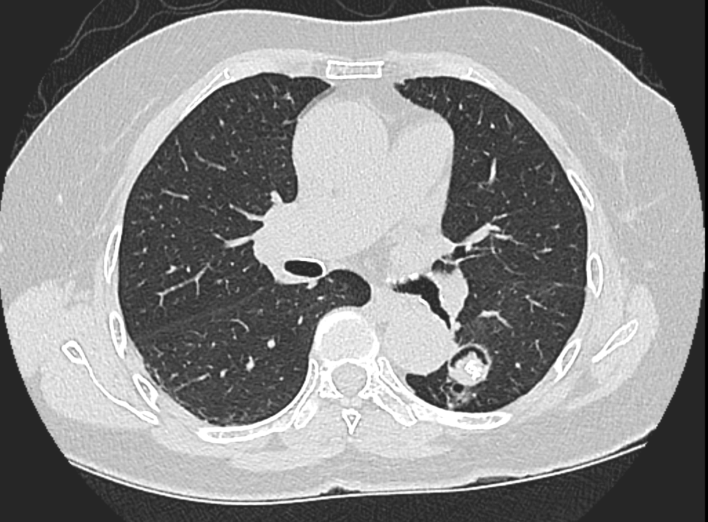
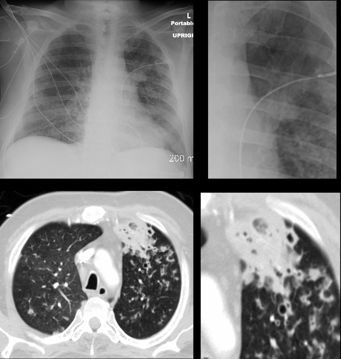
46 year old immunocompromised male with a fever. CT shows small biapical cavitations with internal solid debris .
Diagnosis probable Chronic Pulmonary Aspergillosis with Aspergillomas in TB cavities
Ashley Davidoff TheCommonVein.net

Ashley Davidoff MD
Ashley Davidoff MD
ABPA Segmental Subsegmental and Small Airway Disease

Rossi, SE et al Tree-in-Bud Pattern at Thin-Section CT of the Lungs: Radiologic-Pathologic Overview RadioGraphicsVol. 25, No. 3 2005
ABPA – Dominant Segmental and Subsegmental Airway Disease

47114c01 bronchi lungs fx dilated enlarged impacted with sft tissue finger in glove dx allergic bronchopulomonary aspergillosis ABPA aspergillus dx infection inflammation CTscan Davidoff MD
ABPA – Dominant Small Airway Disease

43 year old man with known aspergillus infection. Note the thickening of the walls of the segmental subsegmental and small airways with extensive tree in bud changes and bronchial wall thickening. There are centrilobular nodules indicating the small airway disease
Ashley Davidoff MD TheCommonVein.net
117816c
Lower Lobe Bronchiectasis and ABPA over 9 years

54 year old female with history of asthma, bronchitis, bronchiectasis, ABPA
CT scan shows initial CT (a) of the lower lobes and part of the middle lobe performed 9 years prior with extensive segmental, fluid/soft tissue filled bronchiectasis within the right lower and to lesser extent the left lower lobe reminiscent of finger in glove morphology. Subsequent imaging 2 (b),5 (c) and 9 years(d), reveals ongoing bronchiectasis (teal arrowhead) subsegmental airway thickening (red arrowhead) and ground glass micronodules indicating small airway disease. The middle lobe was also involved- not shown here.
Ashley Davidoff TheCommonVein.net
ABPA with Atelectasis
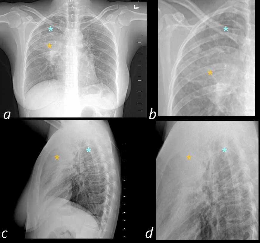
50 year old female with a history of asthma represents with productive cough. CXR in the PA projection (a magnified in b) shows an ill defined density in the right upper lobe of the lung (orange asterisk) and a relatively lucent right apex (blue asterisk) with only minimal elevation of the right hemidiaphragm and minimal rightward mediastinal deviation . The lateral CXR shows a poorly defined density of the atelectatic RUL (orange asterisks c and D) filling in the retrosternal air space, with the hyperinflated right lower lobe reaching the right apex (orange asterisks c and d) . The significantly hyperinflated right lower lobe likely reduces the overall volume loss and hence the subtle compensatory changes of the elevated right hemidiaphragm and the mediastinal shift.
Ashley Davidoff MD TheCommonVein.net

ABPA
The coronal image is relatively anterior and hence presents as a dense consolidation of atelectasis (orange asterisk, a) In the axial images the hyperinflated RLL is seen posteriorly (teal asterisks in b and c) The region of varicose bronchiectasis is noted posteriorly (lime green arrow, c) When the net density of these 3 findings (consolidation, hyperinflated RLL and bronchiectasis in the LUL) are superimposed on CXR they present with a difficult interpretation since it is the overall net density that gets reflected. The CT scan helps us understand the findings
Ashley Davidoff MD TheCommonVein.net
Chronic Pulmonary Aspergillosis
Ashley Davidoff TheCommonVein.ne
ABPA and TB
46 year old immunocompromised male with a fever. CT shows small biapical cavitations with internal solid debris .
Diagnosis probable Chronic Pulmonary Aspergillosis with Aspergillomas in TB cavities
Ashley Davidoff TheCommonVein.net
ABPA and Pneumonia
Note a subsegmental consolidation in the anterior segment of the left upper lobe in a 43 year old man with known aspergillus infection. Note the thickening of the walls of the segmental subsegmental and small airways with bronchiectasis and bronchiolectasis. There are centrilobular nodules indicating the small airway disease
Ashley Davidoff MD TheCommonVein.net 117811c
Invasive Aspergillosis

The coronal CT scan shows a rapidly progressive interstitial infiltrate in the left upper lobe with a focal subsegmental infiltrate in the right lower lobe, both showing features of a reversed halo sign (Atoll sign) There is evidence of bronchial wall thickening.
Diagnosis Aspergillus Infection
Ashley Davidoff TheCommonVein.net
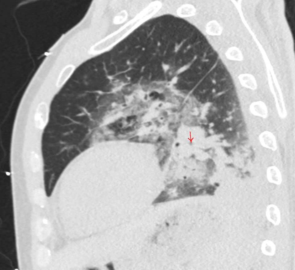
The sagittal CT scan shows a rapidly progressive interstitial infiltrate in the left upper and lower lobes showing features of a reversed halo sign (Atoll sign) There is evidence of basilar consolidation Extensive disease of the segmental and subsegmental airways is noted (red arrow) Fungal Hyphae were identified at bronchoscopy
Diagnosis Aspergillus Infection
Ashley Davidoff TheCommonVein.net
