Mesothelioma is a rare and aggressive type of cancer that affects the lining of the lungs, abdomen, heart, or testicles.
The most common cause of mesothelioma is exposure to asbestos, a naturally occurring mineral that was widely used in construction, shipbuilding, and other industries before its health hazards were recognized.
Symptoms of mesothelioma can vary depending on the location of the tumor, but may include chest pain, shortness of breath, cough, abdominal swelling, nausea, or testicular swelling.
Diagnosis of mesothelioma typically involves imaging studies such as chest X-ray, CT scan, or MRI, as well as a biopsy to confirm the presence of cancer cells and determine the subtype.
Treatment options for mesothelioma may include surgery, radiation therapy, chemotherapy, or a combination of these approaches, depending on the stage and location of the tumor, as well as the overall health of the patient.
The prognosis of mesothelioma is generally poor, as it tends to be diagnosed at an advanced stage and is resistant to many forms of treatment. However, early detection and aggressive therapy may improve survival rates in some cases.
Prevention of mesothelioma involves minimizing exposure to asbestos by following proper safety procedures in the workplace, using protective equipment, and avoiding activities that disturb asbestos-containing materials.
Mesothelioma is a highly specialized field of oncology, and patients may benefit from seeking care at a center that specializes in the diagnosis and treatment of this disease. Clinical trials of new therapies may also offer promising options for eligible patients.
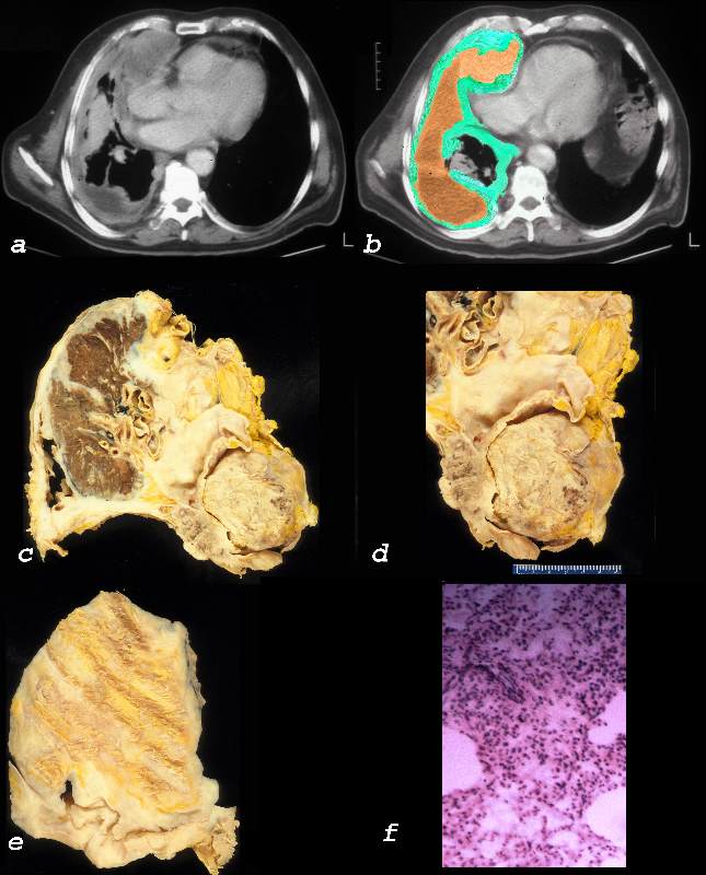
Anterior mediastinal mass and a pleural mass in a 71 year female with mesothelioma Ashley Davidoff MD TheCommonVein.net 29729c.1k This combination of images is from a patient with mesothelioma associated with asbestos related disease. Note the gross anatomy of a pleural plaques in images(f) its imaging appearance on CXR with contracted hemithorax (a,b) and CT appearance c,d,e, as an enhancing rind of soft tissue around the heart (c) and in the posterior costophrenic space. (c,d). The pathology shows a combination of malignant proliferation of stromal and glandular elements (g) abutting relatively normal lung (f) . key words lung pleura mesothelioma mass malignant heart pericardium mesothelioma cardiac imaging radiology CXR plain film CT histopathology CTscan Ashley Davidoff MD TheCommonVein.net 32215c02
This combination of images is from a patient with mesothelioma associated with asbestos related disease. . Note the thickened pleura (green in b) the complex the associated effusion (orange in b). The lung is collapsed under the aggressive tumor. The right atrium is compressed by the tumor. Image c is an autopsy specimen of this patient showing the fibrous rind of tumor (yellow surrounding the lung (brown color) (e) and mediastinal structures magnified in d. Image e shows the external component of the mesothelioma with impressions of the ribs and image f shows the histology of the tumor charactarized by small and monotonous nuclei reminiscent of an epithelioid mesothelioma Ashley Davidoff MD TheCommonVein.net 32640c01L01.8
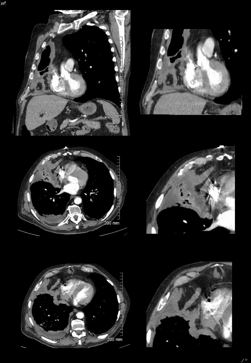
Mesothelioma Shrinking the Thorax and
Invading the Pericardium
CXR shows dense pleural surrounds of a known mesothelioma with decrease in size of the right hemithorax. There are also bands of increase density in the right lower lung zone and shadowing of the right heart boarder indicates involvement of the middle lobe. CTscan confirms the presence of a dense fibrosis surrounding the right lung and compression and atelectasis of the middle lobe Ashley Davidoff MD TheCommonVein.net pleura mesothelioma 0028c CTscan in the coronal and axial planes in this patient with mesothelioma shows dense pleural surrounding the right hemithorax, with decrease in size of the right hemithorax (top row) compressive atelectasis of the middle lobe, (middle row) invasion into the pericardium and pericardial fat and small complex effusion with split pleura sign and thickened pleura (lower row) . Ashley Davidoff MD TheCommonVein.net pleura mesothelioma 0028c01
Mesothelioma Invading the
Posterior Mediastinum and Neural Canal
Biphasic mesothelioma on this CT is characterized by a heterogeneous right apical mass and large effusion(a) that shows invasion into the spinal cord through the neural foramen and into the mediastinum abutting the esophagus (b), with extension into the major fissure and substernal region (c and d) Courtesy Rebecca Schwartz MD TheCommonVein.net 46833c01 CT of a biphasic mesothelioma shows invasion of the mesothelioma into the posterior mediastinum. The malignant soft tissue mass in the axial projection (a,b) is pointed out on the magnified view (b, arrow) anterior to the aorta and esophagus, and in the sagittal view (d, arrow) anterior to the aorta Courtesy Rebecca Schwartz MD TheCommonVein.net 46820cL
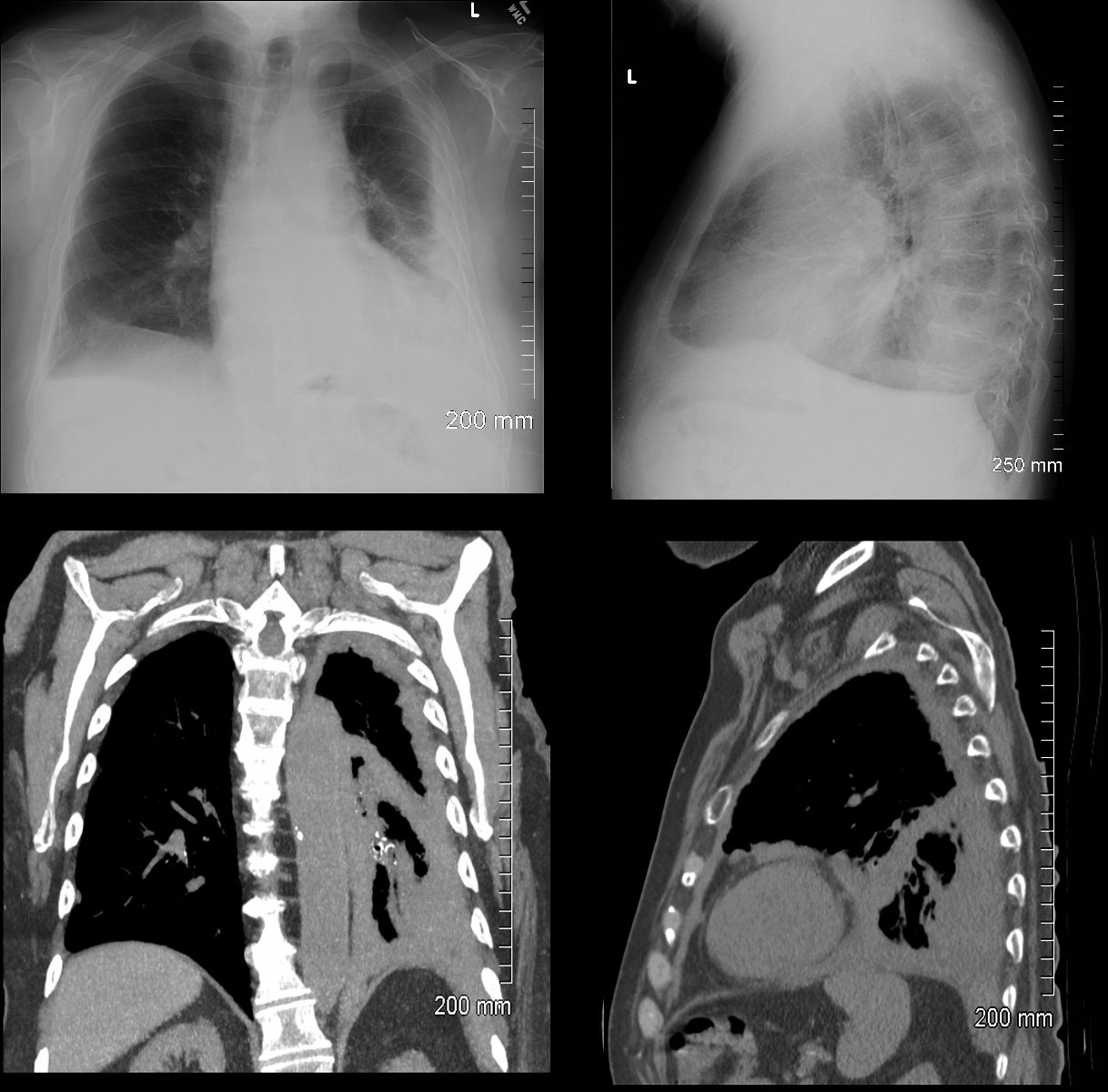
Mesothelioma in a Ship Builder
Pericardial Invasion and Compressive Atelectasis
CXR of an 81 year old shipbuilder shows dense pleural surrounds of a known mesothelioma with decrease in size of the left hemithorax. There is dense multicentric consolidation noted on the lateral CXR CT scan (lower row) confirms the presence of a dense fibrosis surrounding the left lung, dominant in the lung base with compression of the left lower lobe Ashley Davidoff MD TheCommonVein.net pleura mesothelioma 0060c CTscan in the coronal and axial planes in this 81 year old previous shipbuilder with mesothelioma shows dense pleural surrounding the right hemithorax, with decrease in size of the right hemithorax (top row) compressive atelectasis of the lower lobe and probable invasion into the pericardium and pericardial fat (top row) and compression of the lower lobe by thickened malignant pleura (lower row) . Ashley Davidoff MD TheCommonVein.net pleura mesothelioma 0100c