
CT – Left Hilar Carcinoid Tumor Tip of the Iceberg Sign
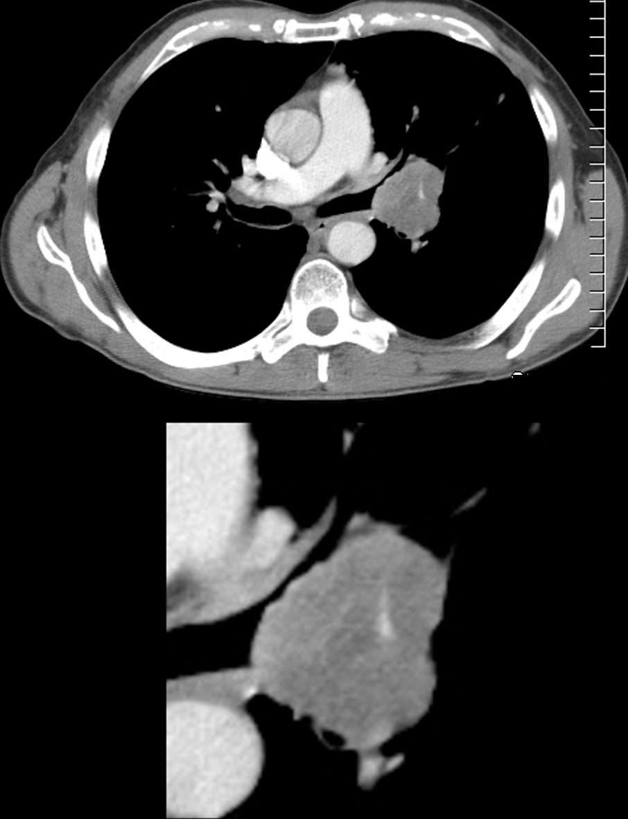
Axial CT scan at the level of the hila in a 56-year-old male shows a 4.2cms vascular mass in the distal left mainstem bronchus impinging on the lumen of the lingula airways. The mass demonstrates the tip of the iceberg sign with the larger portion of the tumor extending beneath the bronchial surface. This is a feature that is characteristic of carcinoid tumors.
Ashley Davidoff MD TheCommonVein.net 261Lu 118381c
CT ? Right Lower Lobe Lung Abscess
Hyperinflation of the Left Lower Lobe
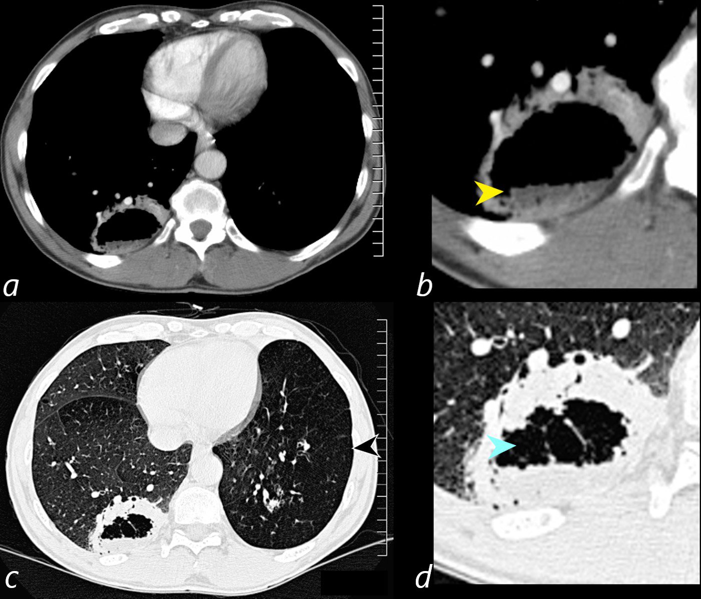
Axial CT scan at the level of the lung bases in a 56-year-old male with an obstructing carcinoid tumor of the lingula shows a cavitating abscess cavity(d, blue arrowhead) with an air fluid level in the right lower lobe (b yellow arrowhead).
The left lower lobe is relatively lucent, reflecting compensatory hyperinflation secondary to the lingula atelectasis (c, black arrowhead)
Ashley Davidoff MD TheCommonVein.net 261Lu 118383cL

56-year-old male with known central carcinoid tumor causing lingula atelectasis, s/p placement of a stent. There is fullness to the left hilum (b, teal arrowhead) with ill-defined abutting infiltrate. A stent in the lingula airway is present (b, black arrowhead)
The lateral examination shows a segmental lingula atelectasis (d, blue arrowhead) and a large cavitating abscess (d, white arrowhead), and air-fluid level (d yellow arrowhead).
Ashley Davidoff MD TheCommonVein.net 261Lu 118379cL