
- Reticular abnormality
- fine network or mesh of
- overlapping linear lines within th
- architectural distortion
- tethering and warping of the
- normal hexagonal appearance
- abnormal
- size or
- shape with
- evidence of volume loss.
- Honeycomb cysts are
- subpleural clustered cystic air spaces
- small in size (3?5 mm). T
- must be contiguous, and
- must touch the pleural surface.
- frequently multilayered, a
Inflammation
NSIP

Ashley Davidoff MD TheCommonVein.net

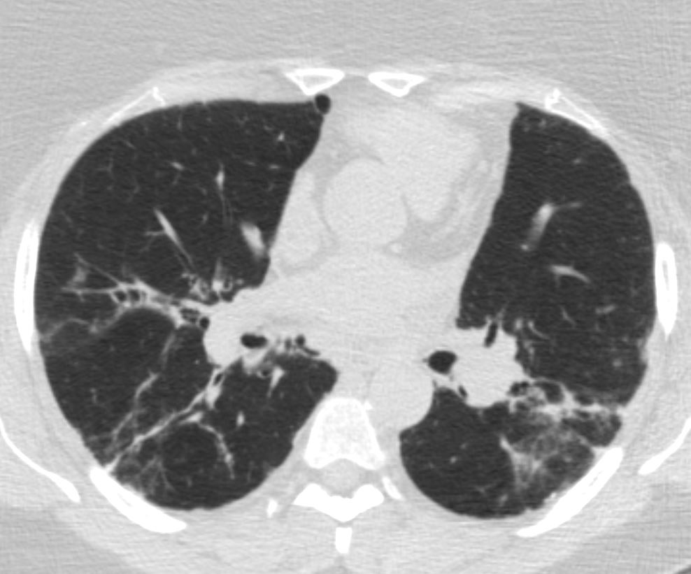
The CT shows mild early peripheral reticular changes, and in this instance additionally characterized by bronchiolectasis abutting the fissure and possibly early honeycomb changes in the LLL posteriorly
Ashley Davidoff MD thecommonvein.net 135079m- lungs UIP

The CT shows mild early peripheral reticular changes in the upper lobe and lung bases
Ashley Davidoff MD thecommonvein.net 135079m01- lungs UIP
UIP
Ashley Davidoff MD thecommonvein.net 134887- lungs UIP

Ashley Davidoff MD thecommonvein.net 134886- lungs UIP
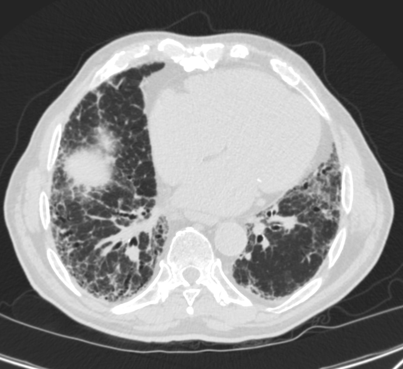
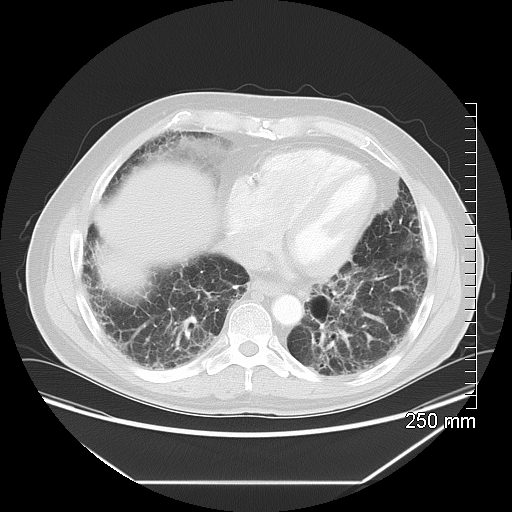
70 year old male with UIP showing extensive dominantly bibasilar and peripheral reticulation
Ashley Davidoff MD TheCommonVein.net uip-008
70 year old male with UIP showing extensive dominantly bibasilar and peripheral reticulation
Ashley Davidoff MD TheCommonVein.net uip-007
70 year old male with UIP showing extensive dominantly bibasilar and peripheral reticulation
Ashley Davidoff MD TheCommonVein.net uip-006
70 year old male with UIP showing extensive dominantly bibasilar and peripheral reticulation
Ashley Davidoff MD TheCommonVein.net uip-005

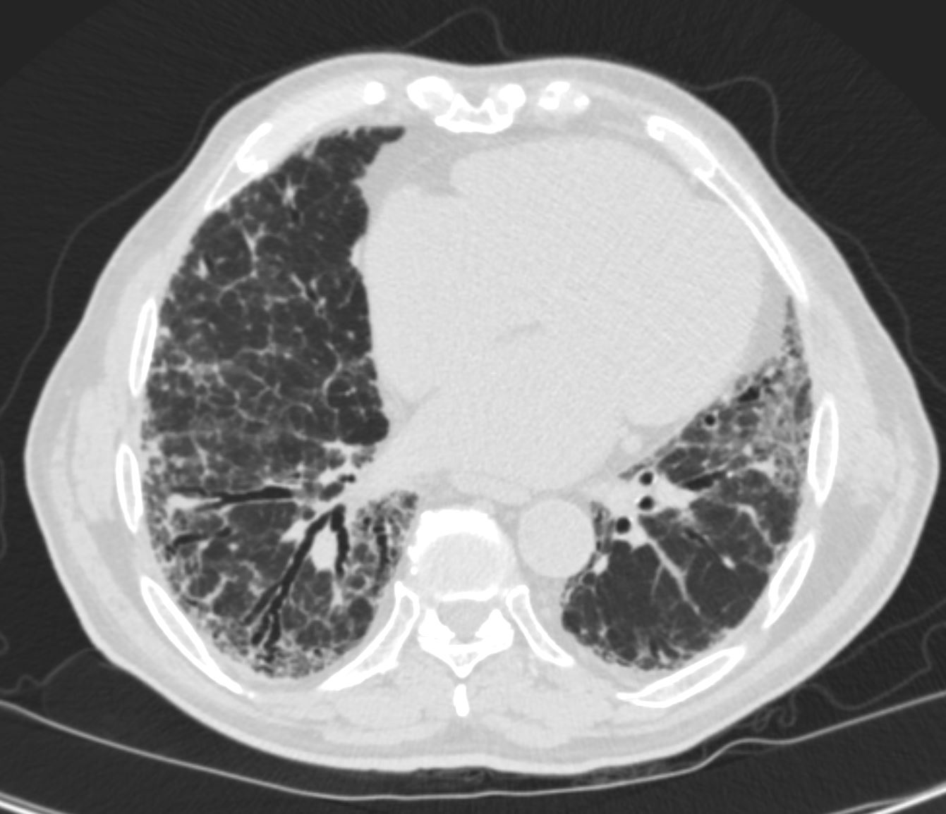
70 year old male with UIP showing extensive, dominantly bibasilar peripheral reticulation, and bronchiolectasis in the medial basal segment of the right lower lobe
Ashley Davidoff MD TheCommonVein.net uip-007

70 year old male with UIP showing extensive dominantly bibasilar and peripheral reticulation
Ashley Davidoff MD TheCommonVein.net uip-002
Ashley Davidoff MD TheCommonVein.net
: A Practical Guide for Radiologists in the New Era of Cancer Care RadioGraphicsVol. 37, No. 5

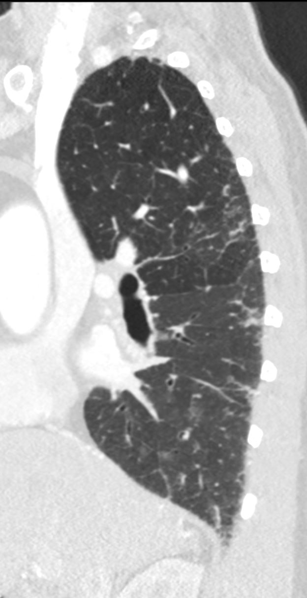
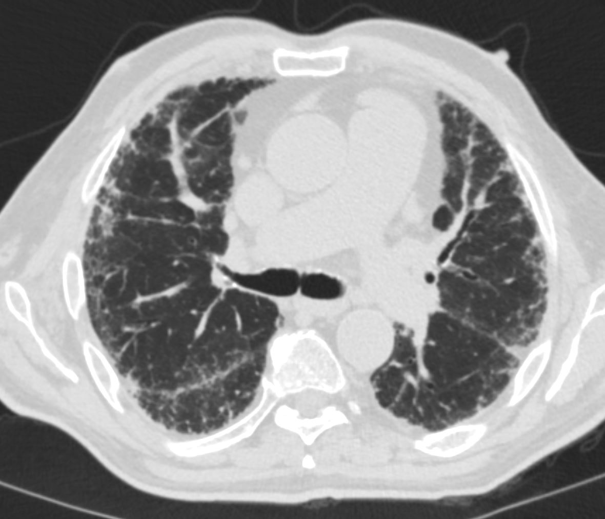
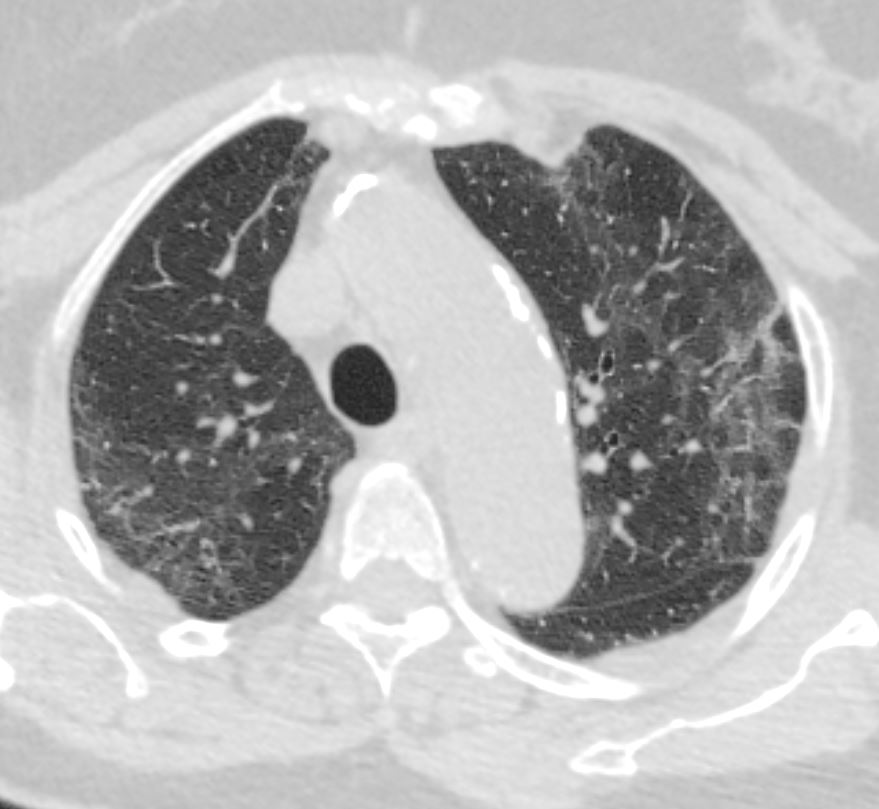
Axial CT shows peripheral reticular changes, ground glass, bronchiolectasis, volume loss with crowding of the bronchovascular bundles posteriorly and subpleural sparing posteriorly. Note air-fluid level in the distended esophagus.
Ashley Davidoff MD TheCommonVein.net

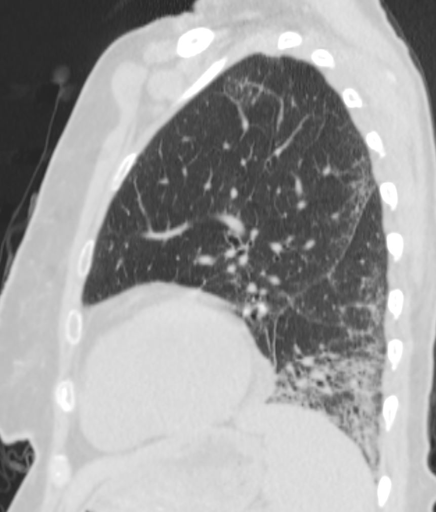
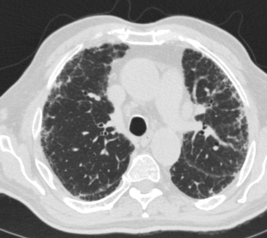
Axial CT shows peripheral reticular changes, and bronchiolectasis in the right upper lobe
Ashley Davidoff MD TheCommonVein.net

Axial CT shows peripheral reticular changes, in the right upper lobe. Note air-fluid level in the distended esophagus.
Ashley Davidoff MD TheCommonVein.net

CT shows mild peripheral ground glass changes in the upper lobes with peripheral sparing and minimal reticular change
Ashley Davidoff MD TheCommonVein.net scleroderma-013
28F-scleroderma-014-2020.jpg
Scleroderma and NSIP (Probable Cellular Form)CT shows mild peripheral ground glass changes in the upper lobes with peripheral sparing and minimal reticular change
Ashley Davidoff MD TheCommonVein.net scleroderma-014
Geometric Distortion of the Secondary Lobules

72 year old female showing reticular changes at the lung bases characterised by irregular thickening of the interlobular septa geometric distortion of the secondary lobules (b,c ringed) Ashley DAvidof TheCommonVein.net
136228cL

72 year old female showing reticular changes at the lung bases characterised by irregular thickening of the interlobular septa geometric distortion of the secondary lobules changes Ashley Davidoff TheCommonVein.net
136229
Changes in the Interlobular Septa
Intralobular Changes
Geometric Distortion
Changes Located in the Subpleural Location
Infection
Neoplasm

Mechanical Trauma Metabolic Circulatory
Immune
Amiodarone Toxicity

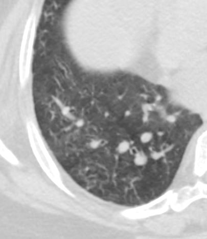
68-year-old male presented with atrial fibrillation during an acute episode of diverticulitis. He was started on amiodarone with good response. He presents 9 months later with dyspnea. CT scan in the axial plane through the lung bases show reticular changes characterised by irregular thickening of the interlobular septa, mild heterogeneous ground glass changes mosaic attenuation centrilobular nodules and bronchiolectasis suggesting a combination of small airway disease and an alveolitis with early fibrotic changes . These findings are consistent with amiodarone toxicity
Ashley Davidoff MD TheCommonVein.277Lu 37873c
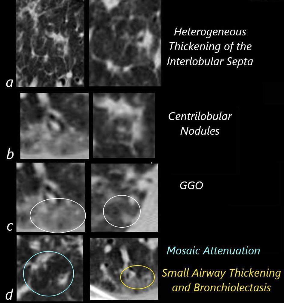
68-year-old male presented with atrial fibrillation during an acute episode of diverticulitis. He was started on amiodarone with good response. He presents 9 months later with dyspnea. CTscan in the axial plane through the lung bases show reticular changes characterised by irregular thickening of the interlobular septa (a), centrilobular nodules, (b)mild heterogeneous ground glass changes (c) mosaic attenuation (d, teal ring) and small airway wall thickening and bronchiolectasis (d yellow ring) suggesting a combination of small airway disease and an alveolitis with early fibrotic changes. These findings are consistent with amiodarone toxicity.
Ashley Davidoff MD TheCommonVein.277Lu 37873c02
Inherited Iatrogenic Idiopathic