
Inflammation
Follicular Bronchiolitis (aka Bronchiolitis Obliterans)
Patient with RA

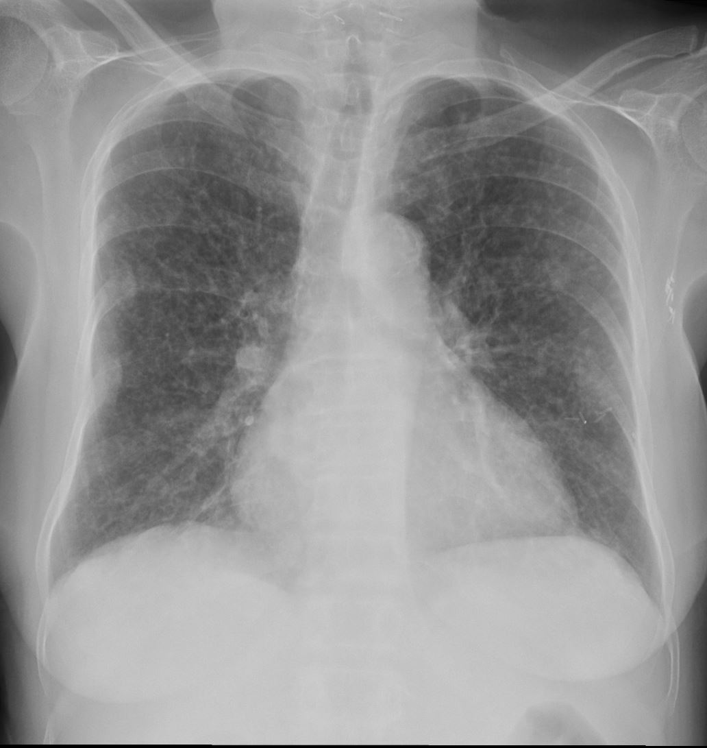
70-year-old female former smoker with long standing history of RA presents with chronic dyspnea. Frontal view of the chest reveals a coarsened nodular interstitial pattern with magnified views showing the micronodularity in the lower panels.
Ashley Davidoff MD TheCommonVein.net 132Lu 136650c01
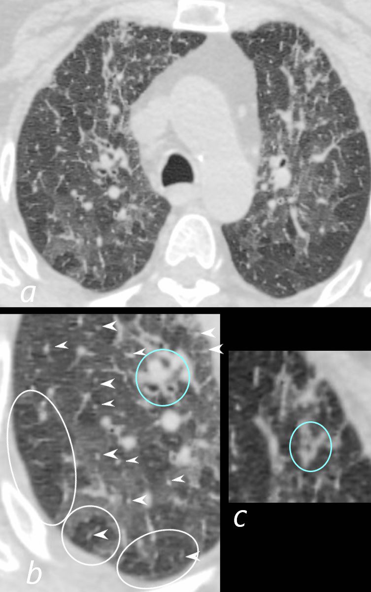
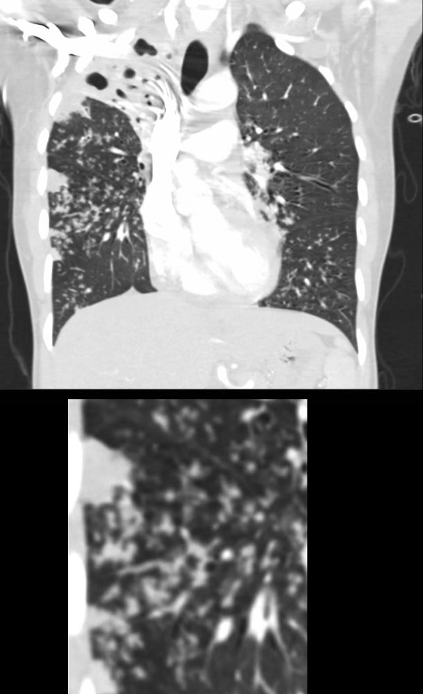
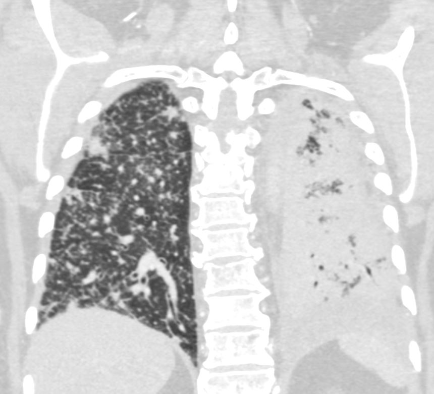
70-year-old female former smoker with long standing history of RA presents with chronic dyspnea.
Axial CT of the chest at the level of the aortic arch reveals centrilobular nodules (b, white arrowheads) , ground-glass opacities, and mosaic attenuation (b, white rings) likely due to air trapping in this context, and bronchial wall thickening (b, c teal rings). There is some irregular thickening of the interlobular septa. In the context of a patient with rheumatoid arthritis a diagnosis of follicular bronchiolitis is likely. However radiologically fibrotic hypersensitivity pneumonitis (HP) is included in the differential diagnosis
Ashley Davidoff MD TheCommonVein.net 132Lu 136652cL
Infection
Transbronchial Spread TB
39-year-old immigrant Vietnamese male presents night sweats, fever, and cough. CT in the coronal plane of the chest shows a large cavitating lesion in the right upper lobe, with innumerable micronodules dominantly in the right midlung field, and to lesser extent in the right upper lung field. Some micronodules are probably present in the left lower lobe as well. Close to the largest subsegmental consolidation there is a bronchus which shows thickening of its wall.
Although it has the appearance has a ?miliary? pattern, this term is usually referred to the hematogenous spread of the disease
Ashley Davidoff MD TheCommonvein.net 135786c
006Lu
Miliary TB
60-year-old immunocompromise female presents with a cough and weight loss CXR shows a diffuse miliary pattern. Final diagnosis was mycobacterium tuberculosis. Associated findings include healed right sided rib fractures and surgical clips in the left axilla
Ashley Davidoff MD TheCommonVein.net 265Lu 136197


60-year-old immunocompromised female presents with a cough and weight loss. Axial CT shows miliary nodules throughout both lung fields. Some of these nodules are centrilobular (c, maroon arrowheads) and others are fissural based (b, pink arrowheads). In some of the secondary lobules there are 2 centrilobular nodules indicating involvement of the airway and arteriole and or the lymphatics (c white rings). One lobule shows centrilobular and interlobular nodules (c gray ring anteriorly). She responded well to treatment and final diagnosis was mycobacterium tuberculosis.
Ashley Davidoff MD TheCommonVein.net 265Lu 136204cL
Acute Miliary Histoplasmosis

22-year-old female presents with flu like symptoms. CT shows extensive diffuse bilateral micronodular miliary disease.
Ashley Davidoff MD Ashley Davidoff MD TheCommonVein.net 131709
Inflammation
Sarcoidosis


Malignancy
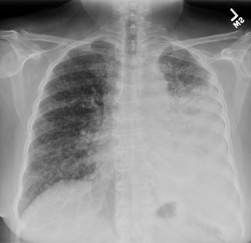
CXR of 50-year-old female with primary adenocarcinoma of the left lung shows extensive consolidation of the left lower lung field with diffuse bilateral reticulonodular changes reminiscent of lymphangitic spread of disease bilaterally
Ashley Davidoff MD TheCommonVein.net 158Lu 131022

50 year old female with primary adenocarcinoma of the left lung presenting with pneumonic consolidation. And diffuse reticulonodular changes bilaterally
Images from the CXR (a, and magnified in b and c) show thickening of the fissures of the right lung (black arrowheads) with diffuse reticulonodular changes.
The coronal CT scan (d, magnified in e) confirm nodular thickening of the fissures (black arrowheads). In addition there are nodular changes with centrilobular location and along the interlobular septa, reminiscent of lymphovascular spread. These findings are consistent with the diagnosis of lymphangitis carcinomatosa
Ashley Davidoff MD TheCommonVein.net 158Lu 131022c
50-year-old female with primary adenocarcinoma of the left lung presenting with pneumonic consolidation, and diffuse reticulonodular changes in the right lung
The right lung is characterized by innumerable large centrilobular nodules (teal arrowheads) and a few foci of intralobular peripheral consolidations. Thickening of the peribronchial tissues likely reflects carcinomatosis of the peribronchial tissues. These findings are consistent with the diagnosis of lymphangitis carcinomatosa
The left lung shows diffuse consolidation of the posterior aspect of the left lower lobe
Ashley Davidoff MD TheCommonVein.net 158Lu 131026
Mechanical/Atelectasis Trauma Metabolic Circulatory- Hemorrhage Immune Infiltrative Idiopathic Iatrogenic Idiopathic