
Necrotizing Pneumonia With Abscesses

54-year-old female presents with a cough, fever and leukocytosis. CT in the axial plane shows a cavitating pneumonia (b, white arrowheads) with large abscess and air-fluid level (b, yellow arrowheads), and multiple small intraparenchymal abscesses (b, orange arrowheads)
Ashley Davidoff MD TheCommonVein.net 31591cL
TB Necrotizing Pneumonia

38-year-old male with HIV presents with cough CT scan in the axial plane shows a focal necrotizing consolidation in the left upper lobe.
Lab tests confirmed a diagnosis of TB
Ashley Davidoff MD TheCommonVein.net 256Lu 136098

38-year-old male with HIV presents with cough CT scan in the axial plane shows a focal necrotizing consolidation in the left upper lobe. Regional lymphadenopathy in the mediastinum is noted
Lab tests confirmed a diagnosis of TB
Ashley Davidoff MD TheCommonVein.net 256Lu 136099
38-year-old male with HIV presents with cough CT scan in the axial plane shows a focal necrotizing consolidation in the left upper lobe. Reginal lymphadenopathy in the mediastinum is noted
Lab tests confirmed a diagnosis of TB
Ashley Davidoff MD TheCommonVein.net 256Lu 136104
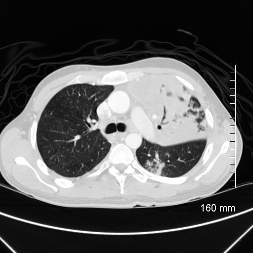
Cavitating Left Lower Lobe Pneumonia with Pseudoaneurysm (PSA)

30-year-old female with a history of IVDU presents with a fever and hemoptysis.
CT in the axial plane shows a cavitating pneumonia in the anterior segment of the left lower lobe. There are groundglass changes surrounding the consolidation representing either edema or hemorrhage
Ashley Davidoff MD TheCommonVein.net 281Lu 136524

30-year-old female with a history of IVDU presents with a fever and hemoptysis.
CT in the sagittal plane shows a cavitating pneumonia in the anterior segment of the left lower lobe. A focally dilated artery in the consolidation (red arrowheads a, and b) represents a mycotic aneurysm. There are groundglass changes surrounding the consolidation representing either edema or hemorrhage
Ashley Davidoff MD TheCommonVein.net 281Lu 136530cL

30-year-old female with a history of IVDU presents with a fever and hemoptysis.
CT in the axial plane shows a cavitating pneumonia in the anterior segment of the left lower lobe (a,b white arrowheads). There are groundglass changes surrounding the consolidation representing either edema or hemorrhage (b pink arrowhead). A focally dilated artery in the consolidation (red arrowheads (c, and d) represents a mycotic aneurysm
Ashley Davidoff MD TheCommonVein.net 281Lu 136526cL
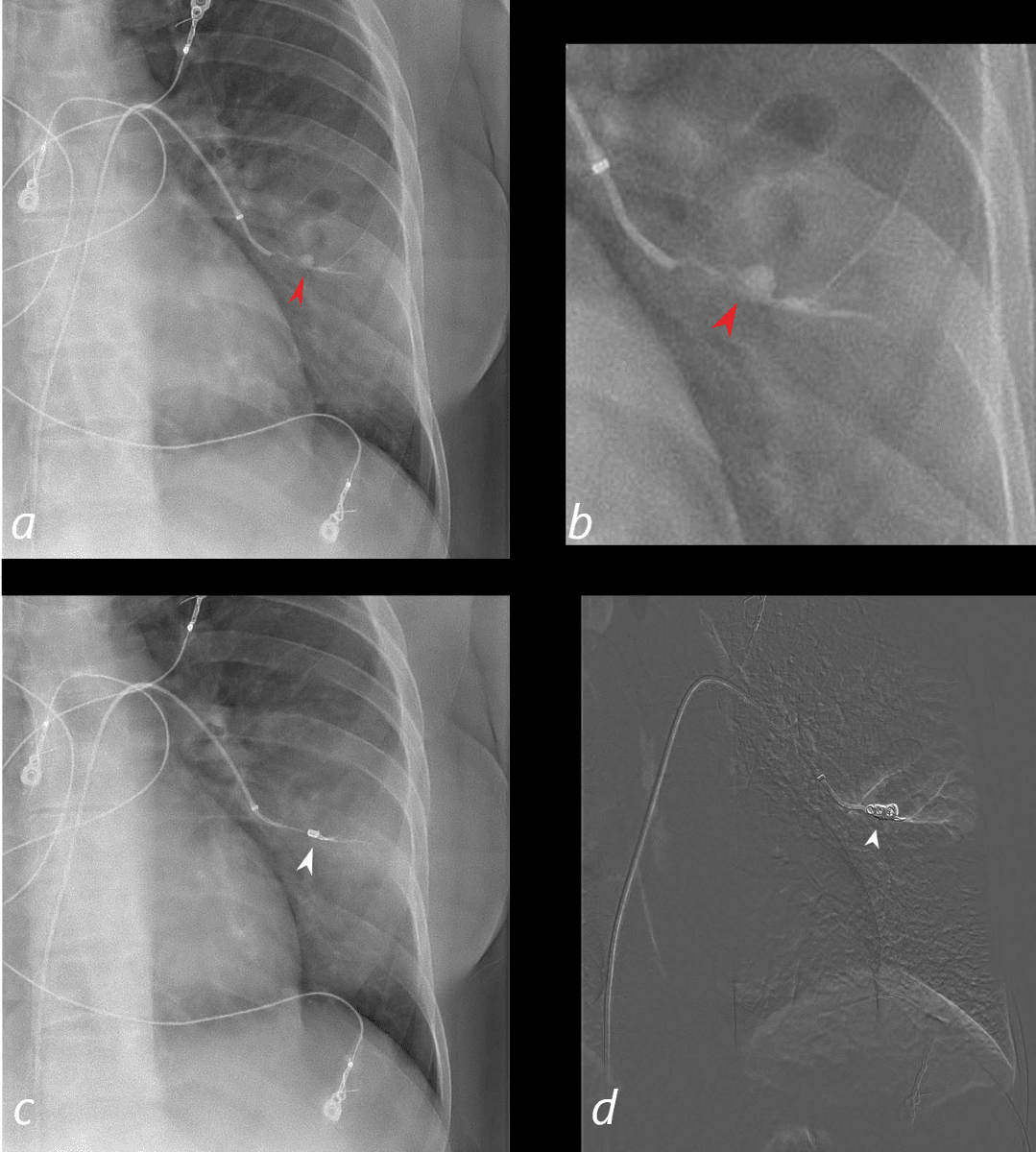
30-year-old female with a history of IVDU presents with a fever and hemoptysis.
Selective angiography of the anterior subsegmental artery of the left lower lobe shows a mycotic pseudoaneurysm (a,b red arrowhead) in the region of a cavitating pneumonia with noted air filled cavity (yellow arrowheads b) reflecting cavitating pneumonia. Images c and d show the embolization coil ((white arrowhead in c and d) with obliteration of the PSA.
Ashley Davidoff MD TheCommonVein.net 281Lu 136532cL