
Infection Inflammation Malignancy Mechanical/Atelectasis Trauma Metabolic Circulatory- Hemorrhage Immune Infiltrative Idiopathic Iatrogenic Idiopathic
Cavitating Nodules in Bacterial Endocarditis
Ashley Davidoff MD TheCommonvein.net 24f PE Hampton’s hump 002
Ashley Davidoff MD TheCommonvein.net 24f PE Hampton’s hump 003
Ashley Davidoff MD TheCommonvein.net 24f PE Hampton’s hump 003
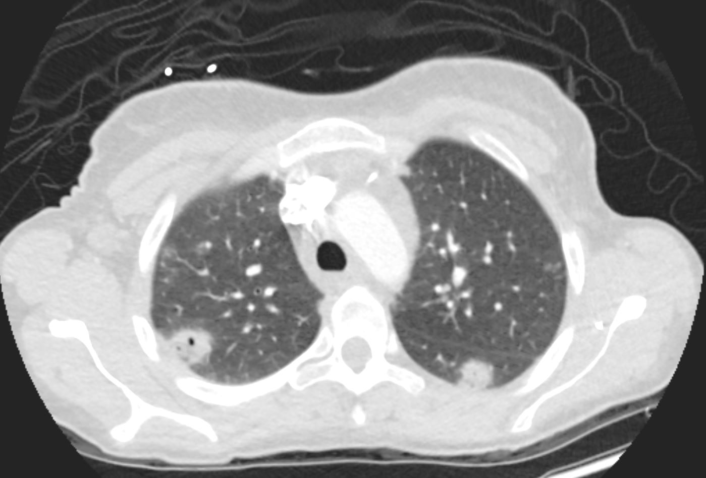
Cavitating Nodules and Mycotic Pseudoaneurysms
Bacterial Endocarditis (BE) and Mycotic Pseudoaneurysm Lung Abscess 39F
Ashley Davidoff TheCommonVein.net b11422b02
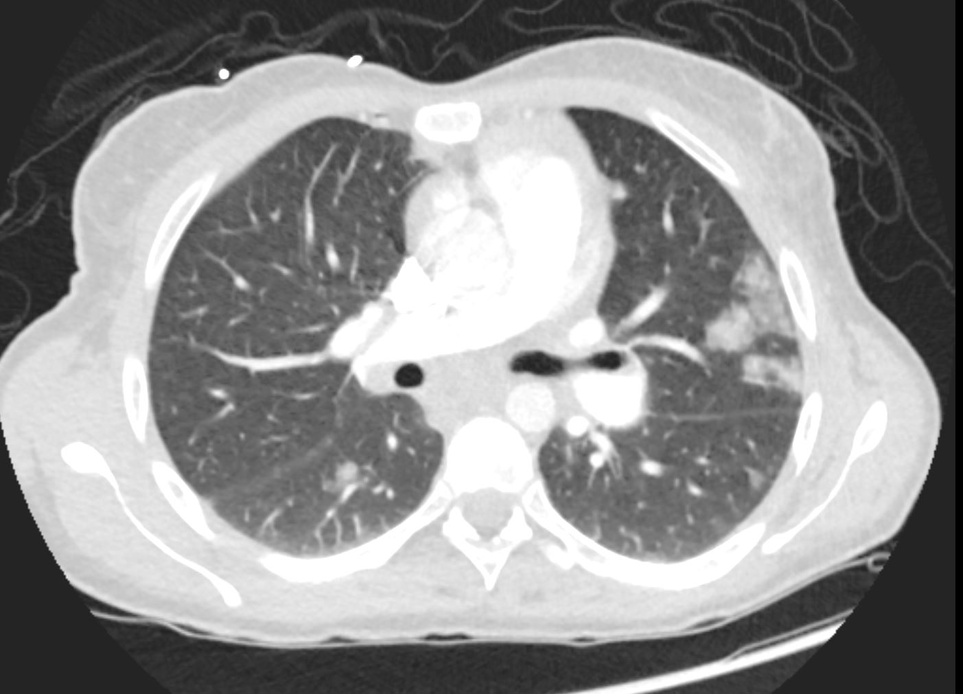
Bacterial Endocarditis (BE) and Mycotic Pseudoaneurysm Lung Abscess 39F
Ashley Davidoff TheCommonVein.net b11422b02
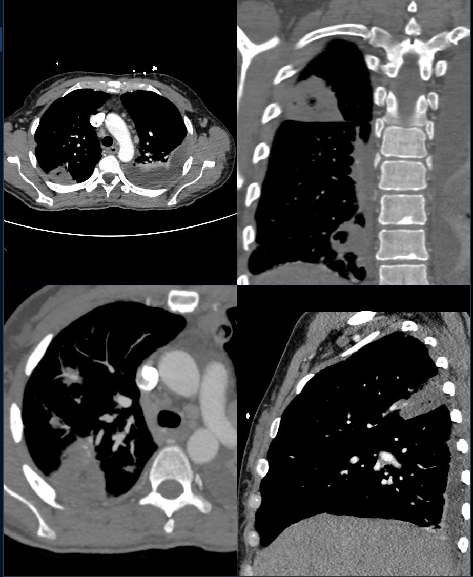
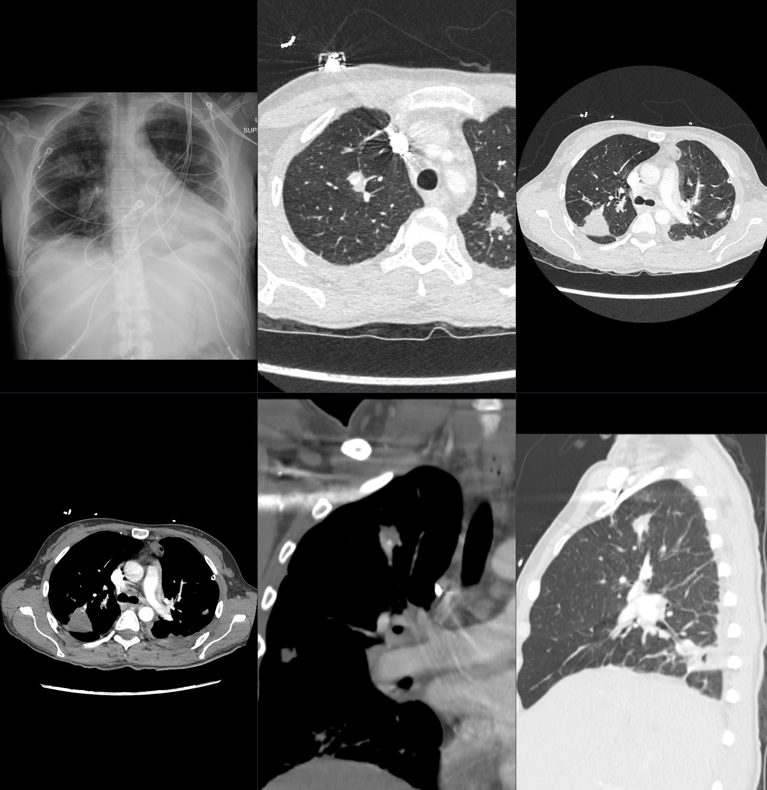
Bacterial Endocarditis (BE) and Mycotic Pseudoaneurysm Lung Abscess 39F
Ashley Davidoff TheCommonVein.net b11422b02
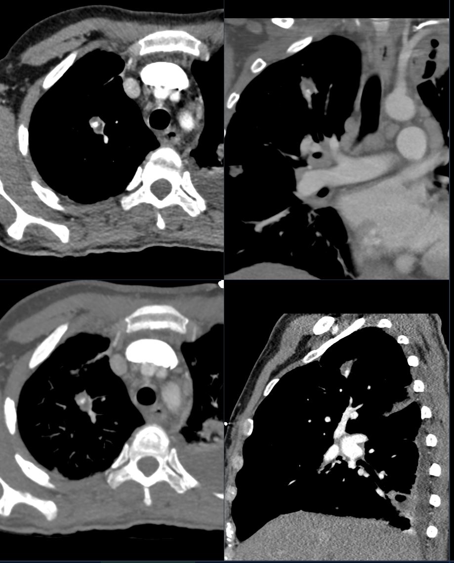
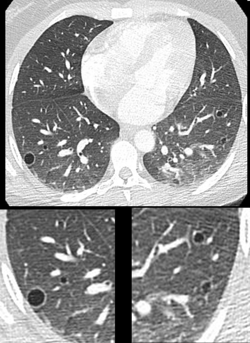
Cavitating Nodule in Cryptococcus

CT scan of a 54-year-old male with HIV presented with chronic headaches. Cryptococcus was identified in his CSF.
Chest CT in the axial projection shows a cavitating nodule in the posterior segment of the left upper lobe
Ashley Davidoff MD TheCommonVein.net 136624
Cavitating Nodules in Non Necrotizing Granuloma

Ashley Davidoff TheCommonVein.net

Ashley Davidoff TheCommonVein.net
Neoplasm Malignant Primary
Neoplasm Malignant Metastases
28year old female presents with vaginal bleeding for 3 days s/p ablation of a vascular molar pregnancy. CT of the chest shows multiple cystic lesions in the lungs bilaterally with slightly thickened walls. Wedge biopsy confirmed a diagnosis of placental site trophoblastic tumor
Ashley Davidoff MD TheCommonVein.net
280Lu 136464.