
Mild CHF Cephalisation
Moderate CHF Interstitial Edema

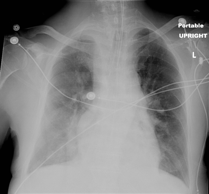
Frontal Chest X-ray in a 69-year-old-male with moderate congestive heart failure (CHF) characterized by a large left atrium, and cephalization with “fuzziness” of the vessels indicating interstitial edema. There is global cardiomegaly and a biventricular pacemaker with a defibrillator on the right ventricular lead.
Ashley Davidoff MD TheCommonVein.net 136546
Thickened Interlobular Septa and Kerley B Lines

50-year-old female with diabetes, chronic renal failure and congestive heart failure. CT in the coronal plane through the posterior aspect of the chest, shows diffuse ground glass changes, thickening of the interlobular septa, thickening of the fissure and centrilobular nodules reflecting arteriolar congestion.
Ashley Davidoff MD TheCommonvein.net 135780 193Lu
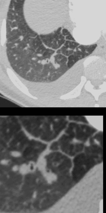
50-year-old female with diabetes, chronic renal failure and congestive heart failure. CT in the axial plane through the right posterior recess, shows thickened interlobular septa at the right base, congested arterioles, alongside the bronchioles, peribronchial cuffing, a congested pulmonary venule in the interlobular septum, ground glass changes and a secondary lobule demonstrating mosaic attenuation. The IVC is dilated and a small complex effusion is present.
Ashley Davidoff MD TheCommonvein.net 135783c 193Lu
Moderate CHF with Interstitial Edema and
Background Centrilobular Emphysema

68 year old male with a history of emphysema presents with increasing dyspnea, Frontal Chest X-ray shows an enlarged left atrium and diffuse interstitial thickening most prominent in the upper lobes.
There is global cardiomegaly, an enlarged main pulmonary artery indicating pulmonary hypertension and a single lead pacemaker.
Ashley Davidoff MD TheCommonVein.net 136556 288Lu

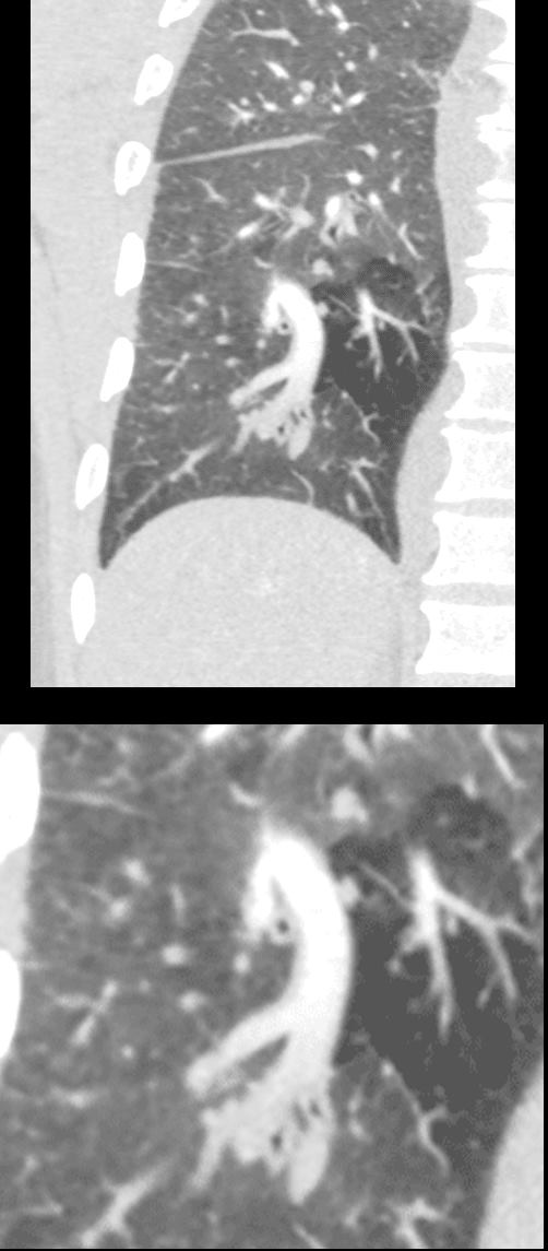
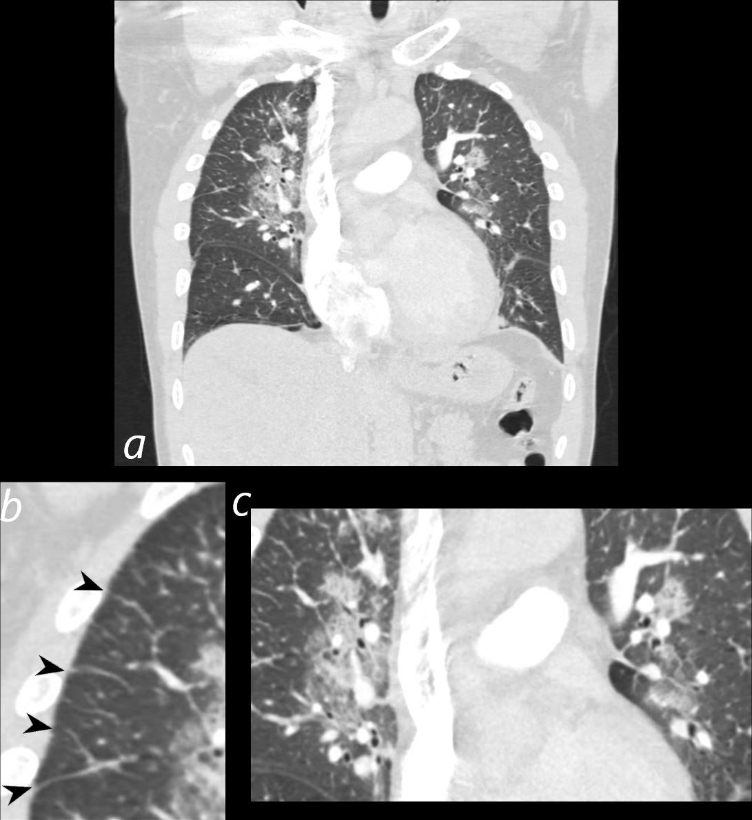
68 year old male with a history of emphysema presents with increasing dyspnea. CT in the coronal plane y shows predominant upper lobe centrilobular emphysema with diffuse thickening of the interlobular septa (b white circle) with evidence of Kerley B lines (c red arrowheads) best exemplified in the left upper lung field, and thickening of the minor fissure (b, pink arrowhead). The left atrium (LA) is enlarged. These findings are consistent with moderate CHF with interstitial edema
Ashley Davidoff MD TheCommonVein.net 288Lu 136557cL
Centrilobular Emphysema

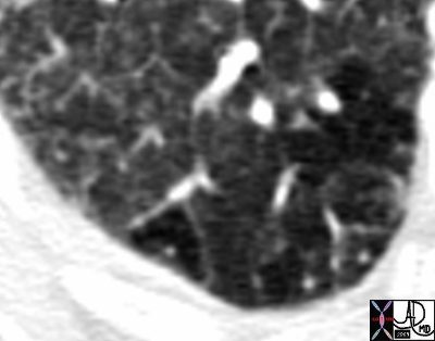
68 year old male with a history of emphysema presents with increasing dyspnea. CT in the axial plane shows predominant upper lobe centrilobular emphysema with diffuse thickening of the interlobular septa .
Ashley Davidoff MD TheCommonVein.net 288Lu 136558
Ground Glass
Mosaic Attenuation
CT Acute Moderate CHF with Interstitial Edema and Mosaic Attenuation
50-year-old female with diabetes, chronic renal failure with congestive heart failure. CT in the coronal plane shows diffuse ground glass changes, Kerley B lines, edema in the fissure, peribronchial cuffing, enlargement of the pulmonary artery, and mosaic attenuation
Ashley Davidoff MD TheCommonvein.net 135778c 193Lu
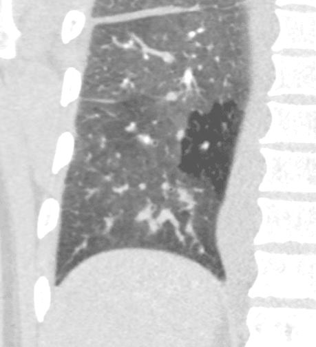
50 year old female with diabetes, chronic renal failure with congestive heart failure. CT in the coronal plane shows diffuse ground glass changes, Kerley B lines, edema in the fissure, and mosaic attenuation
Ashley Davidoff MD TheCommonvein.net 135779c 193Lu
Thickened Interlobular Septa
81F hx atrial fibrillation cardiac failure heart failure CHF RA enlarged LA enlarged ground glass mosaic perfusion Kerley B lines
Ashley Davidoff MD
TheCommonVein.net
44194b01
Engorged Arteries
50-year-old female with diabetes, chronic renal failure with congestive heart failure. CT in the coronal plane shows diffuse ground glass changes, Kerley B lines, edema in the fissure, peribronchial cuffing, enlargement of the pulmonary artery, and mosaic attenuation
Ashley Davidoff MD TheCommonvein.net 135778c 193Lu
Ashley Davidoff MD TheCommonvein.net 135783c 193Lu
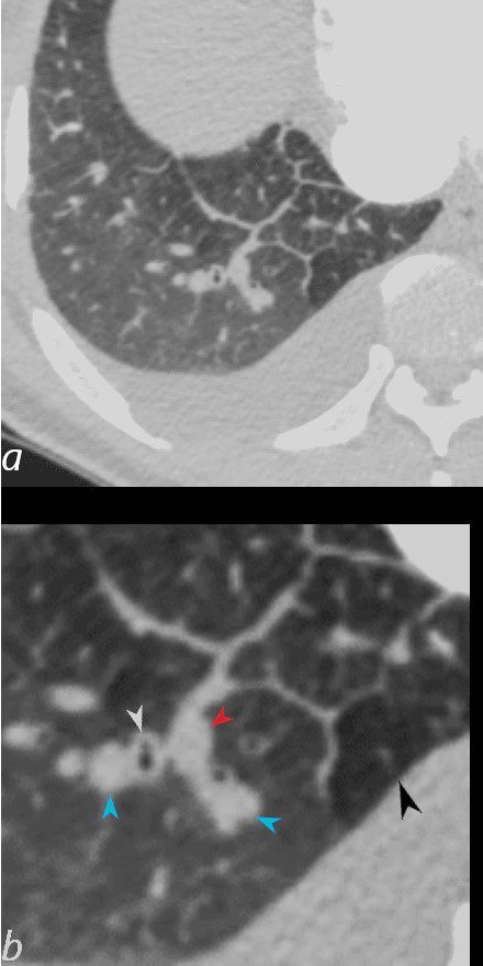
50-year-old female with diabetes, chronic renal failure and congestive heart failure. CT in the axial plane through the right posterior recess, shows thickened interlobular septa at the right base, congested arterioles (light blue arrowheads, b), alongside the bronchioles, peribronchial cuffing (white arrowheads, b), a congested pulmonary venule in the interlobular septum (red arrowhead arrowheads, b), ground glass changes and a secondary lobule demonstrating mosaic attenuation (black arrowhead arrowheads, b). The IVC is dilated and a small complex effusion is present.
Ashley Davidoff MD TheCommonvein.net 135783cL 193Lu
Engorged Veins
Severe CHF
Batwing Pattern

50 year-old male presents with severe dyspnea and orthopnea. CXR in the frontal projection shows perihilar congestion with batwing distribution, left atrial enlargement and left ventricular configuration of the heart. These findings are consistent with severe heart failure with a projected LVEDP of greater than 30mmHg
Ashley Davidoff MD TheCommonVein.net 285Lu 135759
Perihilar Ground Glass Changes and Kerley B Lines
Early Severe CHF
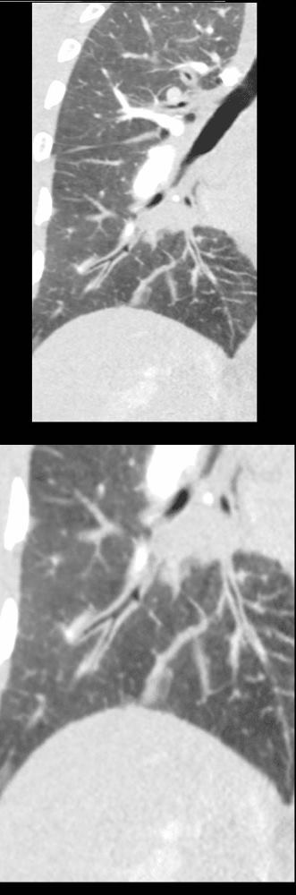
50 year-old male presents with severe dyspnea and orthopnea. CT in the coronal plain shows early severe CHF, with perihilar ground glass changes (magnified in c) and Kerley B lines (b, black arrowheads) These findings are consistent with early severe heart failure with a projected LVEDP of greater than 30mmHg
Ashley Davidoff MD TheCommonVein.net 285Lu 135761cL
Bilateral Effusions Right Greater than left

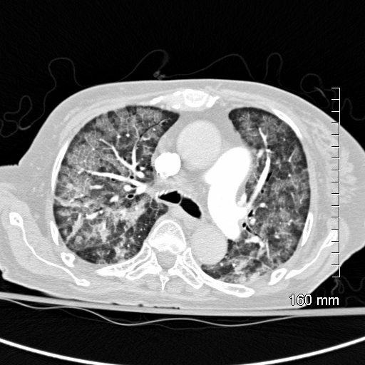
50 year-old male presents with severe dyspnea and orthopnea. CT in the axial plane shows early severe CHF, with perihilar ground glass changes and small bilateral pleural effusions – right greater than left. These findings are consistent with early severe heart failure with a projected LVEDP of greater than 30mmHg
Ashley Davidoff MD TheCommonVein.net 285Lu 135760

57-year-old female with progressive dyspnea.
CXR shows bilateral, diffuse alveolar opacities having a perihilar and basal distribution with sparing of the apices
CT shows diffuse ground glass change with crazy paving morphology characterized by bilateral diffuse ground-glass opacities (GGO) with interlobular and intralobular septal thickening. There is a geographical distribution .
Differential diagnosis
ARDS PCP pneumonia CHF Alveolar Hemorrhage UIP Hypersensitivity Pneumonitis XRT pneumonitis COP Chronic Eosinophilic Lymphangitis Veno-Occlusive Disease
Ashley Davidoff MD
CT scan shows Diffuse ground glass pattern with thickening of the interlobular septa and manifesting as crazy paving pattern
Ashley Davidoff MD TheCommonVein.net 131742
Bronchi
50 year old female with diabetes, chronic renal failure with congestive heart failure. CT in the coronal plane shows diffuse ground glass changes, with prominent peribronchial cuffing in the right upper lobe and and magnified in the lower image in the right lower lobe Prominent Kerley B lines are noted medially – a few laterally and there is with thickening of the right transverse fissure
Ashley Davidoff MD TheCommonvein.net 135777c 193Lu
50-year-old female with diabetes, chronic renal failure and congestive heart failure. CT in the axial plane through the right posterior recess, shows thickened interlobular septa at the right base, congested arterioles (light blue arrowheads, b), alongside the bronchioles, peribronchial cuffing (white arrowheads, b), a congested pulmonary venule in the interlobular septum (red arrowhead arrowheads, b), ground glass changes and a secondary lobule demonstrating mosaic attenuation (black arrowhead arrowheads, b). The IVC is dilated and a small complex effusion is present.
Ashley Davidoff MD TheCommonvein.net 135783cL 193Lu
Unilateral Pulmonary Edema Scimitar PAPVR and CHF
66 year old female presents with dyspnea
Frontal CXR shows unilateral right sided pulmonary edema with cephalization of the left upper lung vessels, with evidence of left atrial enlargement, and enlaged main paulmonary artery suggesting pulmonary hypertension secondary to longstanding left to right shunt. In addition there is a curvilinear structure along the right heart border that is shaped like a sword or a scimitar and the right lung appears smaller than the left. These findings are consistent with scimitar sign and scimitar syndrome
The large shunt results in in increased flow to the right lung and therefore accounts for the unilateral edema
Courtesy Ashley Davidoff MD The CommonVein.net 127H 82595.8