
- Bronchioles are conducting airways
- 2 millimeter or less.
- ciliated,
- peudostratified
- columnar epithelium
- no cartilage
- replaced by y a ring of smooth muscle.
- lack glands.
- Branching
- After the tertiary segmental bronchi, t
- 20-25 branching generations of conducting bronchioles
- three types:
- conducting,
- terminal, and
- respiratory.
- After the tertiary segmental bronchi, t
Club Cells
- Most prominent in the bronchioles –
- diverse function
- synthesis and secretion of the material lining the bronchiolar lumen
- degradation of the mucus
- contribute to the structure of the IGa for surface defences
- engulf airborne toxins
- diverse function
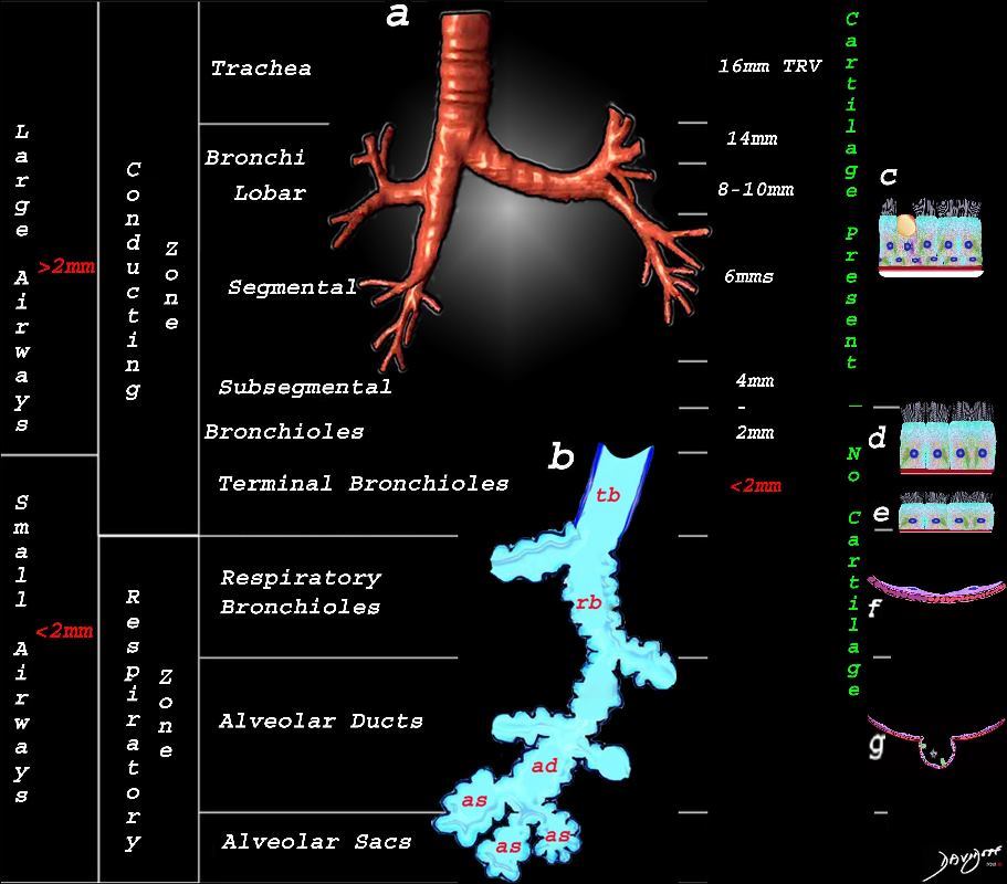
This image shows the division of the airways in the lungs classified as large airways and small airways.
A large airway is considered any airway larger than 2mm, and therefore includes all the airways involved with transport of air except for the terminal bronchiole. Included as seen in image a, are the trachea, mainstem bronchi, lobar bronchi segmental and subsegmental airways and the 3 subsequent divisions of subsegmental bronchi and bronchioles till the last transporting airway ? the respiratory bronchiole which is usually about 2mm and is considered a small airway Image (a) shows the airways starting in the trachea and continuing to the mainstem bronchi, lobar bronchi, segmental bronchi, and subsegmental bronchi.
Image b shows the structures that make up the small airways starting with the terminal bronchiole (tb) followed by the respiratory bronchiole (rb) alveolar duct, (ad) and alveolar sacs (as)
Image (c) shows the histologic makeup of the large airways that include a pseudostratified ciliated columnar epithelium with mucus secreting goblet cells a muscular layer (red) and a prominent cartilage layer (white) In the larger bronchioles (d) the epithelium remains as a pseudostratified, ciliated, columnar epithelium with prominent muscular layer (red). The columnar epithelium transitions to a stratified ciliated cuboidal epithelium by the terminal bronchiole s (f) both still with a muscular layer. The respiratory epithelium transitions from a cuboidal epithelium to a squamous epithelium (f) with alveoli and type I and II pneumocytes starting to branch (g)
Ashley Davidoff MD TheCommonVein.net lungs-0740nL
Cells of the Bronchiole

This upper diagram shows the ciliated columnar epithelium present throughout the 20- 25 generations of branching, until the airways start to transition their function from a transport system to a gas exchange system at the respiratory bronchiole level The ciliated columnar epithelium becomes a ciliated cuboidal epithelium. There are no goblet cells in the bronchioles In addition to the ciliated cells there are 2 other types of cells including the club cell (purple) and the neuroendocrine cell. The club cells Purple with dome shaped superior aspects – formerly Clara Cell) have many functions. The neuroendocrine cell (NE) (dark pink and round ) can be seen as a single cell (NE) and sometimes seen in a cluster, known as a neuroendocrine body (NEB) . The cells rest on a basement membrane, with prominent muscle layer (maroon) as well as elastic tissue (pink). There is no cartilage
Ashley Davidoff MD TheCommonVein.net lungs-0741n
The diagram shows a wall of the bronchiole with transition from a ciliated columnar epithelium to a ciliated cuboidal epithelium over 20-25 branching generations of conducting bronchioles after the tertiary segmental bronchi. In addition to the ciliated cells there are 2 other types of cells including the club cell (purple) and the neuroendocrine cell. The club cells (formerly Clara Cell) are sometimes The neuroendocrine cell (NE) can be seen as a single cell (NE) and sometimes seen in clusters known as a neuroendocrine body (NEB) . There are no mucous secreting goblet cells. The cells rest on a basement membrane, with prominent muscle layer (marron) as wall as elastic tissue (pink). There is no cartilage
Ashley Davidoff MD TheCommonVein.net
NE cells are present in the airway epithelium of human and various animal species and occur singly as well as in clusters called neuroepithelial bodies (NEB).
Links and References
Fleischner Society
bronchiole
Anatomy.?Bronchioles are non?cartilage-containing airways. Terminal bronchioles are the most distal of the purely conducting airways; they give rise to respiratory bronchioles, from which the alveoli arise and permit gas exchange. Respiratory bronchioles branch into multiple alveolar ducts (,30).
Radiographs and CT scans.?Bronchioles are not identifiable in healthy individuals, because the bronchiolar walls are too thin (,4). In inflammatory small-airways disease, however, thickened or plugged bronchioles may be seen as a nodular pattern on a chest radiograph or as a tree-in-bud pattern on CT scans.