
Patterns
Nodule
Diffuse
Patchy
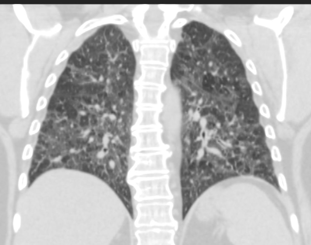
Desquamative Interstitial Pneumonia (DIP)
Patchy Ground Glass Changes,
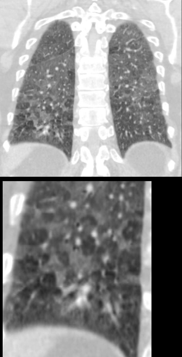
60-year-old male smoker with a history of progressive dyspnea. Coronal CT through the posterior lung fields at the level of the vertebral column shows extensive patchy ground glass changes and mosaic attenuation. A few of the secondary lobules show prominent centrilobular nodules reflecting a small airways component but the predominant pattern is an alveolar pattern
Pathology confirmed a diagnosis of DIP
Ashley Davidoff MD TheCommonVein.net 253Lu 136014c

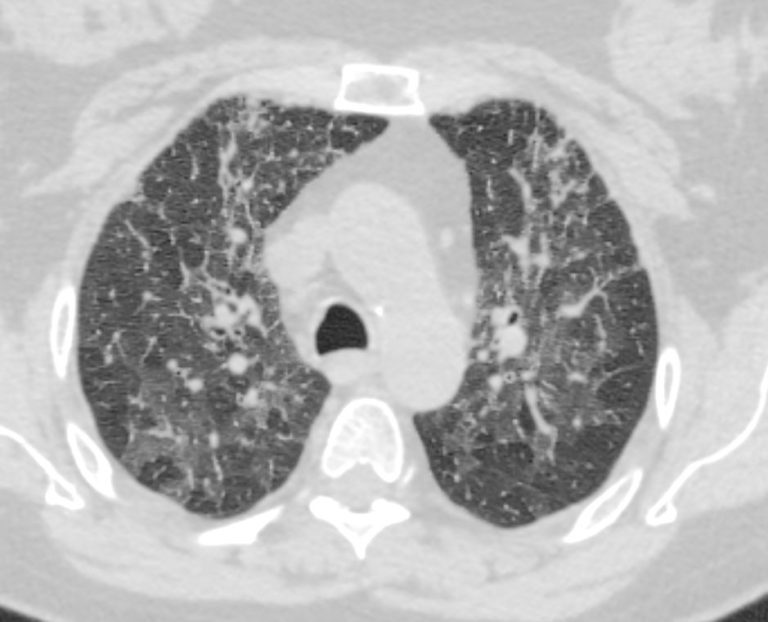
60-year-old male smoker with a history of progressive dyspnea. Coronal CT through the mid lung fields at the level of the left ventricle shows diffuse patchy ground glass changes and mosaic attenuation. There is sparing of the anterior segment of the right upper lobe.
Pathology confirmed a diagnosis of DIP
Ashley Davidoff MD TheCommonVein.net 253Lu 136011
Infection
Inflammation
Desquamative Interstitial Pneumonia (DIP)
Patchy Ground Glass Changes,
60-year-old male smoker with a history of progressive dyspnea. Coronal CT through the posterior lung fields at the level of the vertebral column shows extensive patchy ground glass changes and mosaic attenuation. A few of the secondary lobules show prominent centrilobular nodules reflecting a small airways component but the predominant pattern is an alveolar pattern
Pathology confirmed a diagnosis of DIP
Ashley Davidoff MD TheCommonVein.net 253Lu 136014c
Follicular Bronchiolitis and RA
70-year-old female former smoker with long standing history of RA presents with chronic dyspnea.
Axial CT of the chest at the level of the aortic arch reveals centrilobular nodules, ground-glass opacities, and mosaic attenuation (likely due to air trapping in this context) and bronchial wall thickening. In the context of a patient with rheumatoid arthritis a diagnosis of follicular bronchiolitis is likely. However radiologically fibrotic hypersensitivity pneumonitis (HP) is included in the differential diagnosis
Ashley Davidoff MD TheCommonVein.net 132Lu 136652
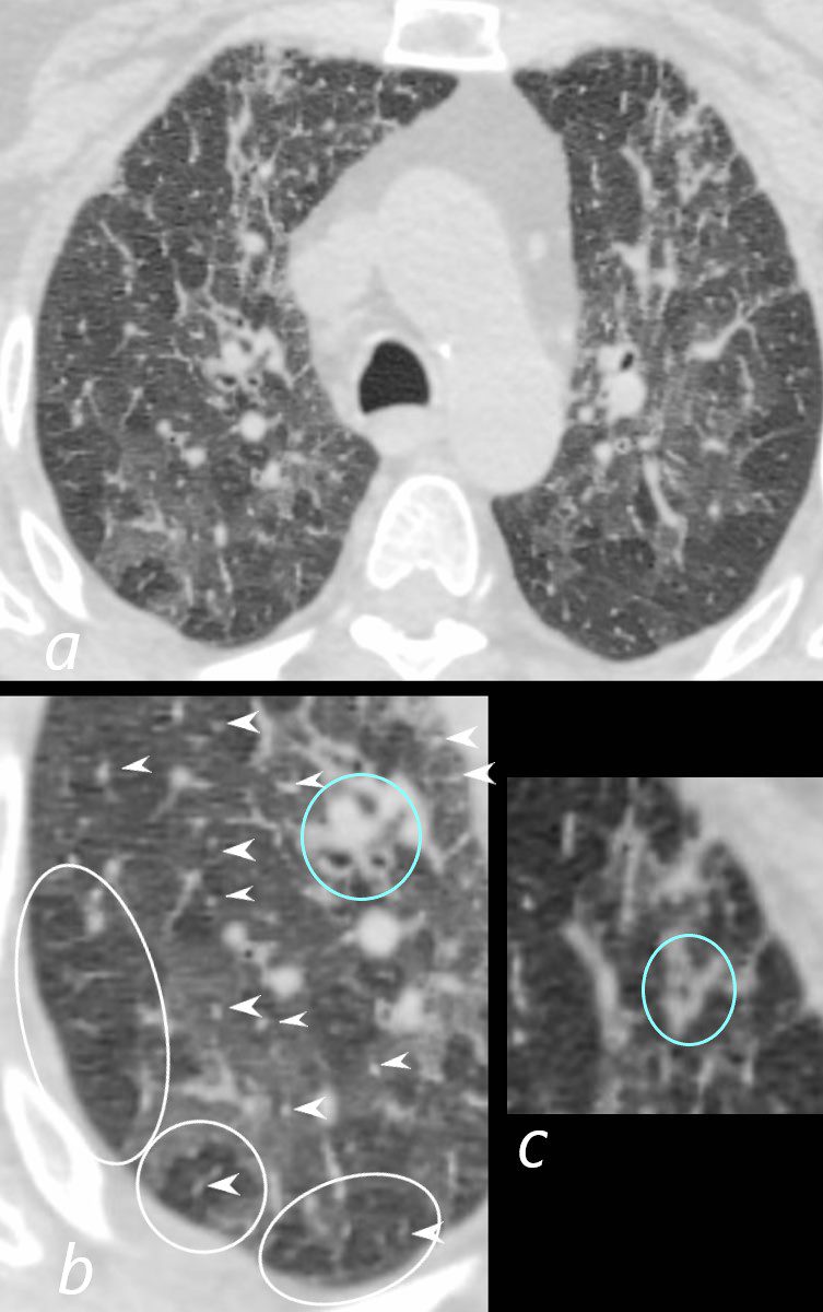
70-year-old female former smoker with long standing history of RA presents with chronic dyspnea.
Axial CT of the chest at the level of the aortic arch reveals centrilobular nodules (b, white arrowheads) , ground-glass opacities, and mosaic attenuation (b, white rings) likely due to air trapping in this context, and bronchial wall thickening (b, c teal rings). There is some irregular thickening of the interlobular septa. In the context of a patient with rheumatoid arthritis a diagnosis of follicular bronchiolitis is likely. However radiologically fibrotic hypersensitivity pneumonitis (HP) is included in the differential diagnosis
Ashley Davidoff MD TheCommonVein.net 132Lu 136652cL
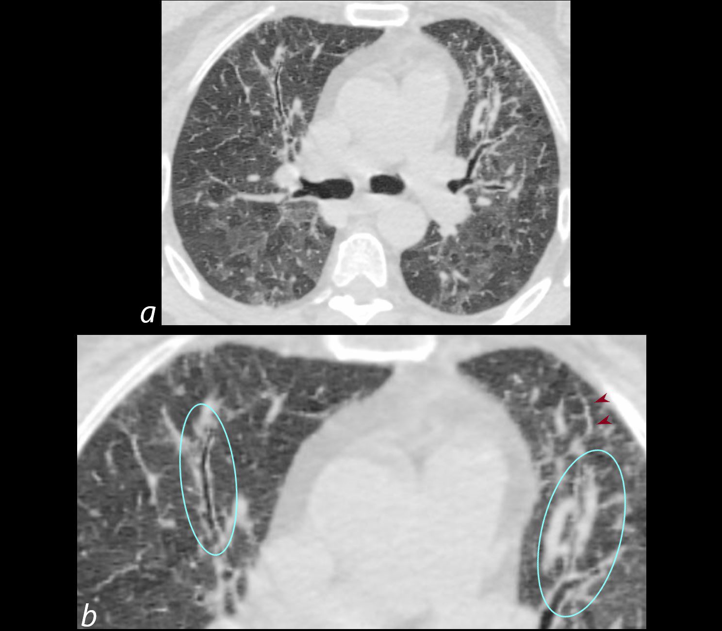
70-year-old female former smoker with long standing history of RA presents with chronic dyspnea.
Axial CT of the chest at the level of the carina reveals centrilobular nodules, ground-glass opacities, and mosaic attenuation (likely due to air trapping in this context) and bronchial wall thickening . Bronchial wall thickening (b, maroon arrowheads) and irregular septal thickening (b maroon arrowheads) are noted.
In the context of a patient with rheumatoid arthritis a diagnosis of follicular bronchiolitis is likely. However radiologically fibrotic hypersensitivity pneumonitis (HP) is included in the differential diagnosis
Ashley Davidoff MD TheCommonVein.net 132Lu 136654cL

70-year-old female former smoker with long standing history of RA presents with chronic dyspnea.
Axial CT of the chest at the level of the lower lung fields reveals centrilobular nodules, ground-glass opacities, and mosaic attenuation (likely due to air trapping in this context). In the context of a patient with rheumatoid arthritis a diagnosis of follicular bronchiolitis is likely. However radiologically fibrotic hypersensitivity pneumonitis (HP) is included in the differential diagnosis
Ashley Davidoff MD TheCommonVein.net 132Lu 136659
70-year-old female former smoker with long standing history of RA presents with chronic dyspnea.
CT in the coronal plane of the chest at the level of the spine reveals bilateral diffuse changes in the lungs characterized by centrilobular nodules, ground-glass opacities, mosaic attenuation (likely due to air trapping in this context) and irregular thickening of the interlobular septa.
In the context of a patient with rheumatoid arthritis a diagnosis of follicular bronchiolitis is likely. However radiologically fibrotic hypersensitivity pneumonitis (HP) is included in the differential diagnosis
Ashley Davidoff MD TheCommonVein.net 132Lu 136663
Trauma
Metabolic
Circulatory-
Hemorrhage
Immune
Infiltrative
Idiopathic Iatrogenic Idiopathic