
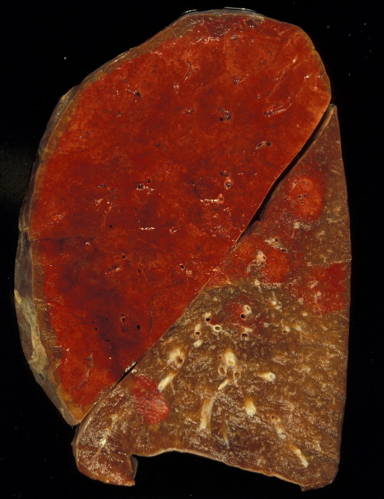
Hemorrhagic Pneumonia
The gross pathology specimen shows a hemorrhagic lobar pneumonia (red hepatisation) in the left upper lobe with patchy subsegmental bronchopneumonic disease in the lower lobe. The relationship to the surface pleura may result in pleuritic pain.
Courtesy Jeffrey Pierce and Ashley Davidoff MD 32320
Parts
Size
Multifocal Pneumonia

45-year-old immunocompromised male presents with a cough fever and shock.
Portable frontal CXR shows a multifocal pneumonic consolidations with air bronchograms in the right upper, left upper right lower and left lower lobes, magnified in the surrounding images. There is silhouetting of the right heart border reflecting middle lobe involvement. The patient is intubated with an intra-aortic balloon pump (IABP) and Swan Ganz line.
Ashley Davidoff MD TheCommonVein.net 136501c
Shape
Position

32 year old female presents with a ever and right posterior pleuritic pain Frontal and lateral chest X-ray shows a pneumonic consolidation localized to the posterior segment of the right upper lobe
Courtesy Ashley Davidoff MD TheCommonVein.net 41800c01

28 year old female presents with a cough and fever
CXR shows a middle lobe consolidation involving the lateral segment.
Ashley Davidoff MD TheCommonVein.net 41816c01
Multifocal Pneumonia
45-year-old immunocompromised male presents with a cough fever and shock.
Portable frontal CXR shows a multifocal pneumonic consolidations with air bronchograms in the right upper, left upper right lower and left lower lobes, magnified in the surrounding images. There is silhouetting of the right heart border reflecting middle lobe involvement. The patient is intubated with an intra-aortic balloon pump (IABP) and Swan Ganz line.
Ashley Davidoff MD TheCommonVein.net 136501c
Character
Cystic Appearing Pneumonia
Pneumonia with Peripheral Cystic Changes
Intralobar Sequestration

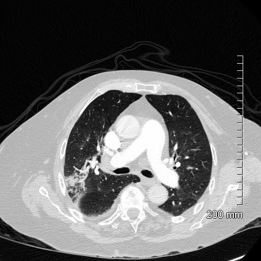
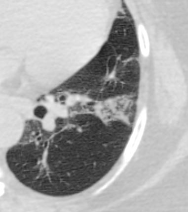
An axial CT scan of a 31-year-old woman with an intralobar sequestration shows a left lower pneumonia with a surrounding region of cystic changes
Ashley Davidoff MD TheCommonVein.net 121374b
Time
Associated Findings
CT Right Upper Lobe Pneumonia with Abscess Formation
CT scan in the axial plane of a 63-year-old female with cough fever and leukocytosis, shows a right upper lobe consolidation with a 2.8cms fluid collection and a dependent bubble of air consistent with a diagnosis of a lung abscess secondary to pneumonia
Ashley Davidoff MD TheCommonVein.net 136170
CT Right Upper Lobe Pneumonia with Abscess Formation

CT scan in the coronal plane of a 63-year-old female with cough fever and leukocytosis, shows a right upper lobe consolidation with a 2.8cms fluid collection and a bubble of air consistent with a diagnosis of a lung abscess secondary to pneumonia
Ashley Davidoff MD TheCommonVein.net 136171
Infection Inflammation
Acute Aspiration Pneumonia on Chronic Interstitial Lung Disease

55-year-old female with shortness of breath. CT (above) is from 4 years prior and shows an interstitial process characterized by ground glass and reticular change. The patient presents 4 years later with fever and white count and the CT (below) shows a pneumonic process in a background of ac chronic interstitial process with cystic air spaces and architectural distortion. Aspiration pneumonia was considered most likely
Ashley Davidoff MD TheCommonVein.net 135536
Malignancy Mechanical/Atelectasis Trauma Metabolic Circulatory- Hemorrhage Immune Infiltrative Idiopathic Iatrogenic Idiopathic
Congenital Disease
Intralobar Sequestration
Cystic Appearing Infiltrate

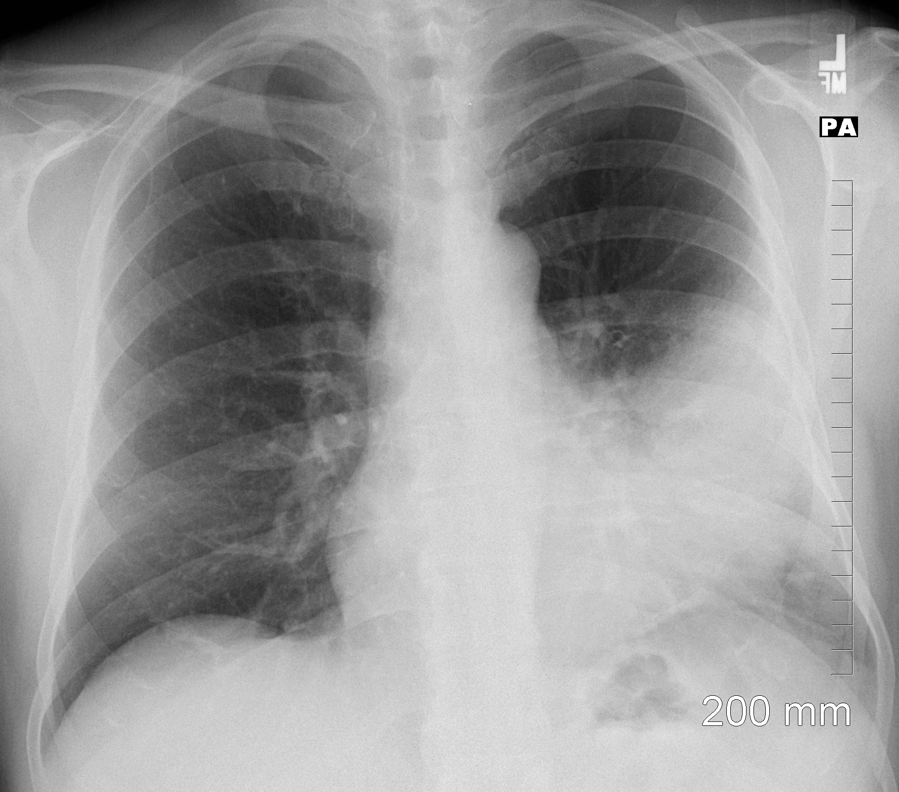
Frontal CXR of a 31-year-old woman with an intralobar sequestration presents with a fever, cough, productive sputum, and an elevated white cell count. The CXR shows a cystic appearing infiltrate of a large part of the upper portion of the left lower lobe consistent with an atypical appearing pneumonia .
Ashley Davidoff MD TheCommonVein.net 121373b.8
An axial CT scan of a 31-year-old woman with an intralobar sequestration shows a left lower pneumonia with a surrounding region of cystic changes
Ashley Davidoff MD TheCommonVein.net 121374b
Coronal CT of a 31-year-old woman with an intralobar sequestration shows a left lower consolidation supplied by an aberrant artery off the aorta Ashley Davidoff MD TheCommonVein.net 121376cL


74 year old male alcoholic with bilateral basilar lobar atelectasis caused by bilateral aspiration
CT scan near the AP window shows reactive lymph nodes, gynecomastia, an infiltrate in the superior segment of the right lobe of the lung and bilateral pleural effusions
Ashley Davidoff MD TheCommonVein.net RnD image
74 year old male alcoholic with bilateral basilar lobar atelectasis caused by bilateral aspiration
CT scan shows airless lower lobes with small bilateral effusions.
Ashley Davidoff MD TheCommonVein.net

74 year old male alcoholic with bilateral basilar lobar atelectasis caused by bilateral aspiration
CT scan shows airless lower lobes with small bilateral effusions.
Ashley Davidoff MD TheCommonVein.net RnD image
74 year old male alcoholic with bilateral basilar lobar atelectasis caused by bilateral aspiration
CT scan shows airless lower lobes with small bilateral effusions.
Ashley Davidoff MD TheCommonVein.net

74 year old male alcoholic with bilateral basilar lobar atelectasis caused by bilateral aspiration
CT scan at the level of the carina shows right main bronchus filled with aspirated content associated with an infiltrate in the right lobe of the lung with both focal consolidations and ground glass infiltrates and bilateral pleural effusions
Ashley Davidoff MD TheCommonVein.net RnD image

52 year old male presents with a cough and fever
CT scan in the axial plane using soft tissue windows, shows a lingular consolidation sign. Both the superior and inferior lingular segments are involved
Ashley Davidoff MD TheCommonVein.net
52 year old male presents with a cough and fever
CT scan in the axial plane using soft tissue windows, shows a lingular consolidation sign. Both the superior and inferior lingular segments are involved
Ashley Davidoff MD TheCommonVein.net

52 year old male presents with a cough and fever
CT scan in the axial plane shows a lingular consolidation. with air bronchograms
Ashley Davidoff MD TheCommonVein.net

52 year old male presents with a cough and fever
CT scan in soft tissue windows in the axial plane shows a lingular consolidation with air bronchograms and reactive mediastinal adenopathy
Ashley Davidoff MD TheCommonVein.net
52 year old male presents with a cough and fever
CT scan in the axial plane shows a lingular consolidation with air bronchograms and a positive silhouette sign. Both the superior and inferior lingular segments are involved
Ashley Davidoff MD TheCommonVein.net
52 year old male presents with a cough and fever
CT scsn in the axial plane shows a lingular consolidation with air bronchograms and a positive silhouette sign. Both the superior and inferior lingular segments are involved
Ashley Davidoff MD TheCommonVein.net
52 year old male presents with a cough and fever
Frontal CXR shows a lingular infiltrate with a positive silhouette sign. Both the superior and inferior lingular segments appear to be involved
Ashley Davidoff MD TheCommonVein.net
Ashley Davidoff TheCommonVein.net RnD Image First program

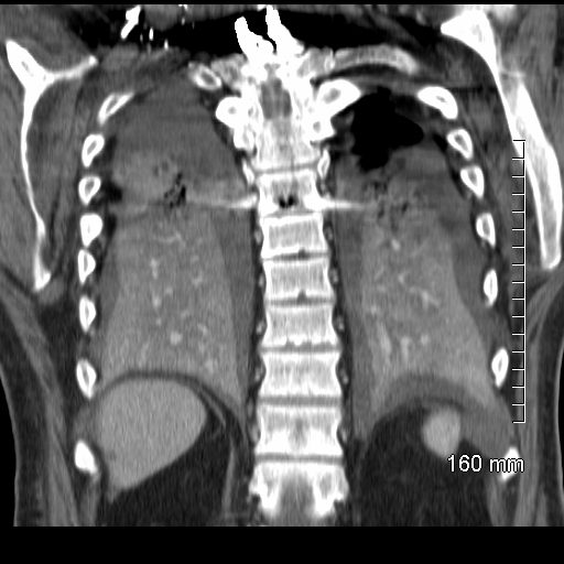
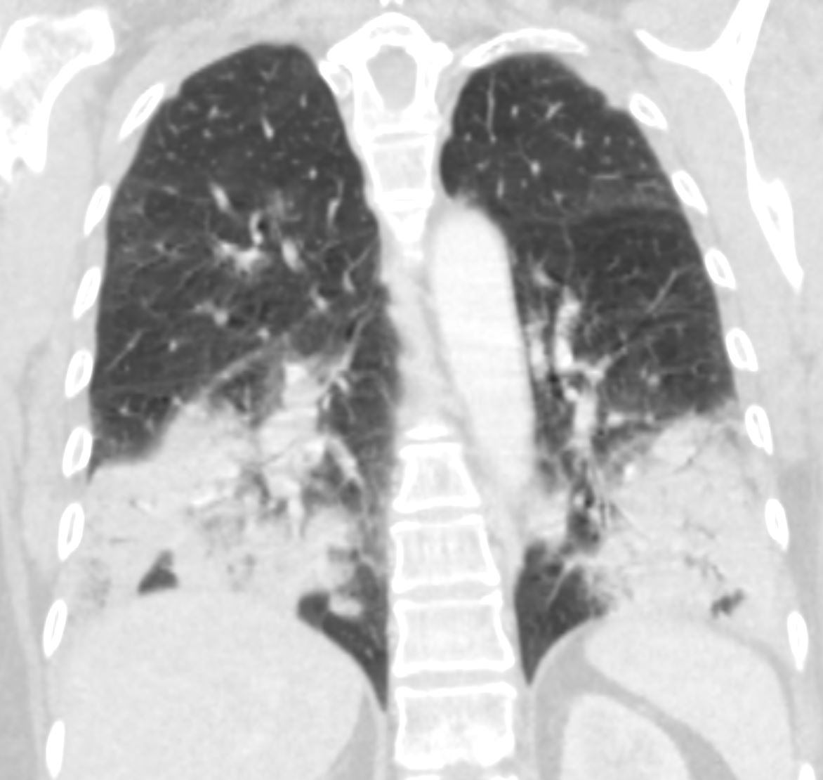
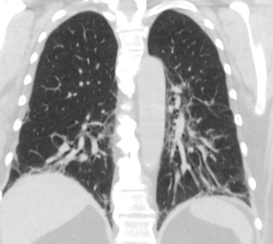
CT scan in the coronal performed 6 months ago at the time of clinical presentation shows upper lobe predominant peripheral infiltrates more prominent in the left upper. Subsequent diagnosis by BAL of chronic eosinophilic pneumonia (CEP) was made
Ashley Davidoff TheCommonVein.net

CT scan in the coronal performed 6 months ago at the time of clinical presentation shows upper lobe predominant peripheral infiltrates more prominent in the left upper lobe. Subsequent diagnosis by BAL of chronic eosinophilic pneumonia (CEP) was made
Ashley Davidoff TheCommonVein.net

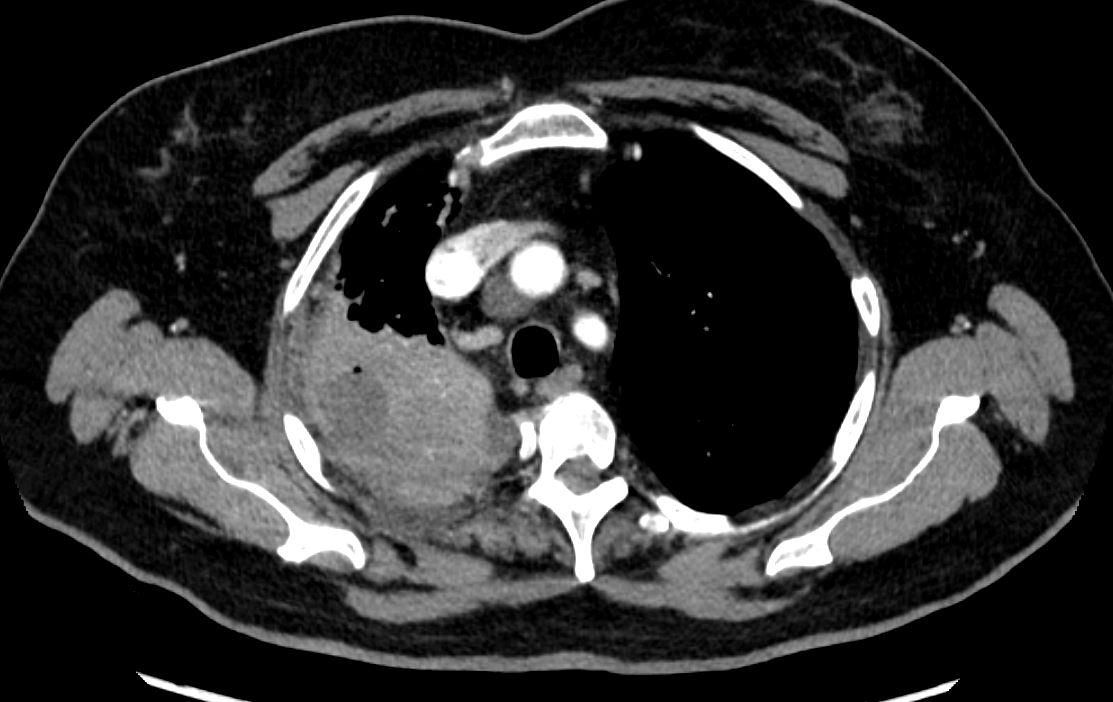
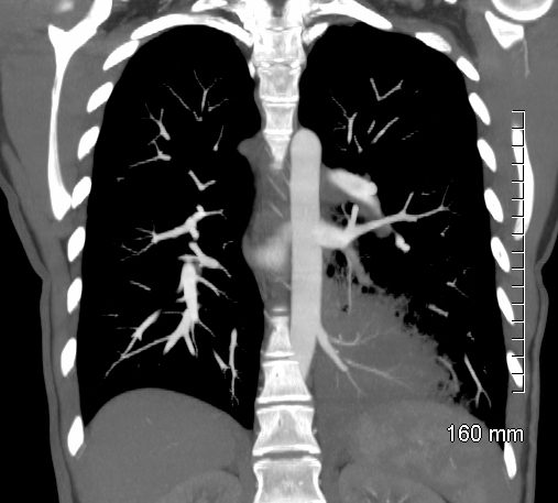
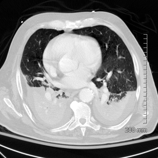
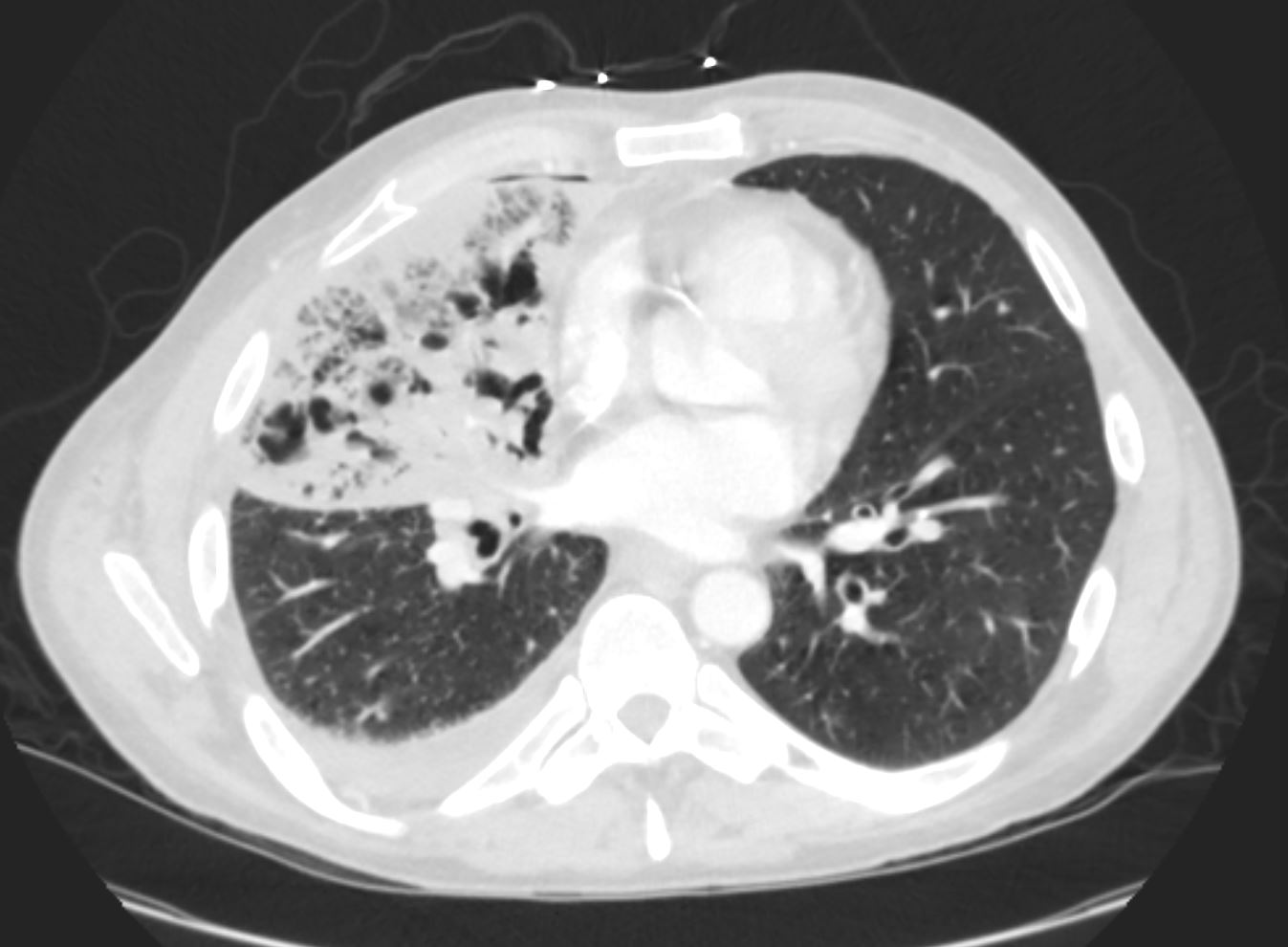
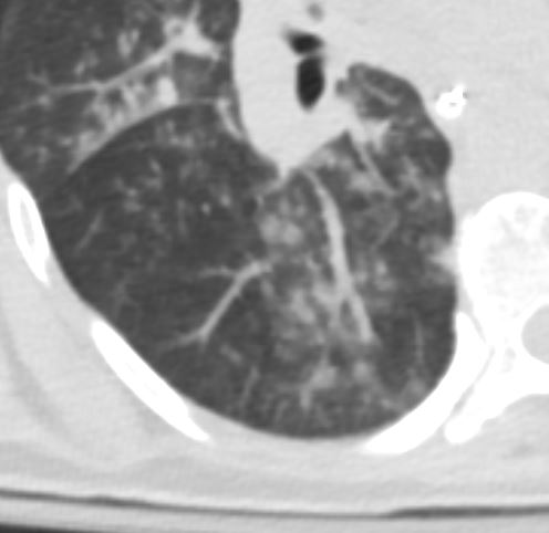
71 yo m w/ a hx of recently diagnosed ANCA-associated vasculitis (diagnosed by kidney biopsy), presents with acute respiratory failure. Bronchoscopic findings were consistent with diffuse alveolar hemorrhage associated with MSSA (Methicillin Sensitive Staph Aureus ) pneumonia/bacteremia. Note Consolidation surrounded by ground glass opacity, the latter likely reflecting a hemorrhagic component
CTscan shows multicentric consolidations likely a combination of alveolar hemorrhage and pneumonia
Ashley Davidoff TheCommonVein.net
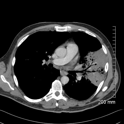

Subsequent diagnosis by BAL of chronic eosinophilic pneumonia (CEP)
Ashley Davidoff TheCommonVein.net
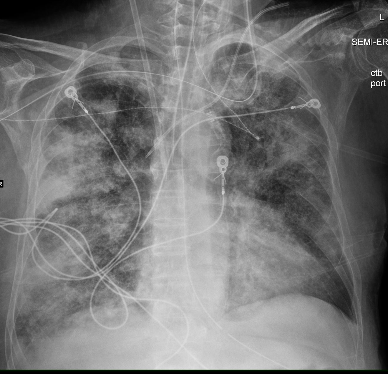
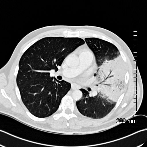
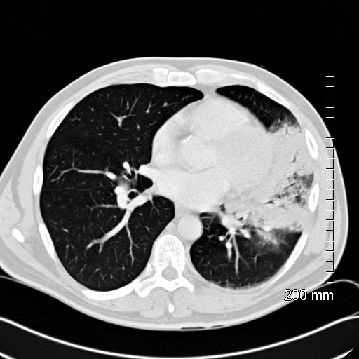
71 yo m w/ a hx of recently diagnosed ANCA-associated vasculitis (diagnosed by kidney biopsy), presents with acute respiratory failure. Bronchoscopic findings were consistent with diffuse alveolar hemorrhage associated with MSSA (Methicillin Sensitive Staph Aureus ) pneumonia/bacteremia
CXR shows multicentric consolidations likely a combination of alveolar hemorrhage and pneumonia
Ashley Davidoff TheCommonVein.net

CT scan in the coronal performed 6 months ago at the time of clinical presentation shows upper lobe predominant peripheral infiltrates more prominent in the left upper lobe. Subsequent diagnosis by BAL of chronic eosinophilic pneumonia (CEP) was made
Ashley Davidoff TheCommonVein.net
Ashley Davidoff MD TheCommonVein.net
eosinophillic-pneumonia-006
Ashley Davidoff MD TheCommonVein.net cavitating pneumonia 59M


Ashley Davidoff TheCommonVein.net Ashley Davidoff TheCommonVein.net RML RLL 004


Cryptogenic Organizing Pneumonia
Ashley Davidoff MD TheCommonVein.net
Cryptogenic Organizing Pneumonia
Ashley Davidoff MD TheCommonVein.net

Cryptogenic Organizing Pneumonia
Ashley Davidoff MD TheCommonVein.net
Cryptogenic Organizing Pneumonia
Ashley Davidoff MD TheCommonVein.net

Cryptogenic Organizing Pneumonia
Ashley Davidoff MD TheCommonVein.net
lungs-COP-004-path-52f-CT.jpg
Lungs COPCryptogenic Organizing Pneumonia
Ashley Davidoff MD TheCommonVein.net

Cryptogenic Organizing Pneumonia
Ashley Davidoff MD TheCommonVein.net

Cryptogenic Organizing Pneumonia
Ashley Davidoff MD TheCommonVein.net