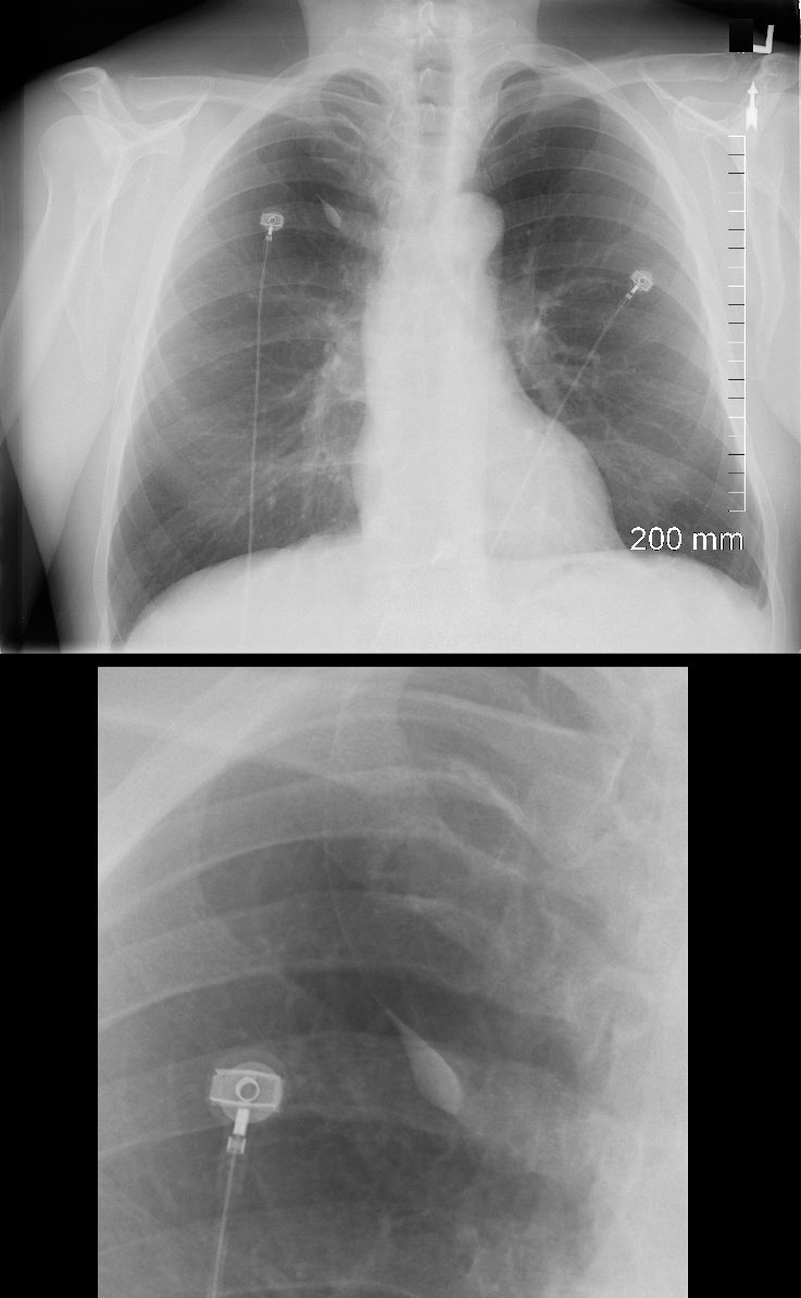
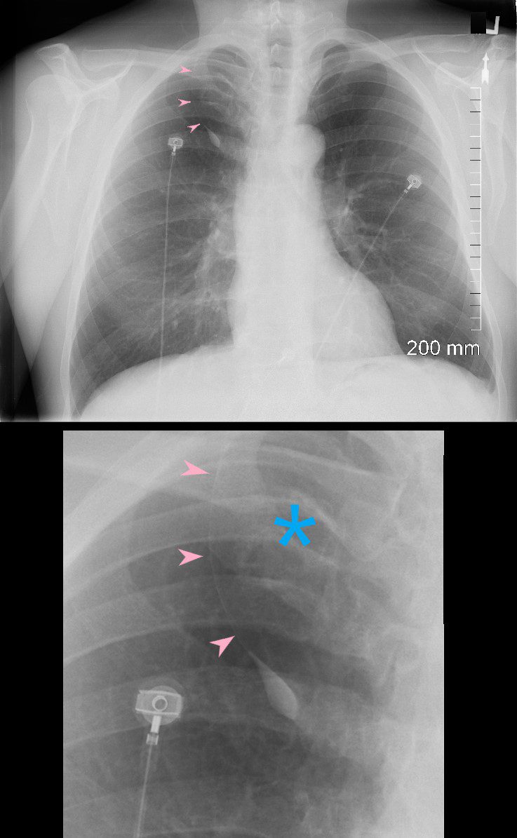
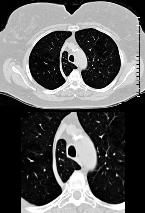
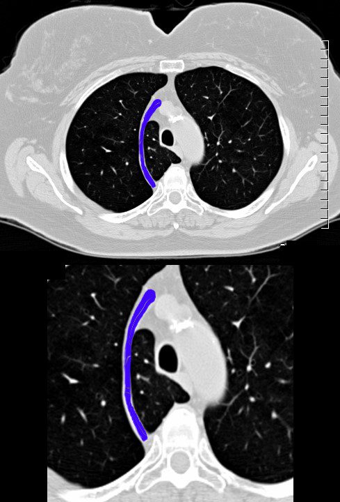
The azygos fissure is a normal anatomical variant seen on chest radiographs and CT scans, resulting from an unusual course of the azygos vein during fetal development. It is characterized by an accessory fissure in the right upper lung lobe that is visible on imaging.
Ashley Davidoff MD
TheCommonVein.net 139142c
Ashley Davidoff MD
TheCommonVein.net 139142cL
139143c-azygos-lobe-fissure-CXR-scaled.jpg
Azygos Fissure and LobeAshley Davidoff MD
TheCommonVein.net 139143c
Ashley Davidoff MD
TheCommonVein.net 139148c
Ashley Davidoff MD
TheCommonVein.net 139148cL
Ashley Davidoff MD
TheCommonVein.net 139149c
Ashley Davidoff MD
TheCommonVein.net 139149cL
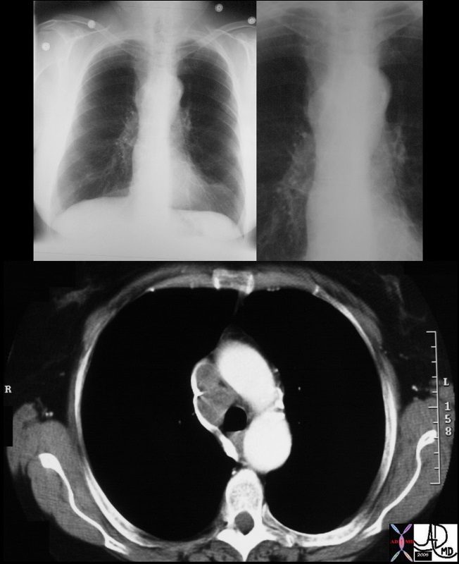
The node in the azygos region is pathologically enlarged but at pathology was shown to be reactive. note the subtle deformity of the azygos region on the CXR
Courtesy Ashley Davidoff MD TheCommonVein.net 33082c01
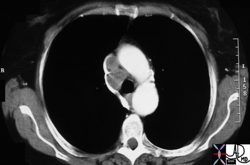
The node in the azygos region is pathologically enlarged but at pathology was shown to be reactive. note the subtle deformity of the azygos region on the CXR
Courtesy Ashley Davidoff MD TheCommonVein.net 33080.800
Radiological Definition and Appearance
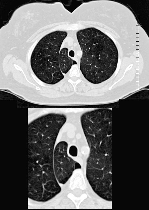
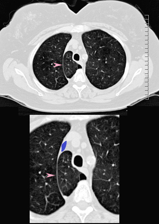
On imaging, the azygos fissure appears as a curved line or arc in the right upper lung zone, typically near the right apex. It is created by four layers of pleura: two layers of visceral pleura from the lung and two layers of parietal pleura from the mediastinum, which surround the azygos vein as it arches over the lung.
- Azygos Lobe: The azygos fissure encloses a small part of the right upper lobe, sometimes referred to as the azygos lobe. This is not a true separate lobe but rather a subdivision of the upper lobe.
- Azygos Vein: The azygos vein typically travels in a more medial path along the right side of the mediastinum, arching over the right main bronchus before joining the superior vena cava. In cases with an azygos fissure, however, the vein takes a more lateral course, creating an accessory fissure as it moves to its final position in the mediastinum.
Key Features on Imaging
- Curved Line: The azygos fissure appears as a thin, curved line in the right upper lung field on X-ray.
- Azygos Vein: A rounded density or ?dot? is seen at the lower end of the fissure, representing the azygos vein within the fissure.
- Location: Typically located in the right upper lobe, the fissure extends from the lung apex down toward the right hilum.
Clinical Relevance
- Incidental Finding: The azygos fissure is usually an incidental finding and is clinically insignificant.
- Surgical Consideration: Surgeons should be aware of this variant to avoid complications during procedures involving the right lung or mediastinum.
In summary, the azygos fissure is a radiologically visible accessory fissure in the right upper lung lobe, formed by the anomalous course of the azygos vein and surrounding pleura. It is usually identified on imaging as a thin, curving line with a small round opacity representing the azygos vein.
