Definition
Atelectasis is the partial or complete collapse of a lung or portion of
the lung. It can occur due to various underlying causes, such as
airway obstruction (mucus plug, tumor) or external compression
(pleural effusion, pneumothorax). As a result, atelectasis can cause
symptoms such as shortness of breath, rapid breathing, chest pain,
and cyanosis, depending on the severity and extent of lung
involvement. Diagnosis is typically made through imaging, with
chest X-rays or CT scans showing areas of lung collapse, often
appearing as increased density or opacity. Clinically, diminished
breath sounds or dullness to percussion may be noted over the
affected area. Treatment depends on the underlying cause and may
involve airway clearance techniques, bronchoscopy, or addressing
the source of compression or obstruction
- Atelectasis implies collapse of part of the lung.

Compressive Atelectasis Secondary Effusion
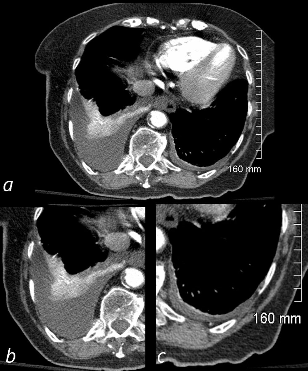
CT with Moderate Right Pleural Effusion, Small Left Effusion and Compressive Atelectasis
92-year-old female presents with dyspnea.
CT scan shows a moderate sized right pleural effusion with compressive atelectasis (magnified in b). There is relative hyperenhancement of the compressed and atelectatic lung due to increased tissue density. At the left lung base, there is a small effusion with a minor degree of atelectasis. (magnified in c)
Ashley Davidoff MD TheCommonVein.net 118437c01
Compressive Atelectasis Secondary Effusion
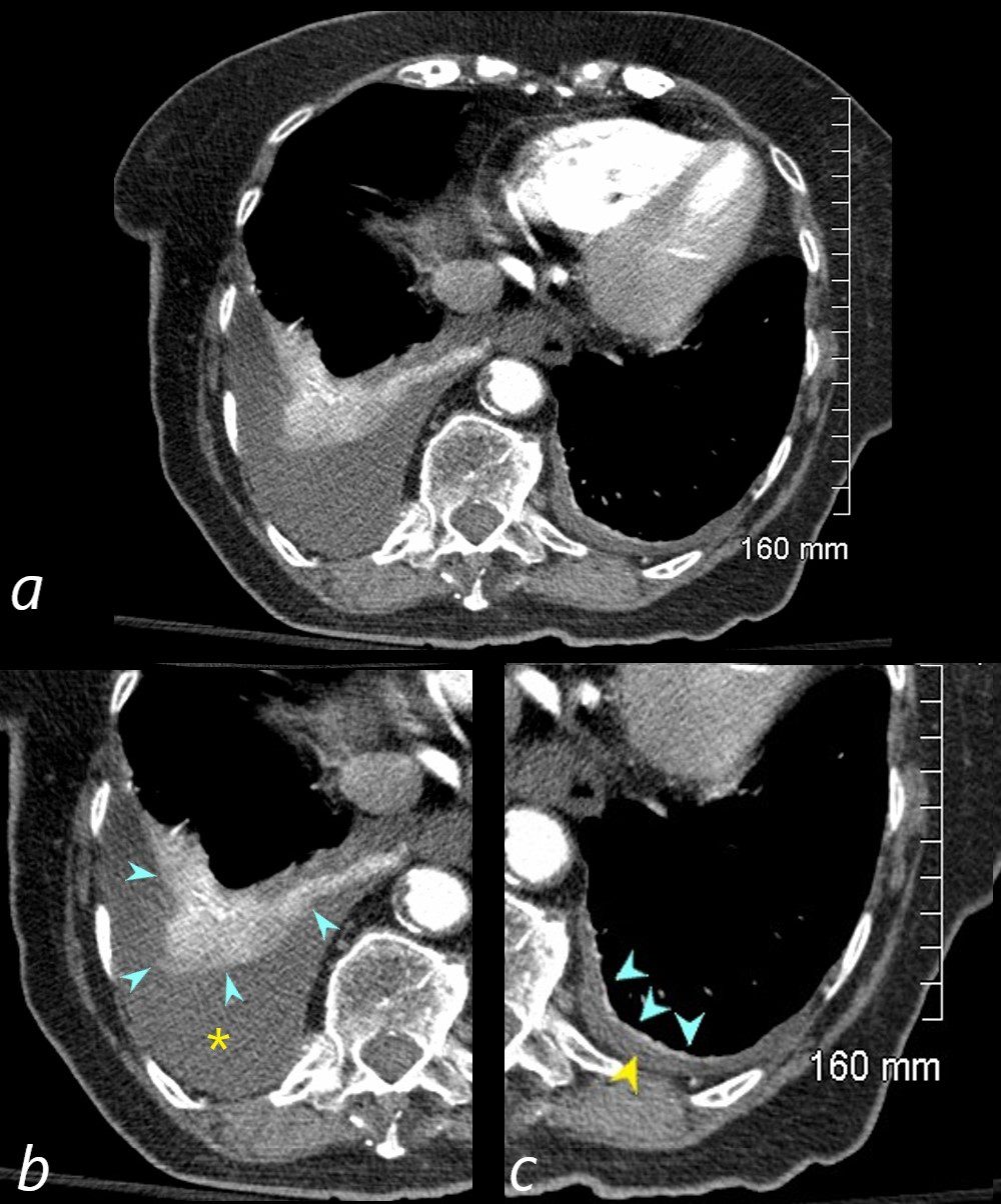
CT with Moderate Right and Small Left Pleural Effusions with Compressive Atelectasis
92-year-old female presents with a dyspnea.
CT scan shows a moderate sized right pleural effusion , magnified in b (yellow asterisk ) with compressive atelectasis (teal arrowheads) . There is relative hyperenhancement of the compressed and atelectatic lung due to increased tissue density. In the left lower lung field , there is a small effusion (yellow arrowhead ,c) with a minor degree of compressive atelectasis (teal arrowheads, c )
Ashley Davidoff MD TheCommonVein.net 118437c01L

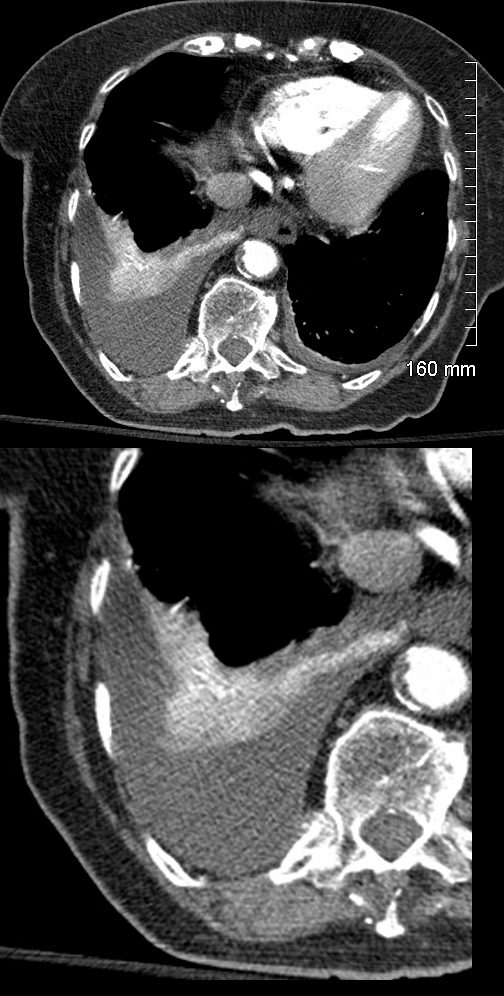
CT with Moderate Right Pleural Effusion and Compressive Atelectasis
92-year-old female presents with a dyspnea.
CT scan shows a moderate sized right pleural effusion with compressive atelectasis and on the left, there is a small effusion with a minor degree of atelectasis. There is relative hyperenhancement of the compressed and atelectatic due to increased tissue density.
Ashley Davidoff MD TheCommonVein.net 118437c
-
- Caused by
- resulting in absence of air in the affected
- sub-segment
- segment,
- lobe
- lung
- When air does not fill the alveoli, the alveoli collapse.
- Diagnosed by
- stethoscope, percussion, x-ray, CT and bronchoscopy.

File source: commons.wikimedia.org/
Types
Post Obstructive (Resorbtive)
- Caused by
- complete obstruction
- neoplasm,
- mucus plugging
- foreign bodies
- complete obstruction
- Result
- air
- no new air can enter lung distal to the obstruction
- trapped air that is absorbed into the capillaries, l
- pleura
- cannot separate
- vacuum and
- traction of mediastinal structures and diaphragm
- mediastinal shift and elevated diaphragm
- air
- Compressive Atelectasis
- Caused by
- pleural effusion,
- pneumothorax and
- diaphragmatic abnormality
- Result
- air
- squeezed out of lung
- pleura
- separated
- potentially only minor or no vaccuum
- air
- Caused by
- Cicatrisation (Traction) Atelectasis
- Caused by
- graulomatous disease,
- necrotizing pneumonia and
- radiation fibrosis
- bronchietasis
- Result
- air
- lung cannot expand
- pleura
-
- cannot separate
- vacuum and
- traction on surrounding structures
-
- air
- Caused by
- Adhesive Atelectasis
- Caused by?
- surfactant deficiency
- diffuse or
- localized
- Result
- surfactant deficiency
- Caused by?
- Gravity Dependant Atelectasis (Dependent Atelectasis)
- Caused by?
- weight of the lungs
- Result
- Crescentic shaped
- ground glass changes
- Crescentic shaped
- Caused by?
- Osteophyte-Induced
- Caused by
- Result
- focal atelectasis
- fibrosis
- bronchiolectasis
Links and References
- TCV
- Discoid Atelectasis on CT

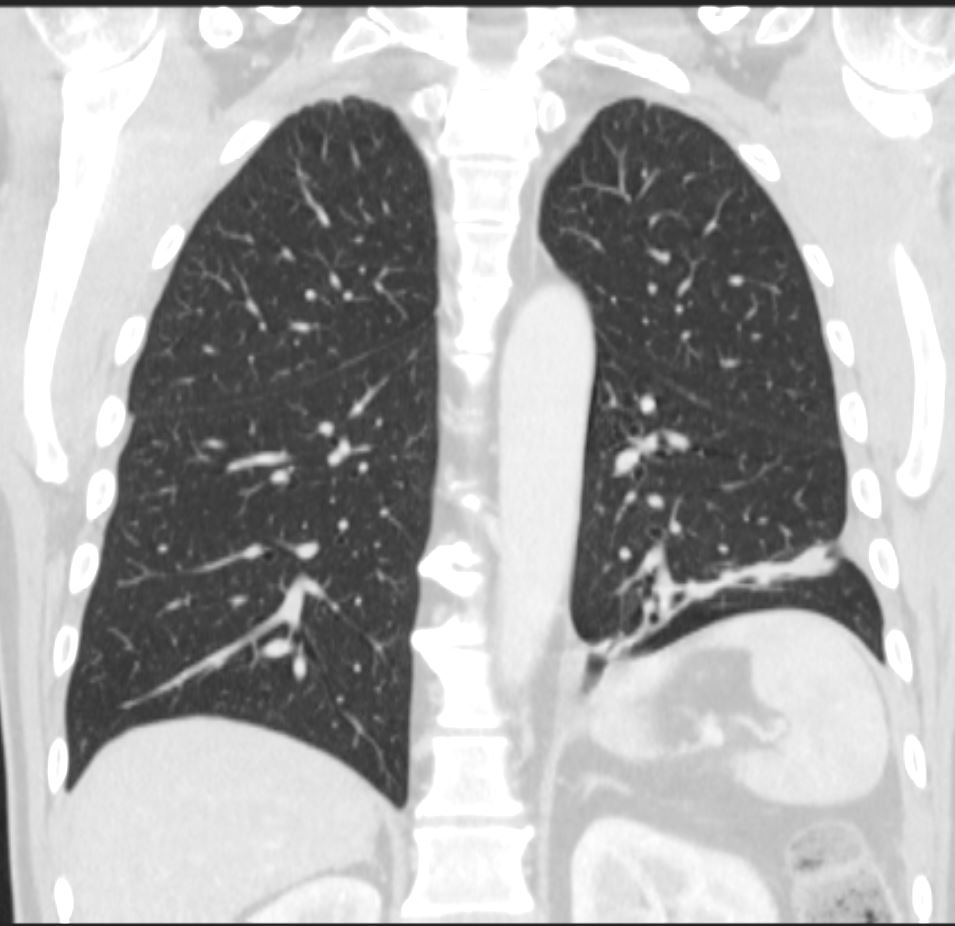
CT Linear Atelectasis 3 Months Later
CT scan in the coronal plane 3 months later shows significant improvement of the atelectasis involving a basal segment of the left lower lobe associated with persistent elevation of the left hemidiaphragm indicating volume loss. The atelectasis now has a discoid, linear, or plate-like appearance
Ashley Davidoff MD TheCommonVein.net 276Lu 136238
aka discoid atelectasis aka plate-like atelectasis
Links and References
TCV
