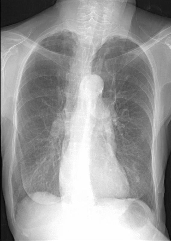
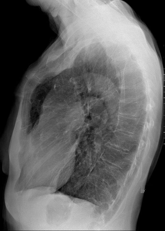
71-year-old female presents with history emphysema
Chest X-ray shows hyperinflated lungs with flattened hemidiaphragms and increase in the retrosternal space and right ventricular enlargement based on the decrease in the retrosternal air space
Ashley Davidoff MD
Ashley Davidoff MD
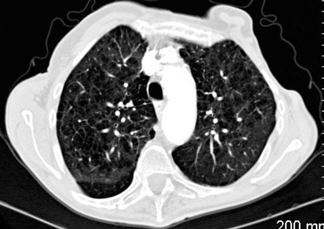
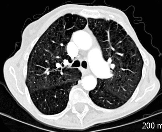
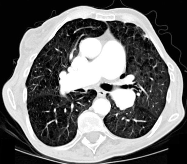
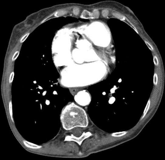
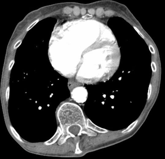
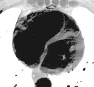
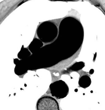
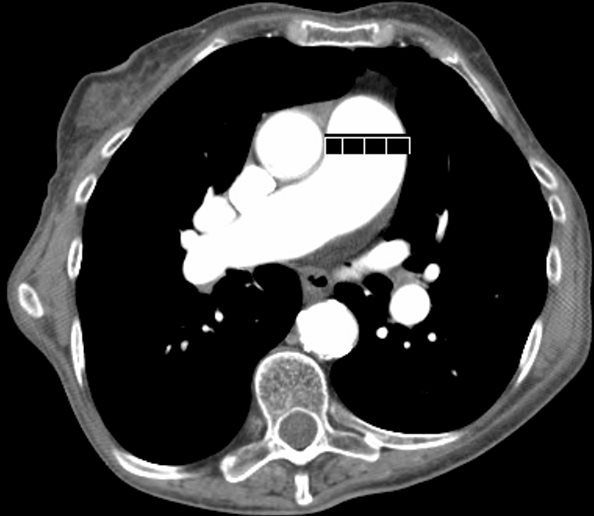
CT scan confirms the presence of centrilobular emphysema, predominantly in the upper lobes with associated right atrial, right ventricular and pulmonary arterial enlargement. The LA and LV are normal
These findings are consistent with cor pulmonale and pulmonary hypertension, secondary to emphysema.

The red arrows point to the soft tissues of the centrilobular emphysema consisting of the arterioles and bronchiolar walls (not usually visible.
Ashley Davidoff MD

Ashley Davidoff MD
Ashley Davidoff MD
Ashley Davidoff MD
Ashley Davidoff MD
115601.8.jpg
EMPHYSEMA, COR PULMONALE and PULMONARY HYPERTENSIONAshley Davidoff MD
EMPHYSEMA, COR PULMONALE and PULMONARY HYPERTENSION
Ashley Davidoff MD
EMPHYSEMA, COR PULMONALE and PULMONARY HYPERTENSION
Ashley Davidoff MD
EMPHYSEMA, COR PULMONALE and PULMONARY HYPERTENSION
Ashley Davidoff MD
115596.8.jpg
ENLARGED MPA . >3CMSEMPHYSEMA, COR PULMONALE and PULMONARY HYPERTENSION
Ashley Davidoff MD
EMPHYSEMA, COR PULMONALE and PULMONARY HYPERTENSION
Ashley Davidoff MD
EMPHYSEMA, COR PULMONALE and PULMONARY HYPERTENSION
Ashley Davidoff MD
