Linear or discoid atelectasis, also known as plate atelectasis, is a form of partial lung collapse that appears as thin, linear opacities on a chest X-ray or CT scan. These opacities are typically parallel to the pleura and are most commonly seen in the lower lobes. Linear atelectasis is often a temporary condition and may resolve with changes in patient positioning or deep breathing.

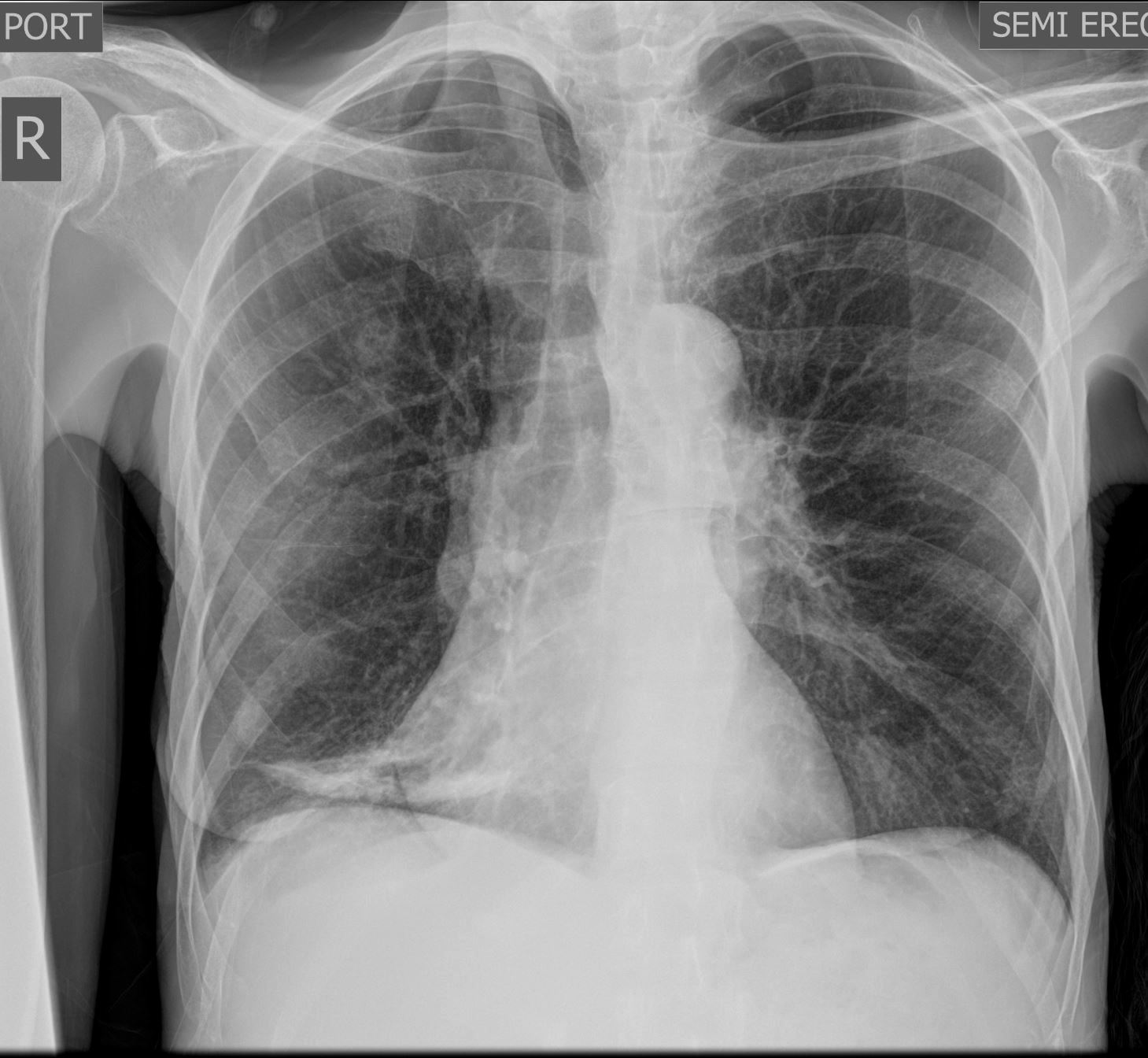
Frontal Chest Xray of a 55-year-old female shows a region of discoid atelectasis (aka linear atelectasis plate atelectasis ) in the right lower lung zone
Courtesy Ashley Davidoff MD TheCommonVein.net 136548

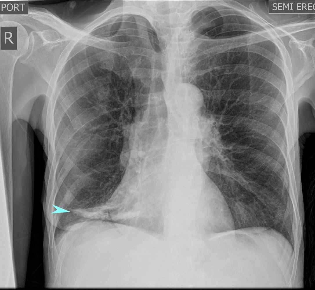
Frontal Chest Xray of a 55-year-old female shows a region of discoid atelectasis (aka linear atelectasis plate atelectasis ) in the right lower lung zone (teal arrow)
Courtesy Ashley Davidoff MD TheCommonVein.net 136548

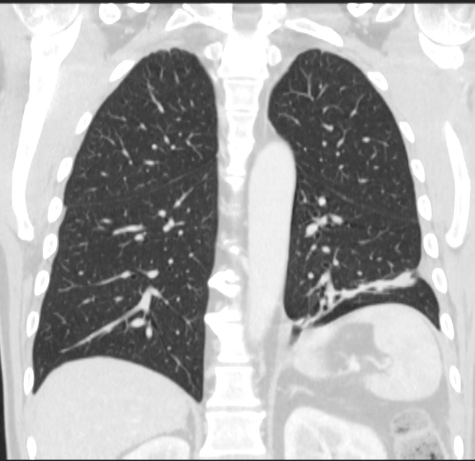
CT scan in the coronal plane 3 months later shows significant improvement of the atelectasis involving a basal segment of the left lower lobe associated with persistent elevation of the left hemidiaphragm indicating volume loss. The atelectasis now has a discoid, linear, or plate-like appearance
Ashley Davidoff MD TheCommonVein.net 276Lu 136238
aka discoid atelectasis aka plate-like atelectasis

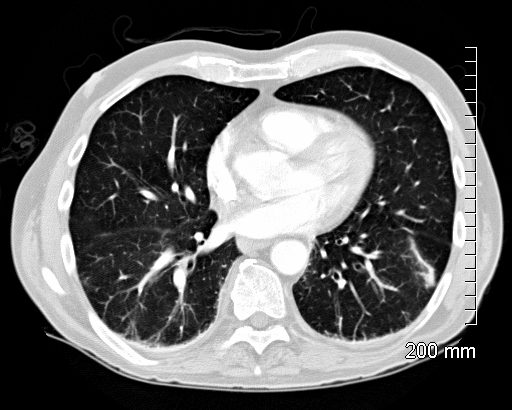
66 year old male with linear (discoid) atelectasis in the left lower lobe on CT
Ashley Davidoff MD TheCommonVein.net

60 year old male with linear (discoid) atelectasis in the middle lobe and the left upper lobe on CT. Note moderate sized bilateral pleural effusion. Minor compressive atelectasis caused by the left effusion.
Ashley Davidoff MD TheCommonVein.net

66 year old male with linear (discoid) atelectasis in the left lower lobe on CT
Ashley Davidoff MD TheCommonVein.net
Radiographic Appearance
- Linear Opacity: Appears as a thin, straight or slightly curvilinear band of increased density, usually 1?3 cm wide.
- Location: Often seen in the lower lobes of the lungs, particularly along the costophrenic angles.
- Orientation: Usually parallel to the pleural surface, often horizontal or oblique in alignment.
Common Causes
Linear or discoid atelectasis can result from various factors that cause localized alveolar collapse, including:
- Hypoventilation: Commonly occurs post-operatively or in patients with prolonged bed rest.
- Shallow Breathing: Seen in patients with pleuritic pain, rib fractures, or abdominal discomfort, as these conditions limit deep inspiration.
- Obstruction: Mucus plugging in smaller airways can cause localized atelectasis.
- Compression: External pressure from pleural effusion, a mass, or pneumothorax can collapse nearby alveoli.
Clinical Significance
Linear or discoid atelectasis is generally a benign finding and often transient. It does not usually indicate significant pathology, although it can sometimes be a marker of poor ventilation or underlying lung disease. It is important to differentiate it from more concerning findings, such as infiltrates or fibrotic bands, which may indicate chronic or progressive disease.
Summary
In radiographic terms, linear or discoid atelectasis is a thin, linear opacity representing areas of localized alveolar collapse, often due to hypoventilation or compression, and is typically a temporary and benign finding.
Links and References
Chat GPT – What is radiographic definition of linear or discoid atelectasis?
