Growth Time
|
|
Aging of the Lungs While the number of bronchi one has at birth is the same as the number in old age, the number of alveoli increases till the age of about 8 years. Thereafter the alveoli increase in size till about 18 years. As the lungs progress through age there is a mild, functionally insignificant increase in size. The respiratory bronchioles and alveolar ducts increase in size in middle age, but there is no destruction of the walls of the alveolar septa. Thus in old age there is mild increase in size of the lungs but without concomitant destruction of lung tissue that is seen in emphysema. There is glandular hypertrophy and progressive calcification of the cartilage of the medium sized bronchi. (London South Bank University) (http://myweb.lsbu.ac.uk/dirt/museum/p6-92.html) |
DOMElement Object
(
[schemaTypeInfo] =>
[tagName] => table
[firstElementChild] => (object value omitted)
[lastElementChild] => (object value omitted)
[childElementCount] => 1
[previousElementSibling] =>
[nextElementSibling] => (object value omitted)
[nodeName] => table
[nodeValue] =>
Atelectasis, bronchiectasis, hyperinflation.
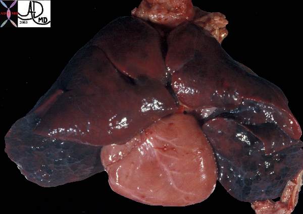
This post mortem specimen of the lungs and heart of a child showing normal lobar pattern of the lungs with 3 lobes on the right and two on the left. The lungs appear hemorrhagic with the interlobular septa and secondary lobules are prominent. A small piece of glistening pleura are seen at the lower portion of the left lung.
Courtesy Ashley Davidoff MD 32558
[nodeType] => 1
[parentNode] => (object value omitted)
[childNodes] => (object value omitted)
[firstChild] => (object value omitted)
[lastChild] => (object value omitted)
[previousSibling] => (object value omitted)
[nextSibling] => (object value omitted)
[attributes] => (object value omitted)
[ownerDocument] => (object value omitted)
[namespaceURI] =>
[prefix] =>
[localName] => table
[baseURI] =>
[textContent] =>
Atelectasis, bronchiectasis, hyperinflation.
This post mortem specimen of the lungs and heart of a child showing normal lobar pattern of the lungs with 3 lobes on the right and two on the left. The lungs appear hemorrhagic with the interlobular septa and secondary lobules are prominent. A small piece of glistening pleura are seen at the lower portion of the left lung.
Courtesy Ashley Davidoff MD 32558
)
DOMElement Object
(
[schemaTypeInfo] =>
[tagName] => td
[firstElementChild] => (object value omitted)
[lastElementChild] => (object value omitted)
[childElementCount] => 1
[previousElementSibling] =>
[nextElementSibling] =>
[nodeName] => td
[nodeValue] => This post mortem specimen of the lungs and heart of a child showing normal lobar pattern of the lungs with 3 lobes on the right and two on the left. The lungs appear hemorrhagic with the interlobular septa and secondary lobules are prominent. A small piece of glistening pleura are seen at the lower portion of the left lung.
Courtesy Ashley Davidoff MD 32558
[nodeType] => 1
[parentNode] => (object value omitted)
[childNodes] => (object value omitted)
[firstChild] => (object value omitted)
[lastChild] => (object value omitted)
[previousSibling] => (object value omitted)
[nextSibling] => (object value omitted)
[attributes] => (object value omitted)
[ownerDocument] => (object value omitted)
[namespaceURI] =>
[prefix] =>
[localName] => td
[baseURI] =>
[textContent] => This post mortem specimen of the lungs and heart of a child showing normal lobar pattern of the lungs with 3 lobes on the right and two on the left. The lungs appear hemorrhagic with the interlobular septa and secondary lobules are prominent. A small piece of glistening pleura are seen at the lower portion of the left lung.
Courtesy Ashley Davidoff MD 32558
)
DOMElement Object
(
[schemaTypeInfo] =>
[tagName] => td
[firstElementChild] => (object value omitted)
[lastElementChild] => (object value omitted)
[childElementCount] => 1
[previousElementSibling] =>
[nextElementSibling] =>
[nodeName] => td
[nodeValue] => Atelectasis, bronchiectasis, hyperinflation.
[nodeType] => 1
[parentNode] => (object value omitted)
[childNodes] => (object value omitted)
[firstChild] => (object value omitted)
[lastChild] => (object value omitted)
[previousSibling] => (object value omitted)
[nextSibling] => (object value omitted)
[attributes] => (object value omitted)
[ownerDocument] => (object value omitted)
[namespaceURI] =>
[prefix] =>
[localName] => td
[baseURI] =>
[textContent] => Atelectasis, bronchiectasis, hyperinflation.
)
DOMElement Object
(
[schemaTypeInfo] =>
[tagName] => table
[firstElementChild] => (object value omitted)
[lastElementChild] => (object value omitted)
[childElementCount] => 1
[previousElementSibling] => (object value omitted)
[nextElementSibling] =>
[nodeName] => table
[nodeValue] =>
Atelectasis, bronchiectasis, hyperinflation.
This post mortem specimen of the lungs and heart of a child showing normal lobar pattern of the lungs with 3 lobes on the right and two on the left. The lungs appear hemorrhagic with the interlobular septa and secondary lobules are prominent. A small piece of glistening pleura are seen at the lower portion of the left lung.
Courtesy Ashley Davidoff MD 32558
Aging of the Lungs
While the number of bronchi one has at birth is the same as the number in old age, the number of alveoli increases till the age of about 8 years. Thereafter the alveoli increase in size till about 18 years. As the lungs progress through age there is a mild, functionally insignificant increase in size. The respiratory bronchioles and alveolar ducts increase in size in middle age, but there is no destruction of the walls of the alveolar septa. Thus in old age there is mild increase in size of the lungs but without concomitant destruction of lung tissue that is seen in emphysema. There is glandular hypertrophy and progressive calcification of the cartilage of the medium sized bronchi. (London South Bank University) (http://myweb.lsbu.ac.uk/dirt/museum/p6-92.html)
[nodeType] => 1
[parentNode] => (object value omitted)
[childNodes] => (object value omitted)
[firstChild] => (object value omitted)
[lastChild] => (object value omitted)
[previousSibling] => (object value omitted)
[nextSibling] => (object value omitted)
[attributes] => (object value omitted)
[ownerDocument] => (object value omitted)
[namespaceURI] =>
[prefix] =>
[localName] => table
[baseURI] =>
[textContent] =>
Atelectasis, bronchiectasis, hyperinflation.
This post mortem specimen of the lungs and heart of a child showing normal lobar pattern of the lungs with 3 lobes on the right and two on the left. The lungs appear hemorrhagic with the interlobular septa and secondary lobules are prominent. A small piece of glistening pleura are seen at the lower portion of the left lung.
Courtesy Ashley Davidoff MD 32558
Aging of the Lungs
While the number of bronchi one has at birth is the same as the number in old age, the number of alveoli increases till the age of about 8 years. Thereafter the alveoli increase in size till about 18 years. As the lungs progress through age there is a mild, functionally insignificant increase in size. The respiratory bronchioles and alveolar ducts increase in size in middle age, but there is no destruction of the walls of the alveolar septa. Thus in old age there is mild increase in size of the lungs but without concomitant destruction of lung tissue that is seen in emphysema. There is glandular hypertrophy and progressive calcification of the cartilage of the medium sized bronchi. (London South Bank University) (http://myweb.lsbu.ac.uk/dirt/museum/p6-92.html)
)
DOMElement Object
(
[schemaTypeInfo] =>
[tagName] => td
[firstElementChild] => (object value omitted)
[lastElementChild] => (object value omitted)
[childElementCount] => 1
[previousElementSibling] =>
[nextElementSibling] =>
[nodeName] => td
[nodeValue] => This post mortem specimen of the lungs and heart of a child showing normal lobar pattern of the lungs with 3 lobes on the right and two on the left. The lungs appear hemorrhagic with the interlobular septa and secondary lobules are prominent. A small piece of glistening pleura are seen at the lower portion of the left lung.
Courtesy Ashley Davidoff MD 32558
[nodeType] => 1
[parentNode] => (object value omitted)
[childNodes] => (object value omitted)
[firstChild] => (object value omitted)
[lastChild] => (object value omitted)
[previousSibling] => (object value omitted)
[nextSibling] => (object value omitted)
[attributes] => (object value omitted)
[ownerDocument] => (object value omitted)
[namespaceURI] =>
[prefix] =>
[localName] => td
[baseURI] =>
[textContent] => This post mortem specimen of the lungs and heart of a child showing normal lobar pattern of the lungs with 3 lobes on the right and two on the left. The lungs appear hemorrhagic with the interlobular septa and secondary lobules are prominent. A small piece of glistening pleura are seen at the lower portion of the left lung.
Courtesy Ashley Davidoff MD 32558
)
DOMElement Object
(
[schemaTypeInfo] =>
[tagName] => td
[firstElementChild] => (object value omitted)
[lastElementChild] => (object value omitted)
[childElementCount] => 1
[previousElementSibling] =>
[nextElementSibling] =>
[nodeName] => td
[nodeValue] => Atelectasis, bronchiectasis, hyperinflation.
[nodeType] => 1
[parentNode] => (object value omitted)
[childNodes] => (object value omitted)
[firstChild] => (object value omitted)
[lastChild] => (object value omitted)
[previousSibling] => (object value omitted)
[nextSibling] => (object value omitted)
[attributes] => (object value omitted)
[ownerDocument] => (object value omitted)
[namespaceURI] =>
[prefix] =>
[localName] => td
[baseURI] =>
[textContent] => Atelectasis, bronchiectasis, hyperinflation.
)
DOMElement Object
(
[schemaTypeInfo] =>
[tagName] => td
[firstElementChild] => (object value omitted)
[lastElementChild] => (object value omitted)
[childElementCount] => 4
[previousElementSibling] =>
[nextElementSibling] =>
[nodeName] => td
[nodeValue] =>
Atelectasis, bronchiectasis, hyperinflation.
This post mortem specimen of the lungs and heart of a child showing normal lobar pattern of the lungs with 3 lobes on the right and two on the left. The lungs appear hemorrhagic with the interlobular septa and secondary lobules are prominent. A small piece of glistening pleura are seen at the lower portion of the left lung.
Courtesy Ashley Davidoff MD 32558
Aging of the Lungs
While the number of bronchi one has at birth is the same as the number in old age, the number of alveoli increases till the age of about 8 years. Thereafter the alveoli increase in size till about 18 years. As the lungs progress through age there is a mild, functionally insignificant increase in size. The respiratory bronchioles and alveolar ducts increase in size in middle age, but there is no destruction of the walls of the alveolar septa. Thus in old age there is mild increase in size of the lungs but without concomitant destruction of lung tissue that is seen in emphysema. There is glandular hypertrophy and progressive calcification of the cartilage of the medium sized bronchi. (London South Bank University) (http://myweb.lsbu.ac.uk/dirt/museum/p6-92.html)
[nodeType] => 1
[parentNode] => (object value omitted)
[childNodes] => (object value omitted)
[firstChild] => (object value omitted)
[lastChild] => (object value omitted)
[previousSibling] => (object value omitted)
[nextSibling] => (object value omitted)
[attributes] => (object value omitted)
[ownerDocument] => (object value omitted)
[namespaceURI] =>
[prefix] =>
[localName] => td
[baseURI] =>
[textContent] =>
Atelectasis, bronchiectasis, hyperinflation.
This post mortem specimen of the lungs and heart of a child showing normal lobar pattern of the lungs with 3 lobes on the right and two on the left. The lungs appear hemorrhagic with the interlobular septa and secondary lobules are prominent. A small piece of glistening pleura are seen at the lower portion of the left lung.
Courtesy Ashley Davidoff MD 32558
Aging of the Lungs
While the number of bronchi one has at birth is the same as the number in old age, the number of alveoli increases till the age of about 8 years. Thereafter the alveoli increase in size till about 18 years. As the lungs progress through age there is a mild, functionally insignificant increase in size. The respiratory bronchioles and alveolar ducts increase in size in middle age, but there is no destruction of the walls of the alveolar septa. Thus in old age there is mild increase in size of the lungs but without concomitant destruction of lung tissue that is seen in emphysema. There is glandular hypertrophy and progressive calcification of the cartilage of the medium sized bronchi. (London South Bank University) (http://myweb.lsbu.ac.uk/dirt/museum/p6-92.html)
)
DOMElement Object
(
[schemaTypeInfo] =>
[tagName] => table
[firstElementChild] => (object value omitted)
[lastElementChild] => (object value omitted)
[childElementCount] => 1
[previousElementSibling] => (object value omitted)
[nextElementSibling] =>
[nodeName] => table
[nodeValue] =>
Neonatal Structures
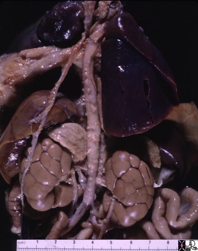
01900.800 adrenal aorta IVC kidney liver lung small bowel ureter diaphragm normal fetal lobation neonate growth time grossanatomy Davidoff MD
[nodeType] => 1
[parentNode] => (object value omitted)
[childNodes] => (object value omitted)
[firstChild] => (object value omitted)
[lastChild] => (object value omitted)
[previousSibling] => (object value omitted)
[nextSibling] => (object value omitted)
[attributes] => (object value omitted)
[ownerDocument] => (object value omitted)
[namespaceURI] =>
[prefix] =>
[localName] => table
[baseURI] =>
[textContent] =>
Neonatal Structures
01900.800 adrenal aorta IVC kidney liver lung small bowel ureter diaphragm normal fetal lobation neonate growth time grossanatomy Davidoff MD
)
DOMElement Object
(
[schemaTypeInfo] =>
[tagName] => td
[firstElementChild] => (object value omitted)
[lastElementChild] => (object value omitted)
[childElementCount] => 1
[previousElementSibling] =>
[nextElementSibling] =>
[nodeName] => td
[nodeValue] => 01900.800 adrenal aorta IVC kidney liver lung small bowel ureter diaphragm normal fetal lobation neonate growth time grossanatomy Davidoff MD
[nodeType] => 1
[parentNode] => (object value omitted)
[childNodes] => (object value omitted)
[firstChild] => (object value omitted)
[lastChild] => (object value omitted)
[previousSibling] => (object value omitted)
[nextSibling] => (object value omitted)
[attributes] => (object value omitted)
[ownerDocument] => (object value omitted)
[namespaceURI] =>
[prefix] =>
[localName] => td
[baseURI] =>
[textContent] => 01900.800 adrenal aorta IVC kidney liver lung small bowel ureter diaphragm normal fetal lobation neonate growth time grossanatomy Davidoff MD
)
DOMElement Object
(
[schemaTypeInfo] =>
[tagName] => td
[firstElementChild] => (object value omitted)
[lastElementChild] => (object value omitted)
[childElementCount] => 2
[previousElementSibling] =>
[nextElementSibling] =>
[nodeName] => td
[nodeValue] =>
Neonatal Structures
[nodeType] => 1
[parentNode] => (object value omitted)
[childNodes] => (object value omitted)
[firstChild] => (object value omitted)
[lastChild] => (object value omitted)
[previousSibling] => (object value omitted)
[nextSibling] => (object value omitted)
[attributes] => (object value omitted)
[ownerDocument] => (object value omitted)
[namespaceURI] =>
[prefix] =>
[localName] => td
[baseURI] =>
[textContent] =>
Neonatal Structures
)
DOMElement Object
(
[schemaTypeInfo] =>
[tagName] => table
[firstElementChild] => (object value omitted)
[lastElementChild] => (object value omitted)
[childElementCount] => 1
[previousElementSibling] =>
[nextElementSibling] => (object value omitted)
[nodeName] => table
[nodeValue] =>
Growth Time
Neonatal Structures
01900.800 adrenal aorta IVC kidney liver lung small bowel ureter diaphragm normal fetal lobation neonate growth time grossanatomy Davidoff MD
[nodeType] => 1
[parentNode] => (object value omitted)
[childNodes] => (object value omitted)
[firstChild] => (object value omitted)
[lastChild] => (object value omitted)
[previousSibling] =>
[nextSibling] => (object value omitted)
[attributes] => (object value omitted)
[ownerDocument] => (object value omitted)
[namespaceURI] =>
[prefix] =>
[localName] => table
[baseURI] =>
[textContent] =>
Growth Time
Neonatal Structures
01900.800 adrenal aorta IVC kidney liver lung small bowel ureter diaphragm normal fetal lobation neonate growth time grossanatomy Davidoff MD
)
DOMElement Object
(
[schemaTypeInfo] =>
[tagName] => td
[firstElementChild] => (object value omitted)
[lastElementChild] => (object value omitted)
[childElementCount] => 1
[previousElementSibling] =>
[nextElementSibling] =>
[nodeName] => td
[nodeValue] => 01900.800 adrenal aorta IVC kidney liver lung small bowel ureter diaphragm normal fetal lobation neonate growth time grossanatomy Davidoff MD
[nodeType] => 1
[parentNode] => (object value omitted)
[childNodes] => (object value omitted)
[firstChild] => (object value omitted)
[lastChild] => (object value omitted)
[previousSibling] => (object value omitted)
[nextSibling] => (object value omitted)
[attributes] => (object value omitted)
[ownerDocument] => (object value omitted)
[namespaceURI] =>
[prefix] =>
[localName] => td
[baseURI] =>
[textContent] => 01900.800 adrenal aorta IVC kidney liver lung small bowel ureter diaphragm normal fetal lobation neonate growth time grossanatomy Davidoff MD
)
DOMElement Object
(
[schemaTypeInfo] =>
[tagName] => td
[firstElementChild] => (object value omitted)
[lastElementChild] => (object value omitted)
[childElementCount] => 2
[previousElementSibling] =>
[nextElementSibling] =>
[nodeName] => td
[nodeValue] =>
Neonatal Structures
[nodeType] => 1
[parentNode] => (object value omitted)
[childNodes] => (object value omitted)
[firstChild] => (object value omitted)
[lastChild] => (object value omitted)
[previousSibling] => (object value omitted)
[nextSibling] => (object value omitted)
[attributes] => (object value omitted)
[ownerDocument] => (object value omitted)
[namespaceURI] =>
[prefix] =>
[localName] => td
[baseURI] =>
[textContent] =>
Neonatal Structures
)
DOMElement Object
(
[schemaTypeInfo] =>
[tagName] => td
[firstElementChild] => (object value omitted)
[lastElementChild] => (object value omitted)
[childElementCount] => 2
[previousElementSibling] =>
[nextElementSibling] =>
[nodeName] => td
[nodeValue] => Growth Time
Neonatal Structures
01900.800 adrenal aorta IVC kidney liver lung small bowel ureter diaphragm normal fetal lobation neonate growth time grossanatomy Davidoff MD
[nodeType] => 1
[parentNode] => (object value omitted)
[childNodes] => (object value omitted)
[firstChild] => (object value omitted)
[lastChild] => (object value omitted)
[previousSibling] => (object value omitted)
[nextSibling] => (object value omitted)
[attributes] => (object value omitted)
[ownerDocument] => (object value omitted)
[namespaceURI] =>
[prefix] =>
[localName] => td
[baseURI] =>
[textContent] => Growth Time
Neonatal Structures
01900.800 adrenal aorta IVC kidney liver lung small bowel ureter diaphragm normal fetal lobation neonate growth time grossanatomy Davidoff MD
)


