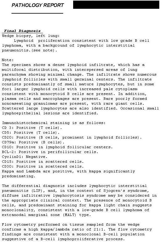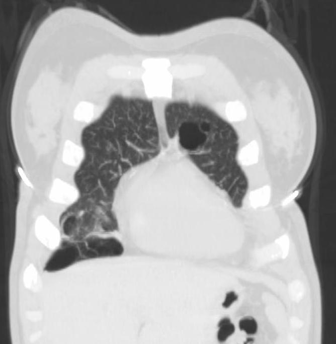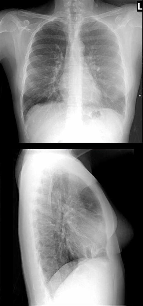
44 year old female presents with a cough.
CXR in 2 views suggest bibasilar cystic changes in the frontal view and cystic changes overlying the heart on the lateral examination
Ashley Davidoff MD TheCommonVein.net
117231b 306Lu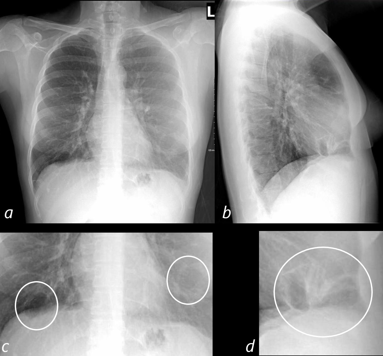
44 year old female presents with a cough.
CXR in 2 views suggest bibasilar cystic changes in the frontal view (c white rings) and cystic changes overlying the heart on the lateral examination (white ring d)
Ashley Davidoff MD TheCommonVein.net 117231b 306Lu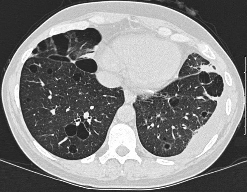
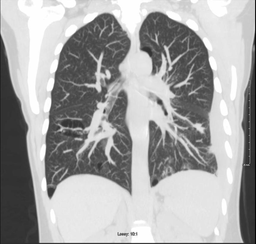
44 year old female presents with a cough.
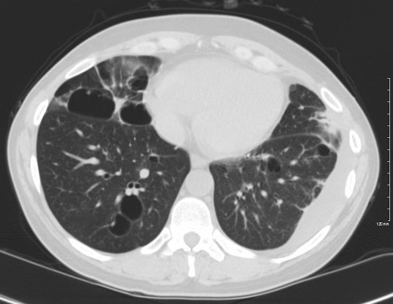
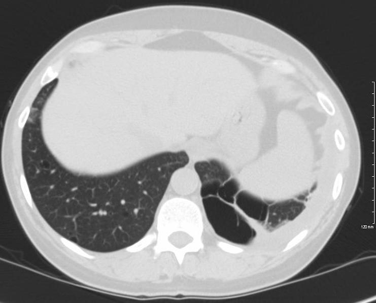
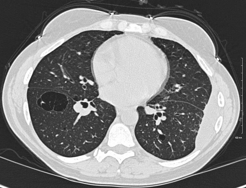
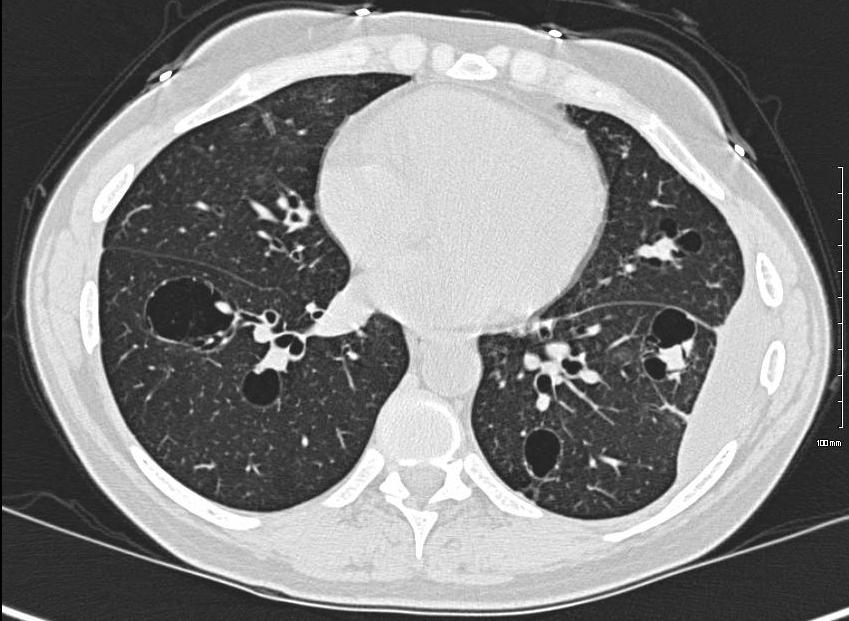
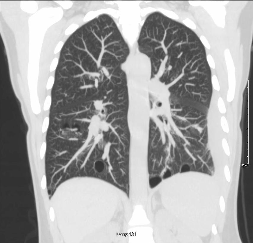
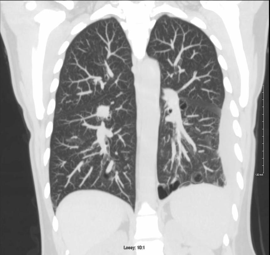
CT of the lung bases confirms the presence bibasilar cystic changes The cysts are thin walled and many are associated with bronchovascular bundles. Nodular changes are noted with the cyst in the anterior aspect of the inferior lingula. The patient has path proven low grade B cell lymphoma and LIP
Ashley Davidoff MD TheCommonVein.net 117231b 306Lu
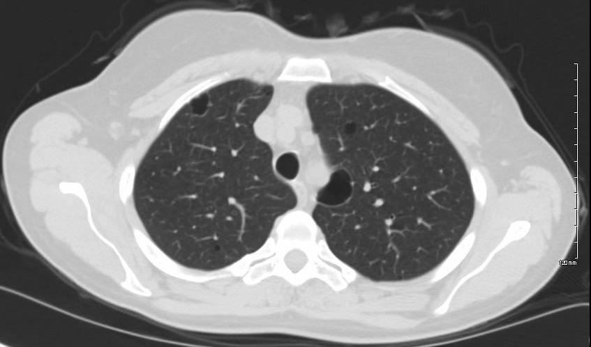
44 year old female presents with a cough.
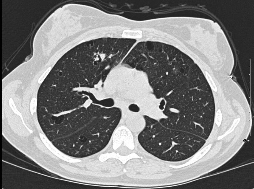
Axial CT through the upper lobes shows multiple thin walled cysts in close association with vessels
Patient has path proven low grade B cell lymphoma and LIP
Ashley Davidoff MD TheCommonVein.net 117232 306Lu
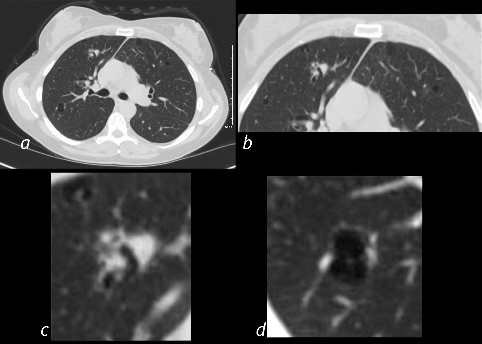
44 year old female presents with a cough.
Axial CT through the upper lobes shows a complex nodular opacity in close association with a bronchovascular bundle a, magnified in b and c). There is also a thin walled cyst in close association with bronchovascular bundles running alongside the cyst (a,and d)
Patient has path proven low grade B cell lymphoma and LIP and these changes are manifestations of the lymphocytic infiltrate alongside the bronchovascular bundle
Ashley Davidoff MD TheCommonVein.net
117234c 306Lu
117234cL.lungs-LIP-lymphocytic-interstitial-pneumonitis-cysts-low-grade-B-cell-lymphoma-CT.bmp
Right Upper Lobe Bronchovascular Nodular Opacity and Bronchovascular Bundle Alongside Thin Walled Cyst44 year old female presents with a cough.
Axial CT through the upper lobes shows a complex nodular opacity in close association with a bronchovascular bundle (red circles a, magnified in b and c). There is also a thin walled cyst in close association with bronchovascular bundles running alongside the cyst (red circle a,and d) and a second smaller cyst with similar features (b, teal arrowhead)
Patient has path proven low grade B cell lymphoma and LIP and these changes are manifestations of the lymphocytic infiltrate alongside the bronchovascular bundle
Ashley Davidoff MD TheCommonVein.net
117234cL 306Lu
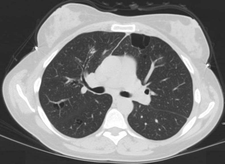
44 year old female presents with a cough.
Axial CT through the upper lobes multifocal irregular thickening surrounding the segmental bronchus with a cluster of micronodules at its distal extent (magnified in b). In the posterior segment of the right upper lobe there is a thin walled cyst in close association with a blood vessel. (magnified in c).
The patient has path proven low grade B cell lymphoma and LIP and these changes are manifestations of the lymphocytic infiltrate alongside the bronchovascular bundle
Ashley Davidoff MD TheCommonVein.net
117235c 306Lu
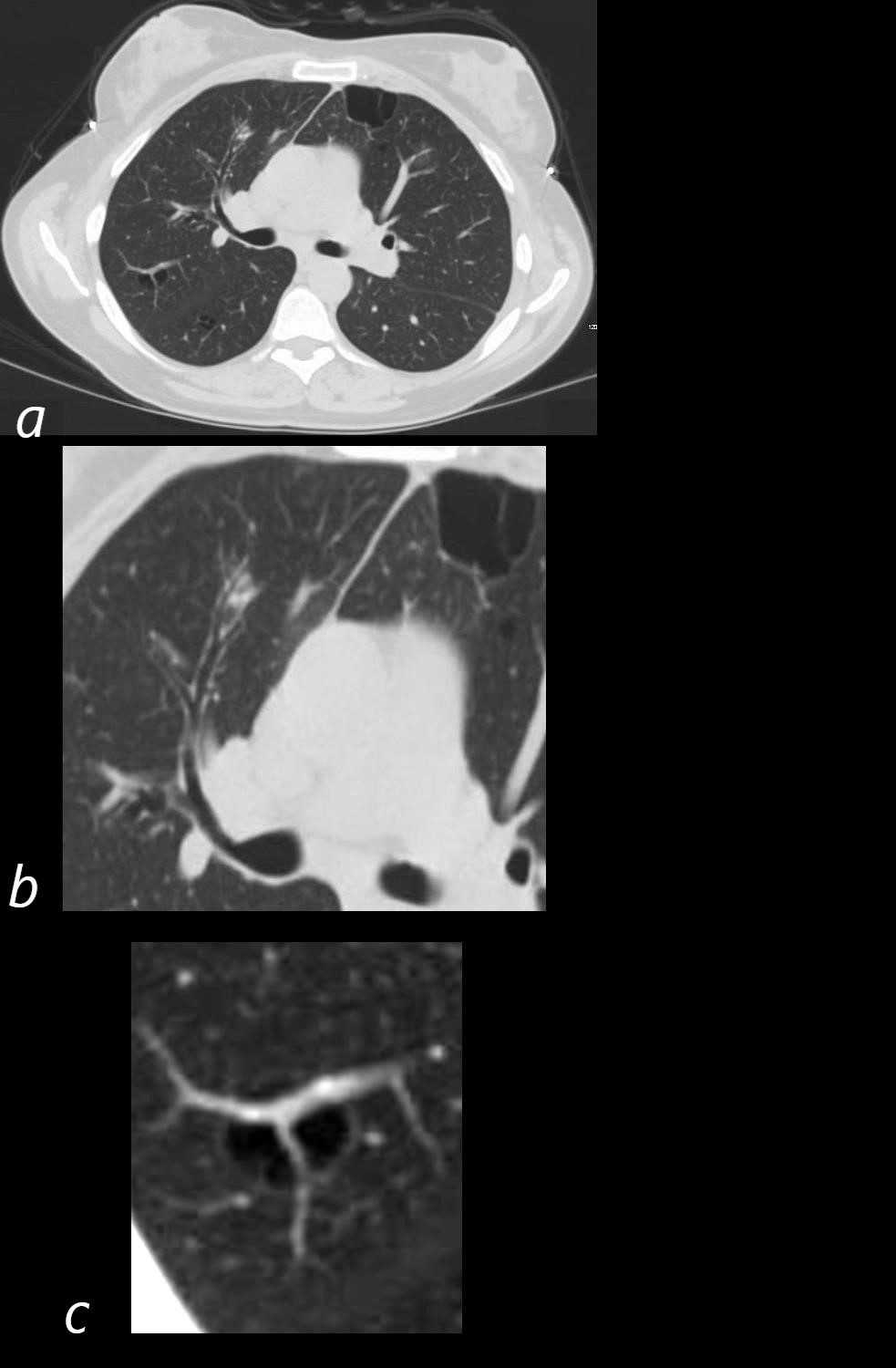
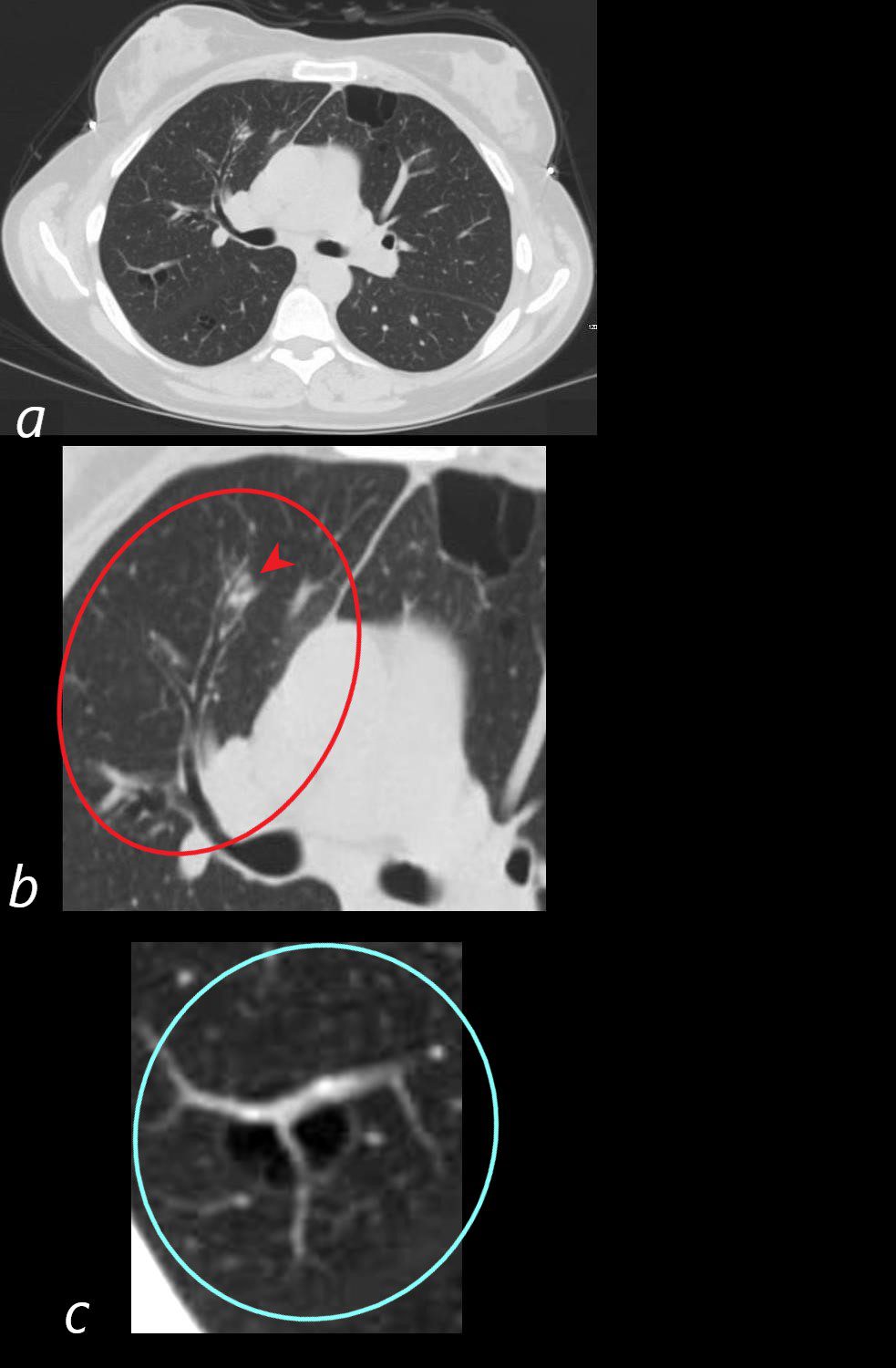
44 year old female presents with a cough.
Axial CT through the upper lobes multifocal irregular thickening surrounding the segmental bronchus (red circle b), with a cluster of micronodules at its distal extent (magnified in b red arrow head).
In the posterior segment of the right upper lobe there is a thin walled cyst in close association with a blood vessel. (magnified in c and teal circle).
The patient has path proven low grade B cell lymphoma and LIP and these changes are manifestations of the lymphocytic infiltrate alongside the bronchovascular bundle
Ashley Davidoff MD TheCommonVein.net
117235cL 306Lu
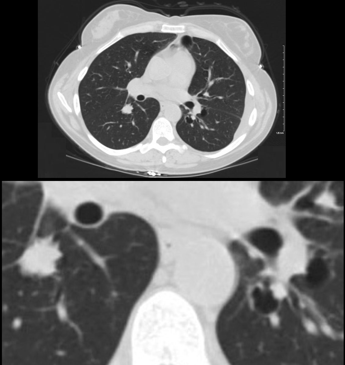
44 year old female presents with a cough.
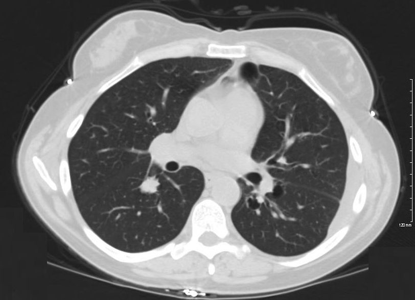
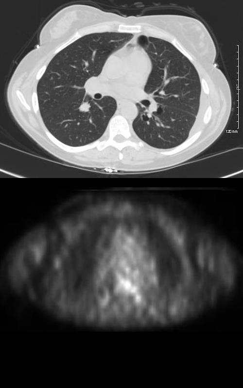
Axial CT through the lower lobes show a spiculated nodule in the right lower lobe that is associated with a bronchus. There is a thin walled cyst in the superior segment of the left lower lobe associated with a bronchovascular bundle running along side the cyst.
The patient has path proven low grade B cell lymphoma and LIP and these changes are manifestations of the lymphocytic infiltrate alongside the bronchovascular bundles
Ashley Davidoff MD TheCommonVein.net
117237c 306Lu
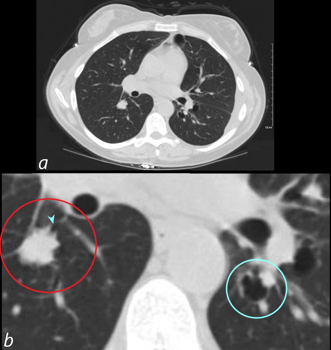
44 year old female presents with a cough.
Axial CT through the lower lobes show a spiculated nodule in the right lower lobe (red circle c) that is associated with a bronchus (teal arrow c).
There is a thin walled cyst in the superior segment of the left lower lobe that is associated with a bronchovascular bundle running along side the cyst (teal circle c).
The patient has path proven low grade B cell lymphoma and LIP and these changes are manifestations of the lymphocytic infiltrate alongside the bronchovascular bundles
Ashley Davidoff MD TheCommonVein.net
117237cL306Lu
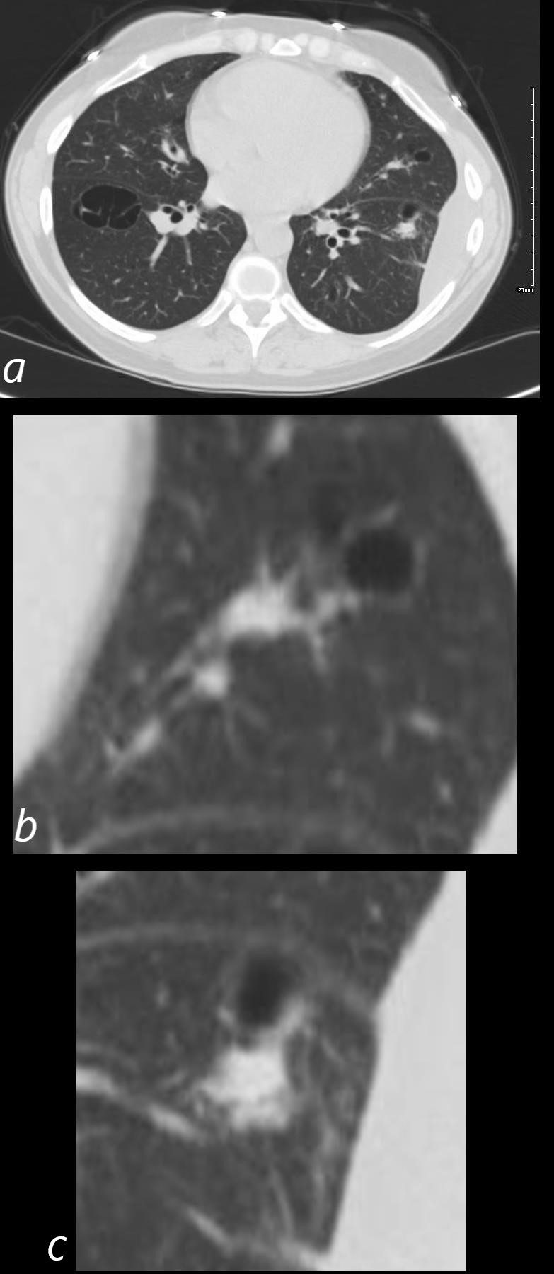
44 year old female presents with a cough.
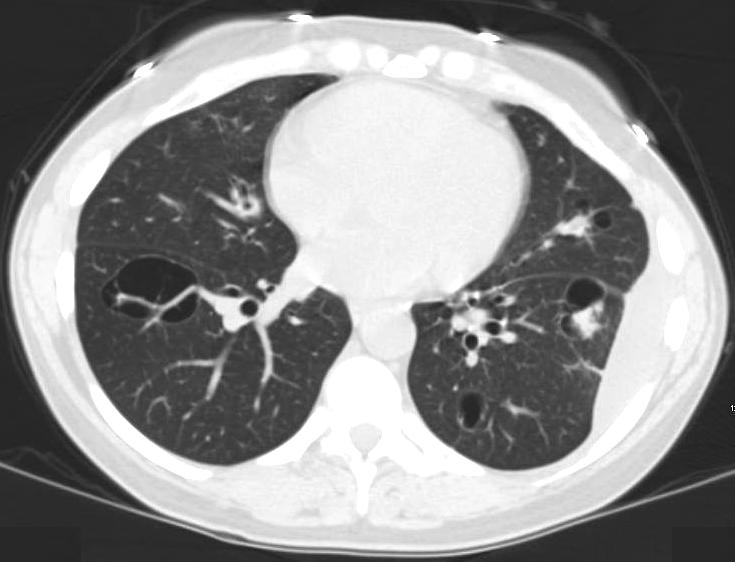
Axial CT through the mid to lower chest shows an ill defined irregular nodule in the lingula associated with a bronchovascular bundle and a cyst (b) A similar ill defined irregular nodule in the LLL associated with a bronchovascular bundle and a cyst (c) Bronchovascuklar bundles are noted alonside the cysts.
The patient has path proven low grade B cell lymphoma and LIP and these changes are manifestations of the lymphocytic infiltrate alongside the bronchovascular bundles
Ashley Davidoff MD TheCommonVein.net
117238cL 306Lu
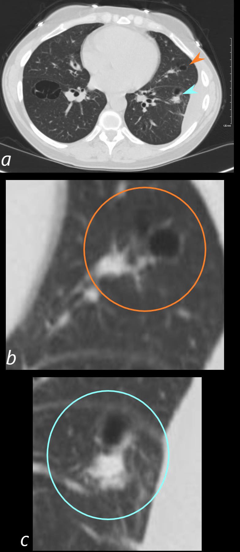
44 year old female presents with a cough.
Axial CT through the mid to lower chest shows an ill defined irregular nodule in the lingula associated with a bronchovascular bundle alongside a cyst (a, orange arrowhead and b orange circle)
A similar ill defined irregular nodule in the LLL is associated with a bronchovascular bundle and a cyst (a teal arrowhead, c teal circle)
The patient has path proven low grade B cell lymphoma and LIP and these changes are manifestations of the lymphocytic infiltrate alongside the bronchovascular bundles
Ashley Davidoff MD TheCommonVein.net
117238cL 306Lu

