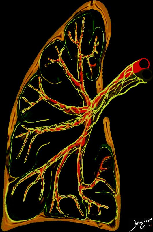
The interstitium of the lungs refers to the structural framework that supports the alveoli, blood vessels, and airways. It is a network of connective tissue that extends throughout the lungs, serving as a scaffold for the functional units of the respiratory system.
Anatomical Distribution
The interstitium is typically divided into three components based on its location:
- Axial Interstitium:
- Surrounds and supports the bronchovascular bundles.
- Includes connective tissue along the bronchi, pulmonary arteries, and veins.
- Extends from the hilum to the periphery of the lung.

The axial interstitium of the lungs that includes a connective tissue network, nerves lymphatics that extend from the hilum to the periphery of the lung.
Ashley Davidoff MD TheCommonVein.net 32187.8
- Parenchymal (Acinar) Interstitium:
- Found within the alveolar walls.
- Includes the fine network of connective tissue that surrounds capillaries and provides structural support to alveoli.
- This component is crucial for gas exchange and is the primary site affected in interstitial lung diseases.
- Peripheral (Subpleural) Interstitium:
- Located at the outermost layer of the lungs.
- Lies beneath the pleura and extends into the septa between lung lobules.
- Includes the interlobular septa and lymphatic vessels.
Significance
The interstitium is not usually visible on imaging unless it is thickened or involved in pathological processes. Diseases such as interstitial lung disease, pulmonary edema, or fibrosis commonly affect specific components of the interstitium, which can help localize the pathology on imaging (e.g., chest CT or high-resolution CT).
