
Lungs NSIP Cellular Form
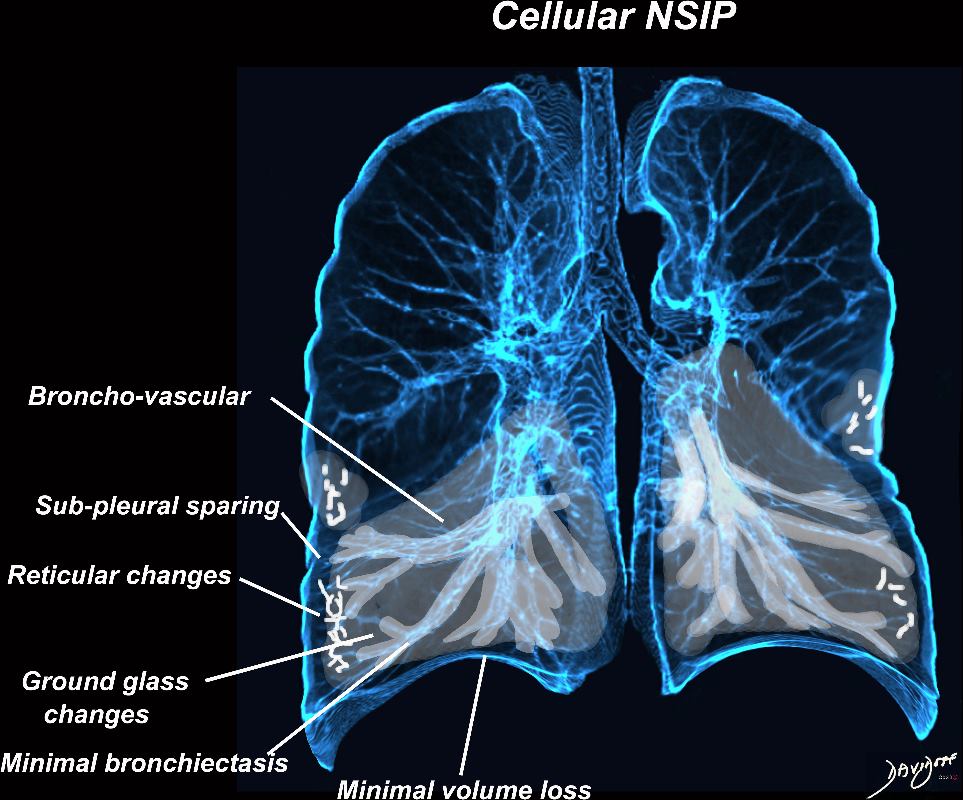
The cellular form of NSIP has a predominance o ground-glass opacities with minimal fibrosis, and is more reversible with treatment, The subpleural sparing is more prominent and the reticulation traction bronchiectasis and volume loss of the lungs is less prominent . Broncho vascular distribution remains characteristic as is the dominance in the lower lung fields with lesser involvement of the middle lobe and lingula.
Ashley Davidoff MD TheCommonvein.net 32679ad
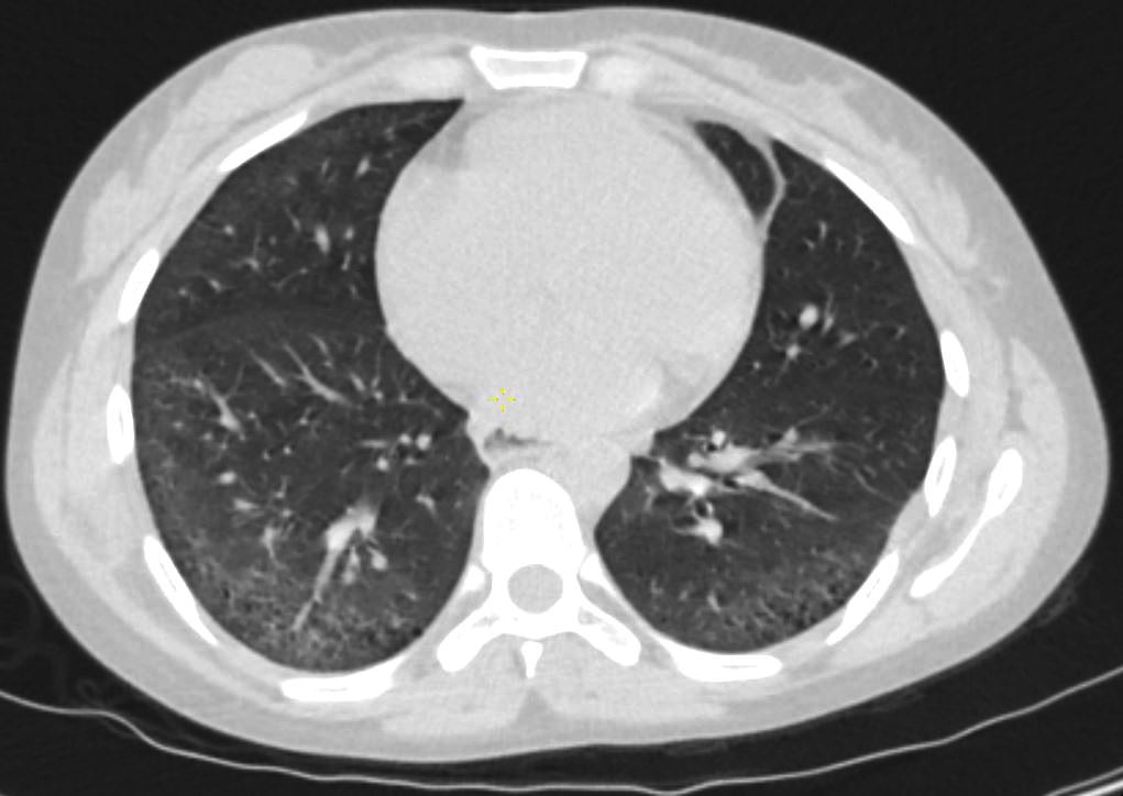
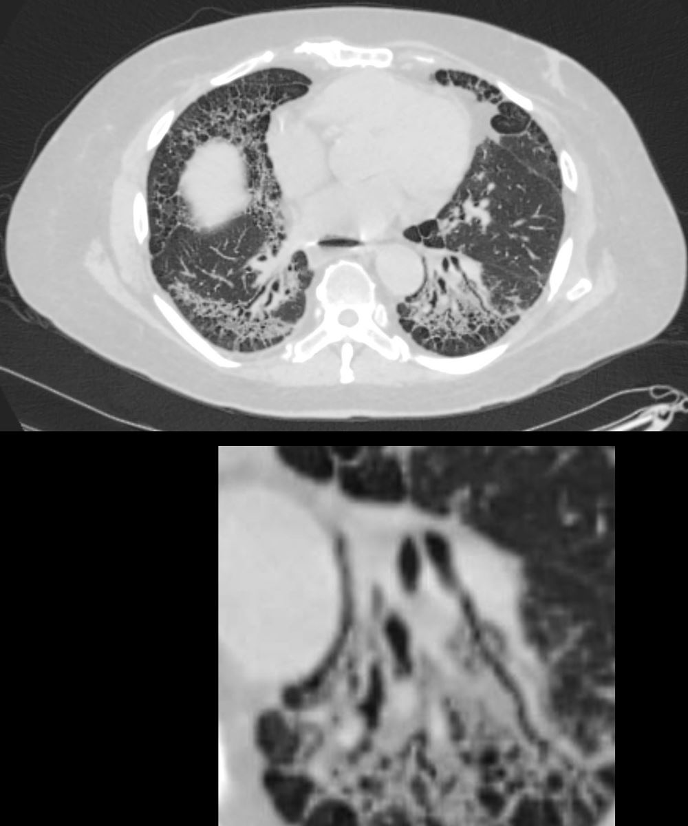
26-year-old female with scleroderma with dyspnea presents for evaluation. Axial CT through the lungs through the lower lungs shows peripherally located, ground glass changes, mild reticulation bronchiolectasis and subpleural sparing. The fissures are normally placed with no obvious loss of volume of the lower lobes. There is no honeycombing
Ashley Davidoff MD TheCommonVein.net 272Lu 136247
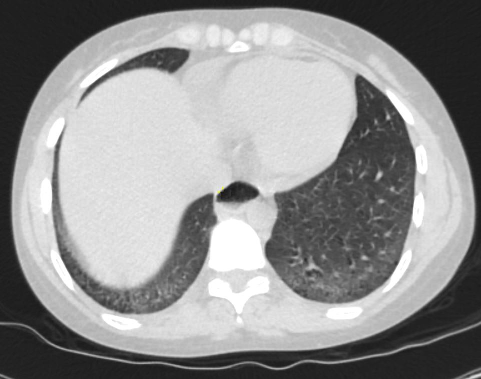
26-year-old female with scleroderma with dyspnea presents for evaluation. Axial CT through the lungs bases shows peripherally located, ground glass changes, bronchiolectasis and subpleural sparing. There is no honeycombing
Ashley Davidoff MD TheCommonVein.net 272Lu 136248

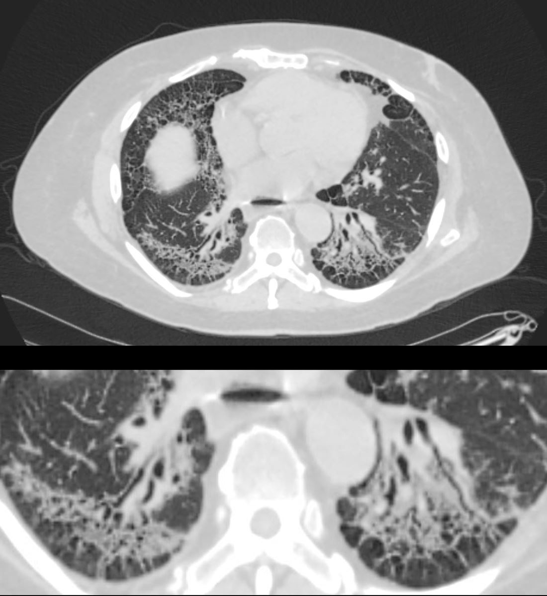
CT shows diffuse ground glass changes lower lobes and to lesser degree in the upper lobes, with minimal reticular change and mild bronchiectasis
Ashley Davidoff MD TheCommonVein.net scleroderma-019
28F-scleroderma-017-2020.jpg
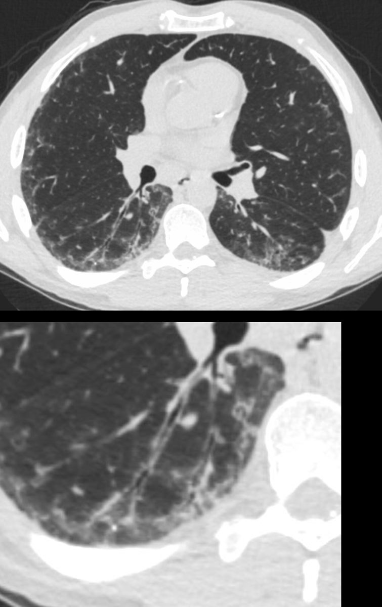
Scleroderma and NSIP (Probable Cellular Form)CT shows diffuse ground glass changes lower lobes with peripheral sparing, minimal reticular change and mild bronchiectasis
Ashley Davidoff MD TheCommonVein.net scleroderma-017
Lungs NSIP Fibrotic Form
Broncho vascular distribution associated with increased reticular changes, more prominent traction bronchiectasis, decreased lung volumes , and decreased lung volumes, dominantly in the lower lobes but to some extent in the middle lobe and upper lobes. Pulmonary hypertension becomes more common.
Ashley Davidoff MD TheCommonvein.net lungs-0771d
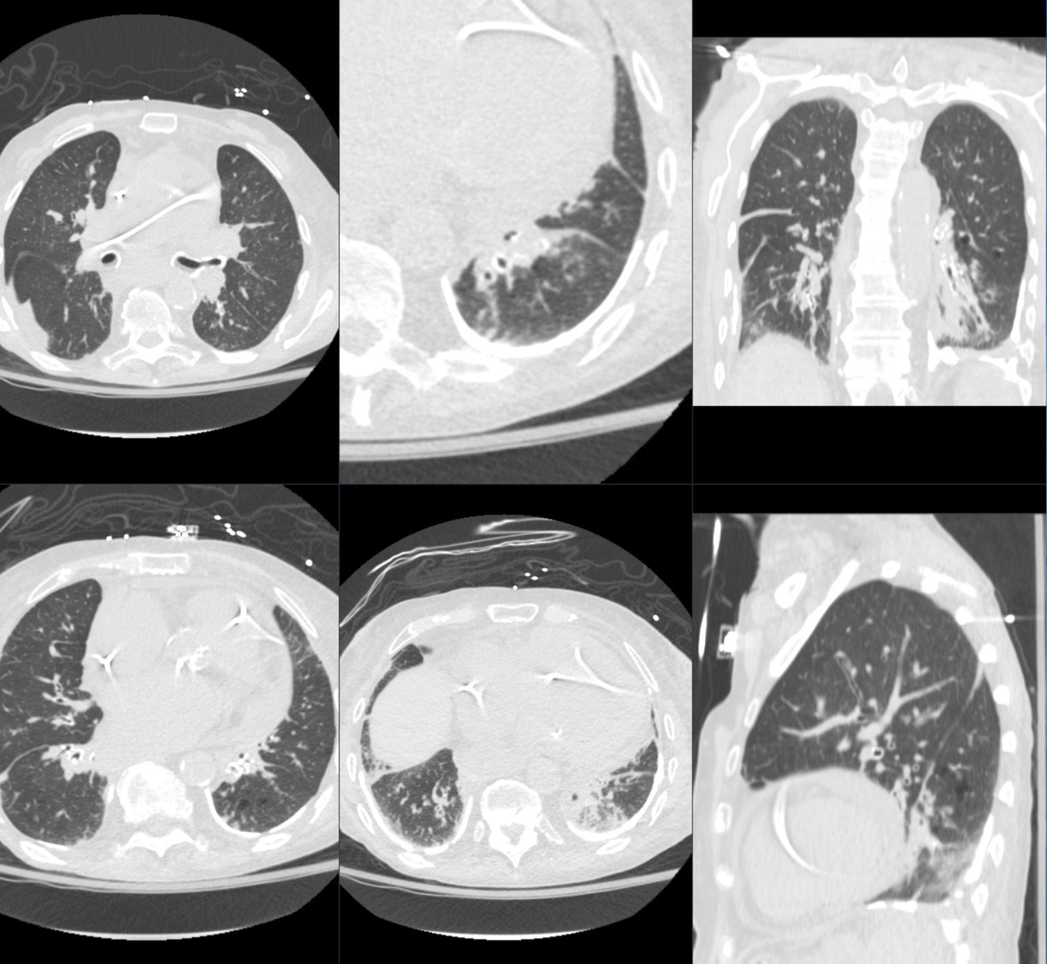
59-year-old male presents with history of scleroderma, Raynaud’s disease, and ILD
Upper Image
Axial CT shows bibasilar peripheral reticular changes, ground glass, bronchiectasis, and bronchiolectasis with volume and with crowding of the bronchovascular bundles posteriorly. There is subpleural sparing posteriorly. Note air-fluid level in the distended esophagus.
Lower Image
The lower image focuses on the traction bronchiectasis caused by the fibrotic process
Ashley Davidoff MD TheCommonVein.net 110Lu 136598c
59-year-old male presents with history of scleroderma, Raynaud’s disease, and ILD
Upper Image
Axial CT shows bibasilar ground glass, bronchiectasis, and bronchiolectasis with volume loss and with crowding of the bronchovascular bundles posteriorly. There is subpleural sparing. Note air-fluid level in the distended esophagus.
The lower image focuses on the peripheral sparing. The spared secondary lobules have also undergone enlargement secondary to the fibrotic process
Ashley Davidoff MD TheCommonVein.net 110Lu 136598c01
Obliterative Bronchiolitis

39-year-old-male with a history of scleroderma associated with ILD and digital vasculopathy with ulcers.
Coronal CT at the level of the spine shows extensive ground glass in the lower lung fields, with subpleural sparing better visualized on the left. Ill defined ground glass centrilobular nodules and mosaic attenuation suggest small airway disease.
In this clinical setting obliterative bronchiolitis (aka bronchiolitis obliterans aka constrictive bronchiolitis) and cellular NSIP are radiological considerations.
Ashley Davidoff MD TheCommonVein.net 132Lu 136667
39-year-old-male with a history of scleroderma associated with ILD and digital vasculopathy with ulcers.
Axial CT shows thickening of the segmental, subsegmental and small airways supplying the posterior basal segment of the right lower lobe. In addition there is a background poorly defined ground glass changes and mild reticulation.
In this clinical setting obliterative bronchiolitis (aka bronchiolitis obliterans aka constrictive bronchiolitis) is suggested. Cellular NSIP is also a radiological consideration.
Ashley Davidoff MD TheCommonVein.net 132Lu 136669c

39-year-old-male with a history of scleroderma associated with ILD and digital vasculopathy with ulcers.
Axial CT at the lung bases shows bibasilar peripheral ground glass changes, with subpleural sparing. Ill defined ground glass centrilobular nodules suggest small airway disease.
In this clinical setting obliterative bronchiolitis (aka bronchiolitis obliterans aka constrictive bronchiolitis) and cellular NSIP are radiological considerations.
Ashley Davidoff MD TheCommonVein.net 132Lu 136676

39-year-old-male with a history of scleroderma associated with ILD and digital vasculopathy with ulcers.
Axial CT through the left posterior recess shows peripheral ground glass changes, with subpleural sparing and bronchiolectasis without wall thickening. Ill defined ground glass centrilobular nodules suggest small airway disease.
In this clinical setting obliterative bronchiolitis (aka bronchiolitis obliterans aka constrictive bronchiolitis) and cellular NSIP are radiological considerations.
Ashley Davidoff MD TheCommonVein.net 132Lu 136678
Esophageal Distension
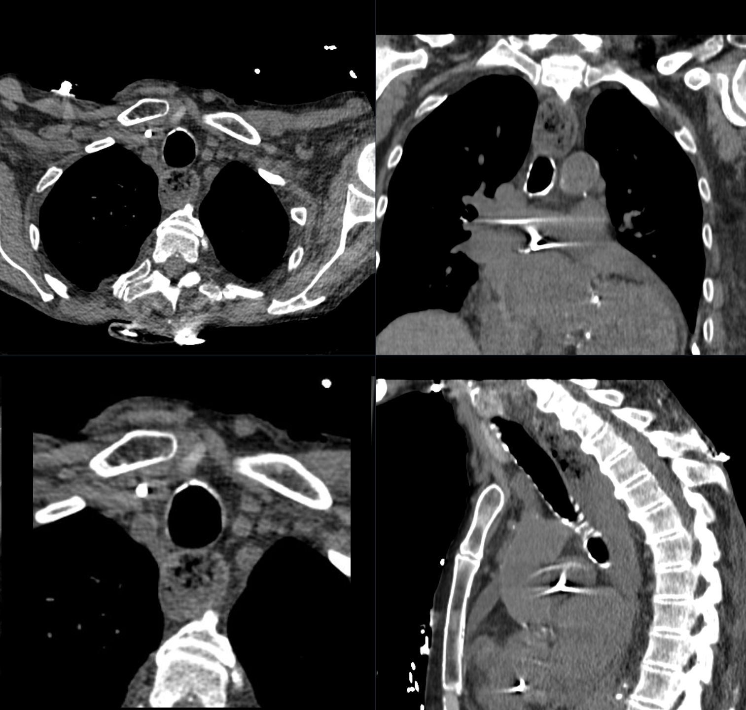
CT scan in axial coronal and sagittal planes in an 83 year old woman with scleroderma shows a food filled dilated esophagus
Ashley Davidoff MD TheCommonVein.net b12096-02 305Lu
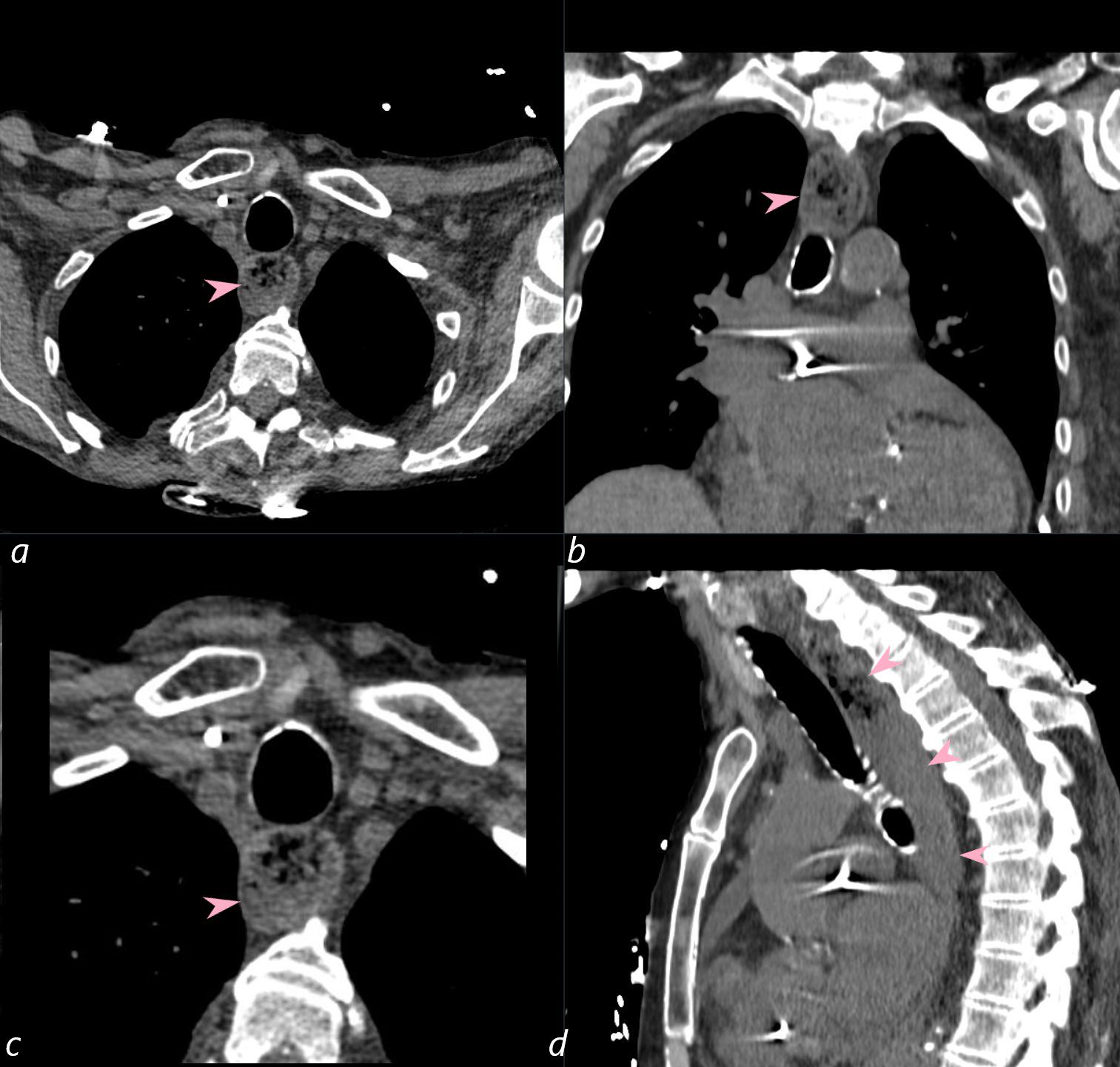
CT scan in axial, coronal, and sagittal planes in an 83 year old woman with scleroderma shows a food filled dilated esophagus (pink arrowheads a,b,c,d)
Ashley Davidoff MD TheCommonVein.net b12096-02L 305Lu
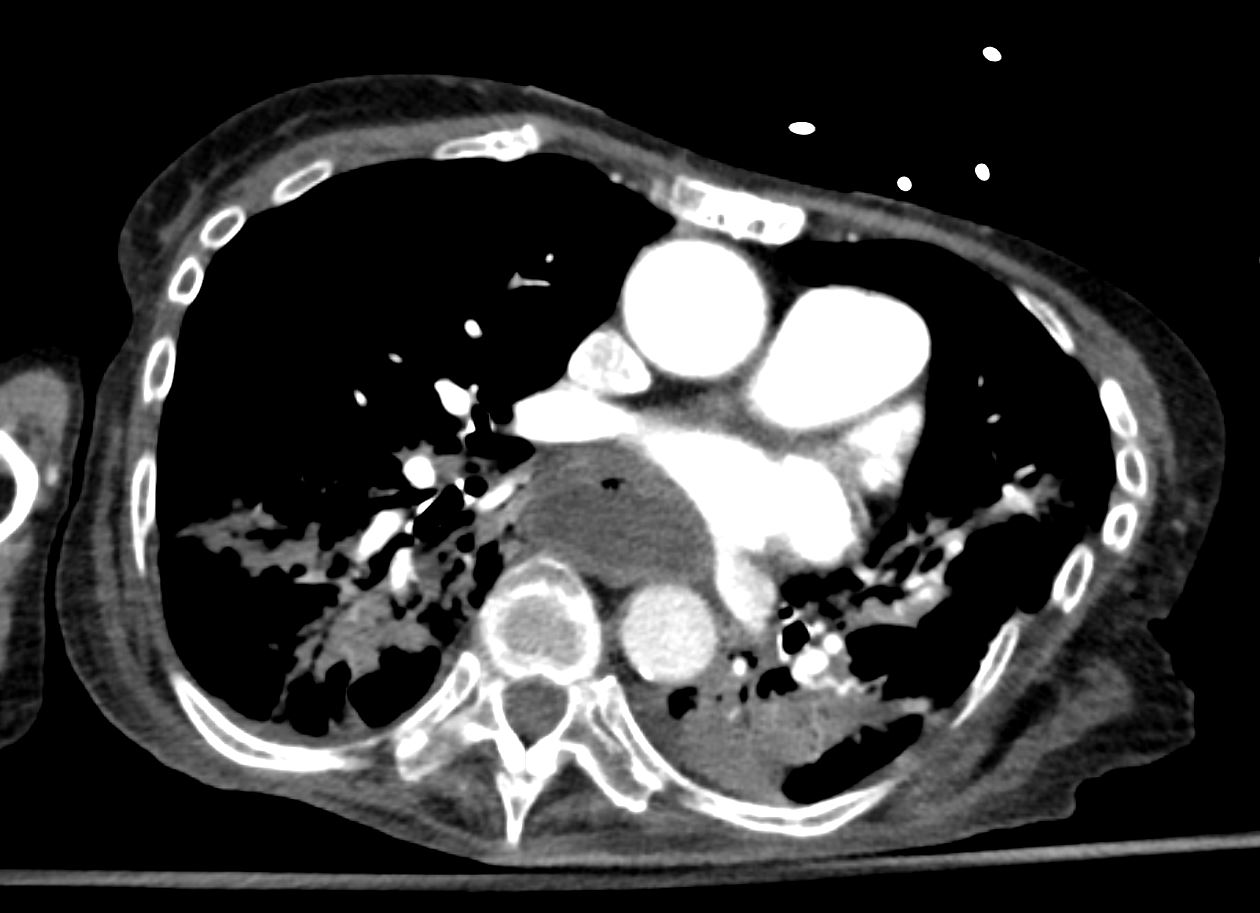
Aspiration
82 year old female with a history of scleroderma presents with dyspnea
CT shows air-fluid levels in the mainstem bronchi, inspissated segmental bronchi of the lower lobes and post obstructive atelectasis secondary to aspiration. Note pleural plaques secondary to asbestos exposure
Ashley Davidoff MD TheCommonVein.net b12096-01 305Lu
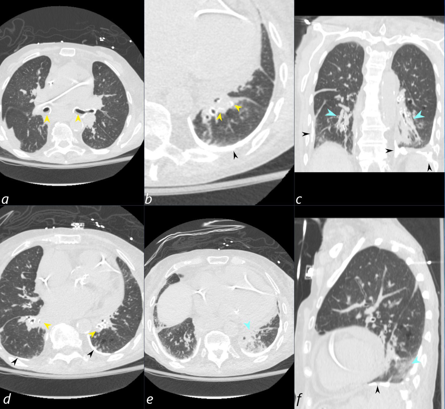
82 year old female with a history of scleroderma presents with dyspnea
CT shows air-fluid levels in the mainstem bronchi, (yellow arrowheads a) inspissated segmental bronchi of the lower lobes (yellow arrowheads b,d) and post obstructive atelectasis secondary to aspiration with solid opacities in the superior segments (teal blue arrowheads c and e) and manifesting as a GGO in f (teal blue arrowhead). Note pleural plaques secondary to asbestos exposure (black arrowheads b,c,d and f)
Ashley Davidoff MD TheCommonVein.net b12096-01L 305Lu
Aspiration Progressing to White Out

74 year old female with scleroderma presents with acute respiratory distress
CT scan shows bilateral basilar consolidation,fluid filled and significantly dilated esophagus, with fluid seen in the right lower lobe bronchus? consistent with aspiration pneumonia
Ashley Davidoff MD TheCommonVein.net
74F scleroderma reflux aspiration pneumonia 002 esoph
74 year old female with scleroderma presents with acute respiratory distress
CT scan shows bilateral basilar consolidation, fluid filled and significantly dilated esophagus, with fluid seen in the right lower lobe bronchus? consistent with aspiration pneumonia
Ashley Davidoff MD TheCommonVein.net
74F scleroderma reflux aspiration pneumonia 003 esophagus
1 Day Later White Out
74 year old female with scleroderma presents with hyperacute respiratory distress
A CXR shows an acute white out of the left hemithorax and a right lower lobe infiltrate. There is mediastinal shift to the left suggesting left sided volume loss consistent with obstructive atelectasis and likely die t ongoing aspiration
Ashley Davidoff MD TheCommonVein.net
74F scleroderma reflux aspiration pneumonia 001 white out
Carcinoma
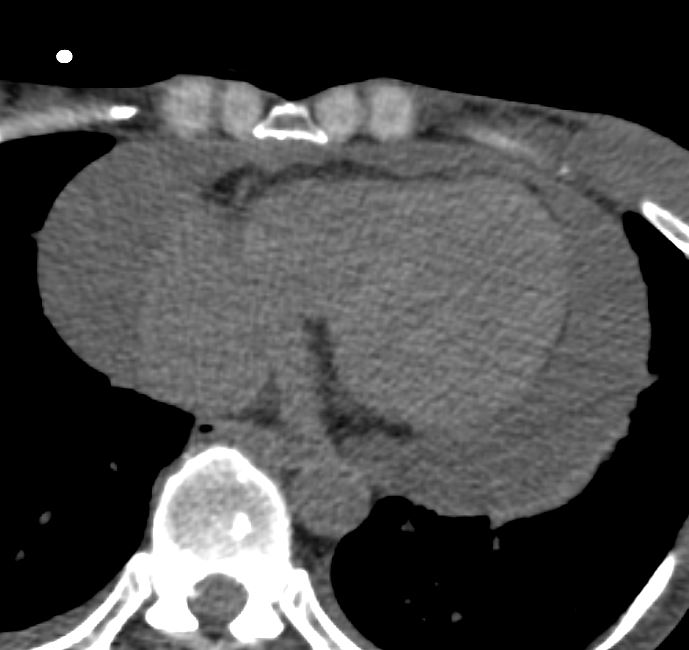
Pericardial Effusion
Ashley Davidoff MD
The CommonVein.net
Pulmonary Hypertension Calcifications and Ossifications
References and Links
- Scleroderma
-
-
- 000 Scleroderma
- Faces of Scleroderma
- Cases
- 001Lu Scleroderma NSIP and Calcinosis of the Spine
- 050Lu 28f Scleroderma NSIP Acro osteolysis
- 110Lu NSIP and Scleroderma
- 118Lu Scleroderma and Aspiration Pneumonia
- 126Lu Scleroderma ILD Acroosteolysis Pericardial Effusion
- 142Lu scleroderma no lung disease ST calcification
- 153Lu Myopathy Arthritis ST Calcifications and Possible Scleroderma Overlap Fibrotic NSIP
- 195Lu NSIP probably Cellular Scleroderma
- 196Lu Scleroderma NSIP 5 years Improved
- 198Lu Scleroderma Early NSIP Mild Over 3 Years
- 272Lu Scleroderma Cellular NSIP Acro-osteolysis
- 305Lu Scleroderma Asbestos Related Calcified Pleural Plaques Aspiration Lymphocytic Interstitial Pneumonia LIP
-
-